
ANTERIOR and POSTERIOR TRIANGLES of NECK Gross HSB
ANTERIOR and POSTERIOR TRIANGLES of NECK Gross HSB Reviewer Courtesy of Katherine Joy N. Lumucso 1 -A 2 FEU-NRMF SY 2005 -2006
Anterior Triangle of Neck
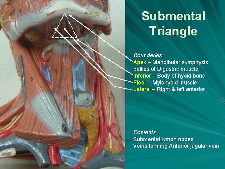
Submental Triangle Boundaries: Apex – Mandibular symphysis bellies of Digastric muscle Inferior – Body of hyoid bone Floor – Mylohyoid muscle Lateral – Right & left anterior Contents: Submental lymph nodes Veins forming Anterior jugular vein
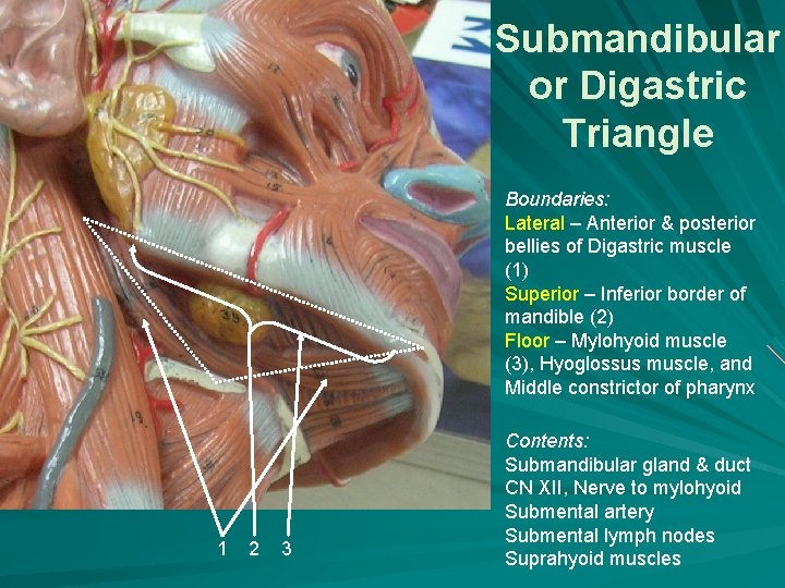
Submandibular or Digastric Triangle Boundaries: Lateral – Anterior & posterior bellies of Digastric muscle (1) Superior – Inferior border of mandible (2) Floor – Mylohyoid muscle (3), Hyoglossus muscle, and Middle constrictor of pharynx 1 2 3 Contents: Submandibular gland & duct CN XII, Nerve to mylohyoid Submental artery Submental lymph nodes Suprahyoid muscles
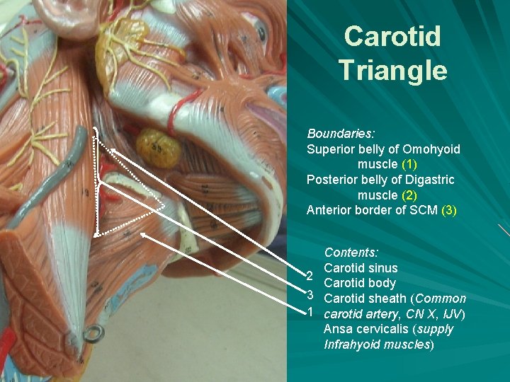
Carotid Triangle Boundaries: Superior belly of Omohyoid muscle (1) Posterior belly of Digastric muscle (2) Anterior border of SCM (3) Contents: Carotid sinus 2 Carotid body 3 Carotid sheath (Common 1 carotid artery, CN X, IJV) Ansa cervicalis (supply Infrahyoid muscles)
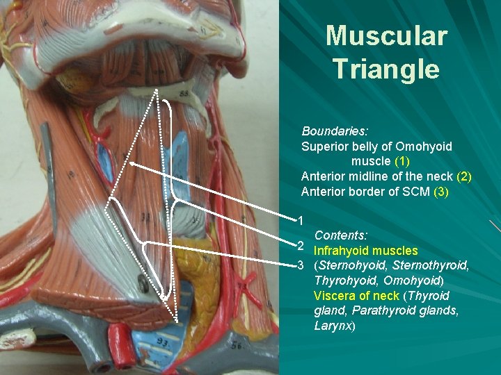
Muscular Triangle Boundaries: Superior belly of Omohyoid muscle (1) Anterior midline of the neck (2) Anterior border of SCM (3) 1 Contents: 2 Infrahyoid muscles 3 (Sternohyoid, Sternothyroid, Thyrohyoid, Omohyoid) Viscera of neck (Thyroid gland, Parathyroid glands, Larynx)
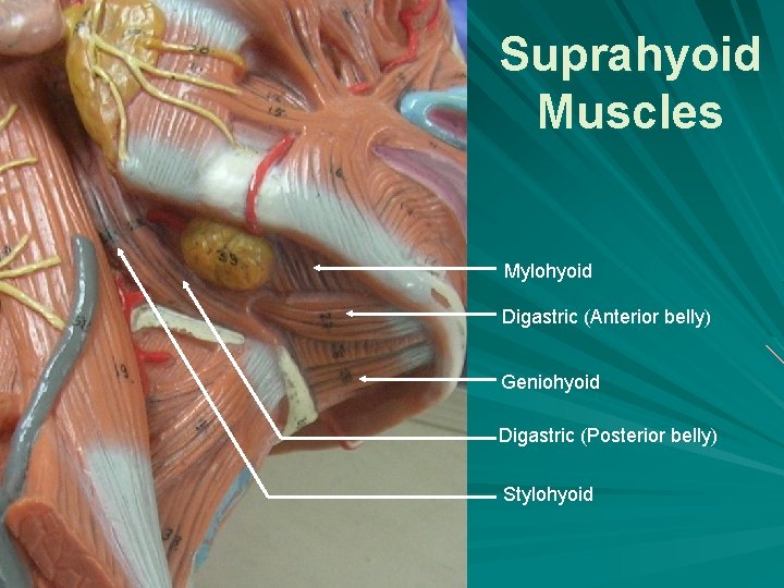
Suprahyoid Muscles Mylohyoid Digastric (Anterior belly) Geniohyoid Digastric (Posterior belly) Stylohyoid
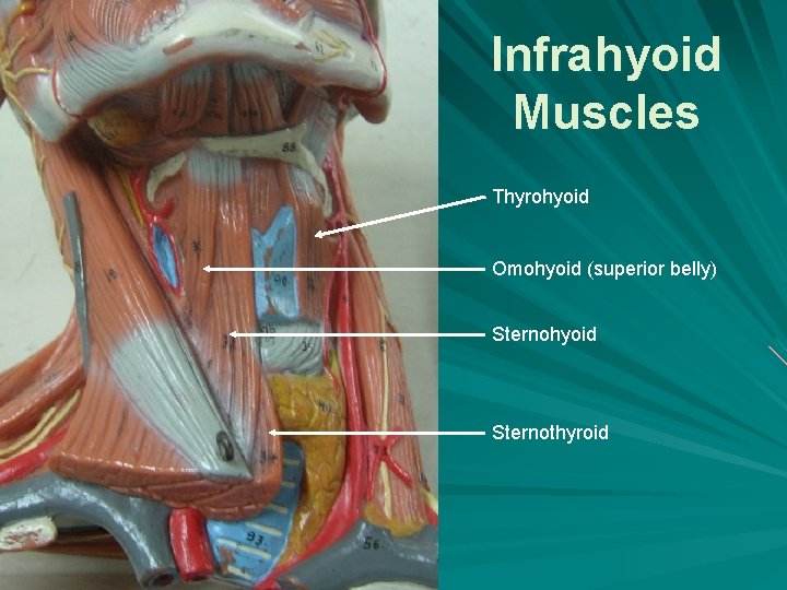
Infrahyoid Muscles Thyrohyoid Omohyoid (superior belly) Sternohyoid Sternothyroid
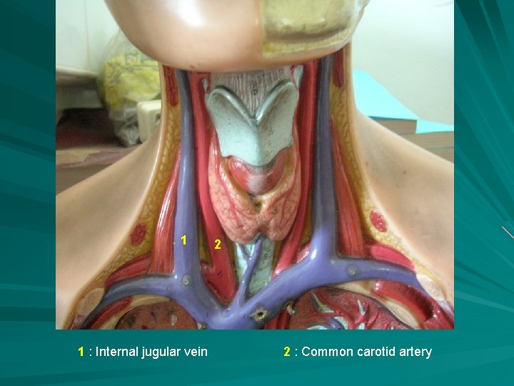
1 1 : Internal jugular vein 2 2 : Common carotid artery
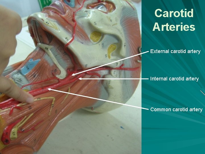
Carotid Arteries External carotid artery Internal carotid artery Common carotid artery
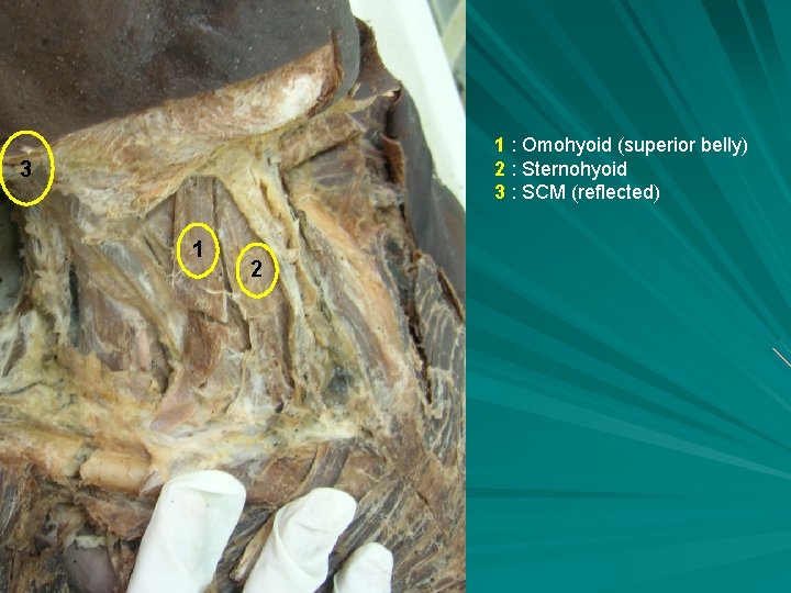
1 : Omohyoid (superior belly) 2 : Sternohyoid 3 : SCM (reflected) 3 1 2
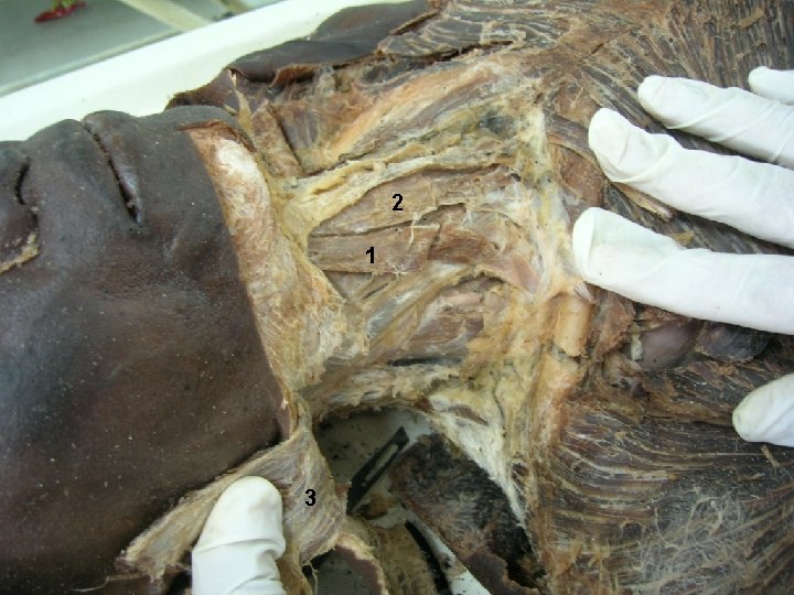
2 1 3
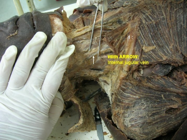
1: With ARROW: Internal jugular vein
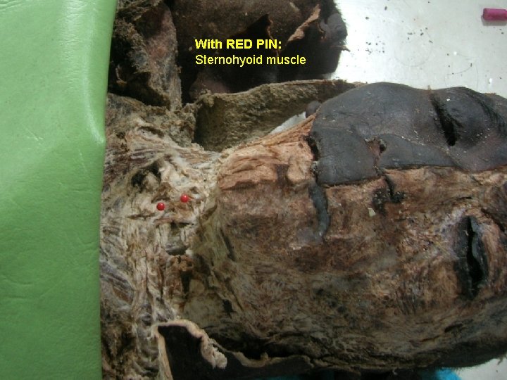
With RED PIN: Sternohyoid muscle
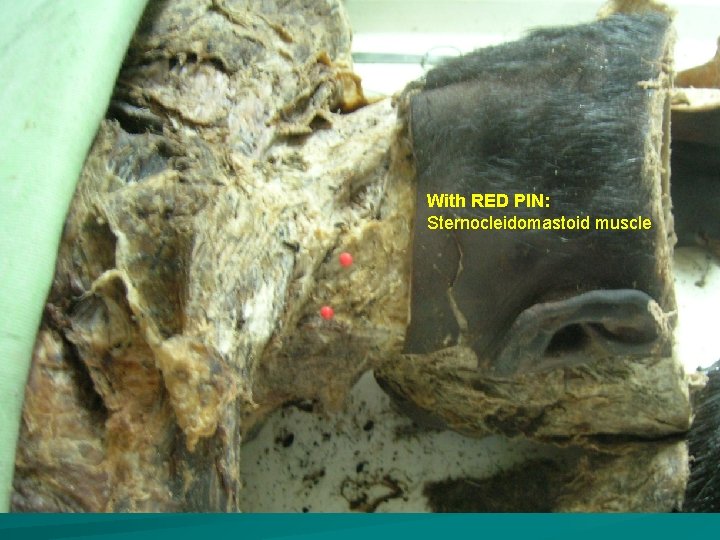
With RED PIN: Sternocleidomastoid muscle
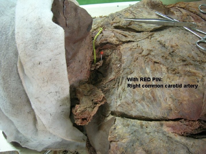
With RED PIN: Right common carotid artery
Posterior Triangle of Neck
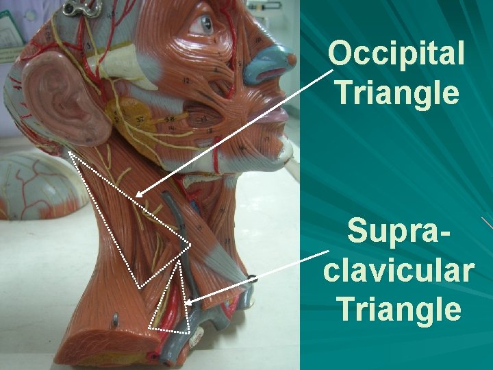
Occipital Triangle Supraclavicular Triangle
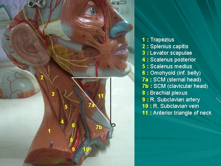
2 3 11 7 a 5 4 1 6 7 b 8 9 10 1 : Trapezius 2 : Splenius capitis 3 : Levator scapulae 4 : Scalenus posterior 5 : Scalenus medius 6 : Omohyoid (inf. belly) 7 a : SCM (sternal head) 7 b : SCM (clavicular head) 8 : Brachial plexus 9 : R. Subclavian artery 10 : R. Subclavian vein 11 : Anterior triangle of neck
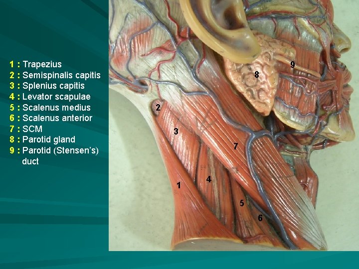
1 : Trapezius 2 : Semispinalis capitis 3 : Splenius capitis 4 : Levator scapulae 5 : Scalenus medius 6 : Scalenus anterior 7 : SCM 8 : Parotid gland 9 : Parotid (Stensen’s) duct 8 2 3 7 1 4 5 6 9
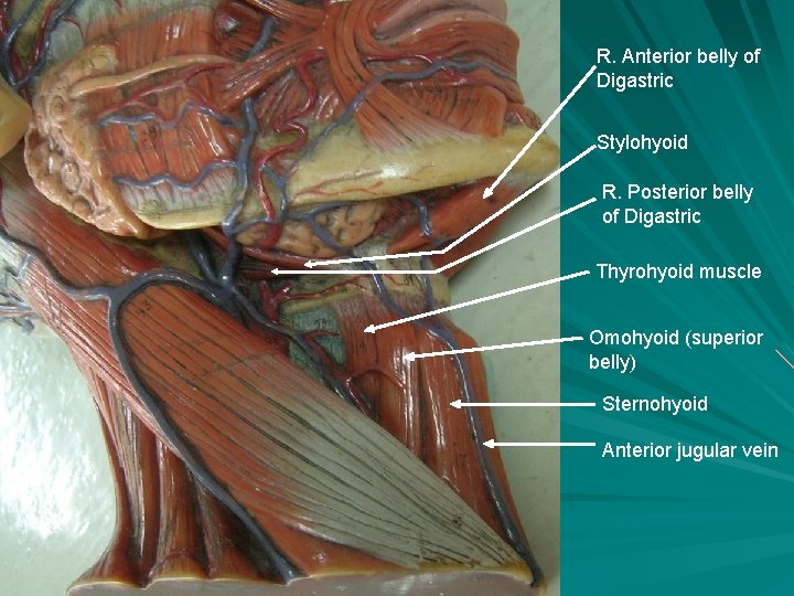
R. Anterior belly of Digastric Stylohyoid R. Posterior belly of Digastric Thyrohyoid muscle Omohyoid (superior belly) Sternohyoid Anterior jugular vein
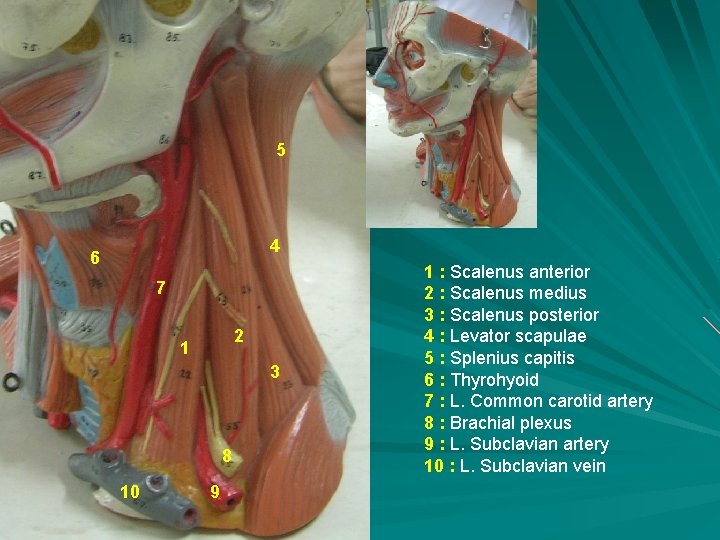
5 4 6 7 2 1 3 8 10 9 1 : Scalenus anterior 2 : Scalenus medius 3 : Scalenus posterior 4 : Levator scapulae 5 : Splenius capitis 6 : Thyrohyoid 7 : L. Common carotid artery 8 : Brachial plexus 9 : L. Subclavian artery 10 : L. Subclavian vein
- Slides: 22