ANTERIOR ABDOMINAL WALL Dr Manjula Vastrad Asst Professor

ANTERIOR ABDOMINAL WALL Dr. Manjula Vastrad Asst Professor Dept of Rachana Shareera SMVVS RKM AMC VIJAYAPURA

INTRODUCTION Anterior abdominal wall usually includes both the front as well as the side walls of the abdomen and needs to be called anterolateral abdominal wall Abdomen is the lower part of the trunk lies below the diaphragm It is bounded by large extent of muscles Anterolateral abdominal wall Anterior wall Right lateral wall (Right Flank) Left lateral wall (Left Flank)

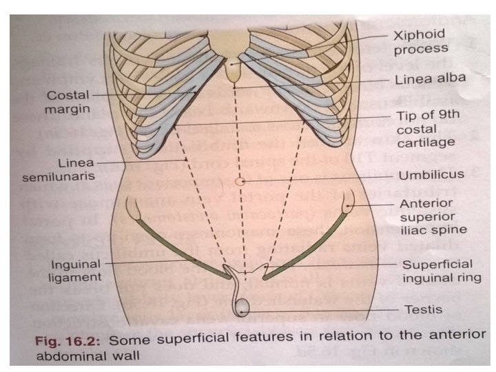
This extended from the thoracic cage to the pelvis and bounded : Superiorly 7 th to 10 th costal cartilages and xiphoid process Inferiorly Inguinal ligaments and the pelvic bones.

BONY LANDMARKS AROUND ABDOMEN Iliac crest Ant Sup iliac spine Pubic crest Inguinal ligament Costal margin Xiphoid process

CONTENTS SKIN SUPERFICIAL FASCIA 1. 2. FATTY LAYER MEMBRANOUS LAYER DEEP FASCIA MUSCULAR LAYER TRANSVERSALIS FASCIA PARIETAL PERITONEUM

THE SKIN Capable of undergoing enormous stretching eg: - pregnancy, obesity, ascitis …. . etc Lineae albicantes – whitish streakes in the skin

THE UMBLICUS A little below the middle of the median furrow is an irragular depressed or elevated area called umblicus It is the site at which the umbilical cord is attached in fetal life. Position: - Disc between the 3 rd & 4 th lumbar vertebrae ANATOMICAL IMPORTANCE: - Ø Water shed line Ø Caput medusae

Embryological importance Meeting point of four folds of embryonic plate Meeting point of 3 systems

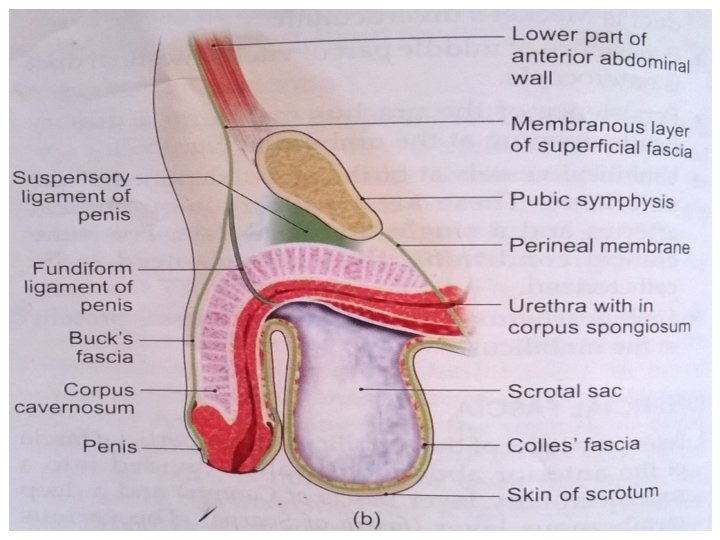
SUPERFICIAL FASCIA CAMPER’S FASCIA: Superficial fatty layer Continuous with the fascia of thorax and thigh Penis devoid of fat In scrotum it is replaced by dartos muscle SCARPA’S FASCIA: Deep membranous layer Continuous as colles’ fascia

ANATOMICAL IMPORTANCE The attachment of Scarpa’s fascia of the abdomen and Colles’ Fascia of the perineum are such that they prevent the passage of extravasated urine backwards into the ischiorectal fossa and downwards into the thigh.

In the median plane, the membranous layer is thickened to form the suspensory ligament and fundiform ligament of penis or clitoris. a) b) c) d) The fascia contains An extremely variable quantity of fat, which tends to accumulate in the lower part of the abdomen after puberty. Cutaneous nerves cutaneous vessels Superficial lymphatics

CUTANEOUS NERVE SUPPLY Is derived from the anterior rami of the lower six thoracic and first lumbar nerves Thoracic nerves are the lower five intercostal and the subcostal nerves First lumbar nerve is represented by the iliohypogastric and ilioinguinal

CUTANEOUS ARTERY SUPPLY Skin near the midline is supplied by branches of the superior epigastric artery (br. of int. thoracic artery) and the inferior epigastric artery ( br. of external iliac artery) Skin of the flanks is supplied by branches from the intercostal, lumbar, and deep circumflex arteries

CUTANEOUS VEINS Venous blood is collected into a network of veins that radiate from the umbilicus The network is drained above into the axillary vein via the lateral thoracic vein Below into the femoral vein via the superficial epigastric and the great saphenous veins Few small veins, the paraumbilical veins form a clinically important portal-system venous anastomosis

SUPERFICIAL LYMPHATICS Lymph drainage of the skin of the anterior abdominal wall above the umbilicus is upward to the anterior axillary (pectoral group of nodes) Below the level of umbilicus drains downward and laterally to the superficial inguinal nodes Swelling in the groin is may be due to enlarged superficial inguinal node

DEEP FASCIA Deep fascia in the anterior abdominal wall is merely a thin layer of connective tissue covering the muscles It lies immediately deep to the membranous layer of the superficial fascia

ANT ABD WALL FUNCTIONS Supports the abdominal viscera Expulsive act –During micturation, parturition, defaecation, vomiting etc Forcefull expiratory act – coughing, sneezing, shouting etc Move the trunk and help to maintain posture. Helps to maintain or increase the intra abdominal pressure.

APPLIED ANATOMY Abdominal Regions Abdomen is divided into 9 regions via four planes: – Two horizontal [sub-costal (10 th) and trans tubercules plane] (L 5). – Two vertical (midclavicular planes). They help in localization of abdominal signs and symptoms.

LOCATION OF THE ORGANS Right and left hypochondriac: Contain liver Epigastric: Contains: liver, stomach, pancreas Right and left lateral (lumbar): Right contains ascending colon. Left contains descending colon. Umbilical: Contains small intestine and transverse colon. Right and left inguinal: Right contains ileocecal junction and appendix. Left contains sigmoid colon. Hypogastric: Contains small intestine, urinary bladder (full), pregnant uterus.
 – Fat, – Faeces,")
PROTUBERANCE OF THE ABDOMEN The five common causes (5 F) – Fat, – Faeces, – Fetus, – Flatus – Fluid

ASCITIS FINDINGS IN ASCITIS: - Smiling Umbilicus Shiny skin Engorgement of superficial veins

LIPOSUCTION Surgical method of removing unwanted subcutaneous fat using a subcutaneously placed suction tube and high vaccum pressure

THANK YOU
- Slides: 25