Anterior abdominal wall and the inguinal region Dr

Anterior abdominal wall and the inguinal region Dr. Selda Önderoğlu
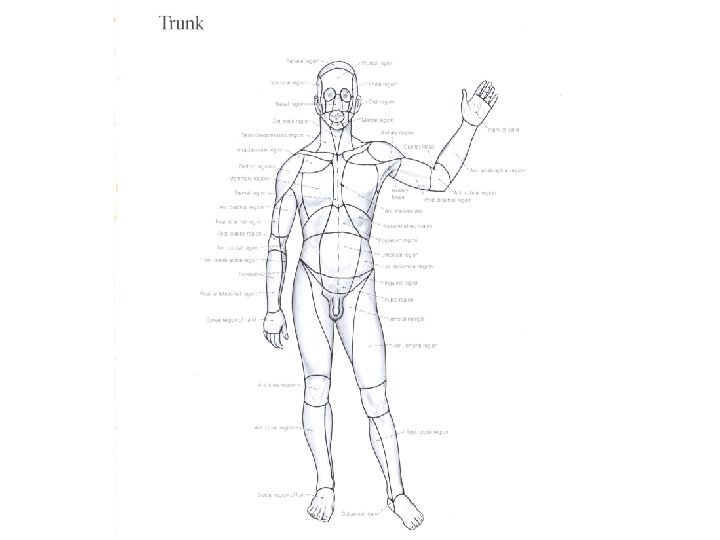

ABDOMEN • The region between: Diaphragm and pelvis. • Boundaries: Roof: Diaphragm Posterior: Lumbar vertebrae+ Mm. Of the posterior abd. wall Infrerior: No boundary, continuous with the pelvic cavity, Superior Pelvic aperture Anterior and lateral: Anterior Abdominal Wall , Muscles
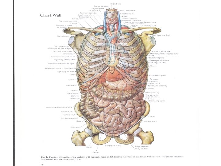

Bony structures of the abdomen

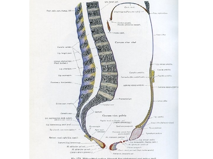
Posterior abdominal wall
 TRANSVERSE PLANES • Transpyloric plane : tip of 9")
Topography of the Abdomen (PLANES) TRANSVERSE PLANES • Transpyloric plane : tip of 9 th costal cartilages ; pylorus of stomach , L 1 vertebra level. • Subcostal plane: tip of 10 th costal cartilages , L 3 vertebra. • Transtubercular plane: tubercles if iliac crests ; L 5 vertebra level. • Interspinous plane: anterior superior iliac spines ; promontory of sacrum VERTICAL PLANES • Mid-clavicular plane: midpoint of clavicle- mid-point of inguinal ligament. • Semilunar line: lateral border of rectus abdominis muscle.

Planes of abdomen
 epigastric; hypogastric L")
Regions of the Abdomen • 9 regions: umbilical (around the umbilicus) epigastric; hypogastric L hypochondriac ; R hypochondriac L inguinal ; R inguinal L lumbar ; R lumbar region.

Regions of abdomen

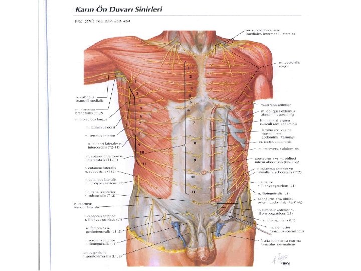
Cutaneous nn. Of the anterior abdominal wall Skin innervation: lower 5 intercostal nerves+ subcostal nerve+ L 1 spinal nerve (ilioinguinal+iliohypogastric nn. ). Umbilical region skin inn. : T 10.
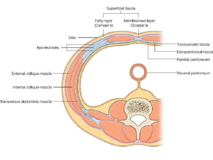
 superficial fatty layer(CAMPER’S")
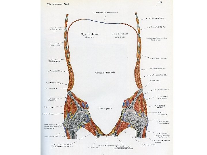
Anterior Abdominal Wall • Skin • Superficial fascia ( two layers) superficial fatty layer(CAMPER’S fascia) deep membranous layer(SCARPA’S fascia) • No deep fascia • External oblique muscle • Internal oblique muscle • Transversus abdominis muscle • Transversalis fascia • Lateral to midline(linea alba)- rectus abdominis muscle. • Extraperitoneal tissue layer- peritoneum.

 2 -deep membranous layer")
Superficial fascia two layers 1 -superficial fatty layer (CAMPER’S fascia) 2 -deep membranous layer (SCARPA’S fascia) –
 Internal oblique")
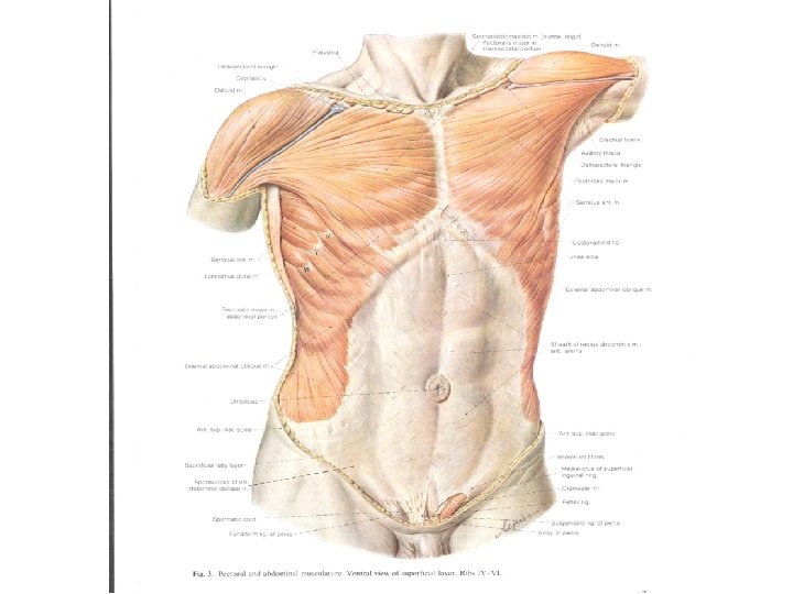
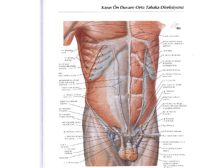
Muscles of the ant. abd. wall External oblique muscle (most superf. m) Internal oblique muscle Transversus abdominis muscle Those 3 mm are laterally located On both sides of the midline(linea alba) Rectus abdominis muscle inferiorly: Pyramidalis muscle
 • • Most superficial muscle. O: 5")
External Oblique Muscle (M. Obliquus externus abdominis) • • Most superficial muscle. O: 5 -12 ribs I: Linea alba+ inguinal ligament Parts of inguinal lig. : reflected part +lacunar ligament+ pectineal Lig. • Inn: lower 5 intercostal nn. + subcostal n. + L 1. • Superficial inguinal ring: opening in the aponeurosis of external Oblique Muscle. has: Lateral crus-medial crus- intercrural fibres.

inguinal ligament from anterior sup. iliac spine- to pubic symphsis lacunar ligament pectineal ligament

Superficial inguinal Ring -Lat. crus -Medial crus -intercrural fibers
 • -middle layer muscle -fibers are 90")
Internal Oblique Muscle (Musculus Obliquus internus abdominis) • -middle layer muscle -fibers are 90 degrees to external oblique m. fibers • O: thoracolumbar fascia+iliac crest+inguinal lig. • I: linea alba+conjoint tendon ( common tendon with the transversus abdominis muscle)+ Pubic crest+pecten pubis. • Inn. : lower 5 intercostal nn+subcostal n. +L 1. (same with external oblique).
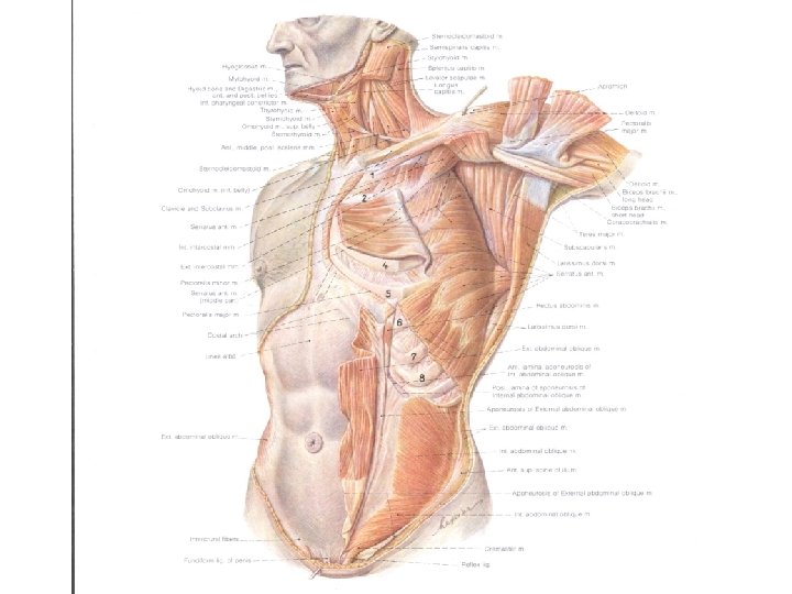
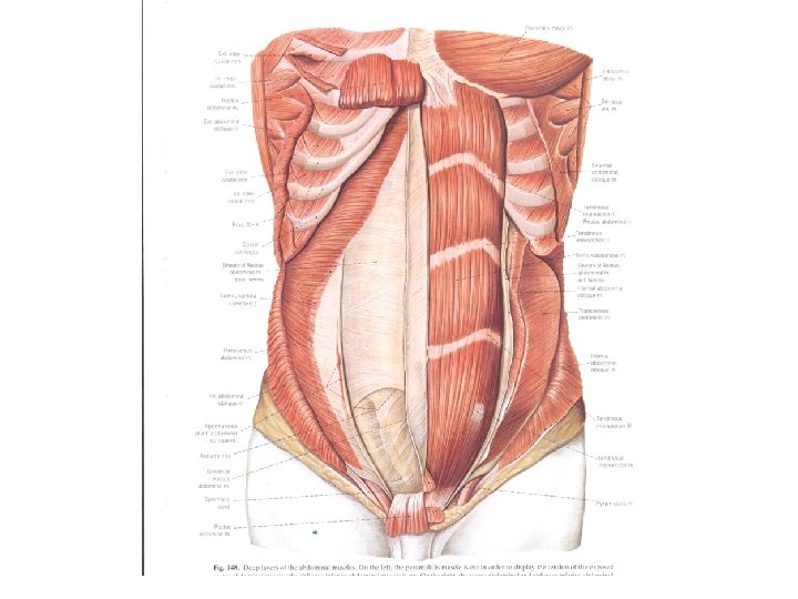

TRANSVERSUS ABDOMINIS MUSCLE • located Innermost • O: inf. 6 Costal cartilages+Thoracolumbar fascia+inguinal ligament • I: conjoint tendon+linea alba. • Innerv. : lower 5 intercostal nn. + subcostal n. + L 1. (same with external oblique)

• TRANSVERSALIS FASCIA • Located post to transv. Abd. m. deep inguinal ring. • CONJOINT TENDON ( FALX INGUINALIS): common tendon of internal oblique+ transversus abdominis mm.

Functions of anterior Abdominal muscles • support+protection+movements of trunk ( external oblique- turns the trunk to the other side); internal oblique( turns the trunk to the same side). • During coughing, sneezing, vomiting, parturition ( during birth of a child) all of these muscles contract( increase intraabdominal pressure. )
 O: Xiphoid")
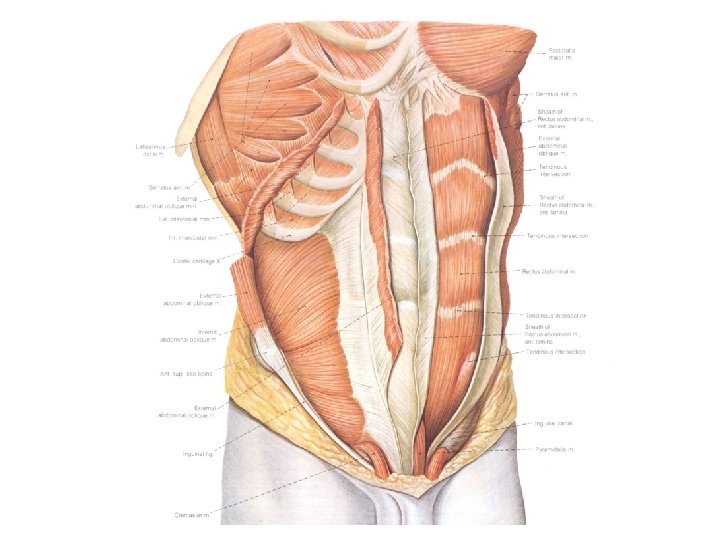
Rectus Abdominis Muscle • • Located on both sides of midline(linea alba) O: Xiphoid process I: symphysis pubis Inn: lower 5 intercostal nn. + subcostal nn!! ( different from the previous 3 Mm. ) • F: flexes the trunk. • Has tendinous intersections: 3 -4 in number • enveloped by a sheath: RECTUS SHEATH. • Lat. Border: semilunar line

Rectus abdominis muscle

Rectus sheath
 : ABOVE")
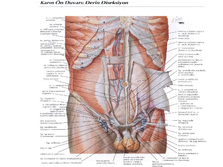
RECTUS SHEATH • 4 -5 cm below UMBILICUS -ARCUATE LINE (SEMICIRCULAR LINE) : ABOVE this line: Anterior layer : external oblique apon. + anterior lamina of internal oblique’s aponeurosis. ) Posterior layer: Posterior lamina of internal oblique apon. + transversus abdominis apon. BELOW this line: Anterior layer : external oblique apon. +internal oblique apon. +transversus abdominis aponeurosis. Posterior layer: Only Transversalis fascia. • Structures within the rectus sheath: rectus abdominis muscle+ superior epigastric artery+ inferior epigastric artery+ lower 5 intercostal nn. + subcostal n.

Rectus sheath

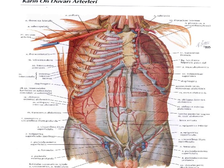
Arteries of anterior Abdominal wall -musculophrenic -Sup epigastric -İnferior epigastric -Deep circumflex iliac -Superficial circ. iliac

INGUINAL CANAL • Surgically an important canal because it is the site of inguinal hernias • obliquely located; tubelike • 3 -4 cm. in length. • Has two openings : • Superficial inguinal ring external oblique apon. -medial • Deep ingunal ring: transversalis fascia - Lateral

inguinal canal • • • superficial inguinal ring Anterior wall Post. Wall Superior wall inferior wall deep inguinal ring 4 -6 cm
+ internal oblique")
INGUINAL CANAL • WALLS: • anterior wall: skin+ superficial fascia+external oblique (medially)+ internal oblique ( laterally). • Posterior wall: Reflected ingunal lig. + conjoint tendon+ transversalis fascia. • Inferior wall: inguinal lig. + lacunar lig. • Superior wall: inferior margins of internal oblique+ transversus abdominis mm.
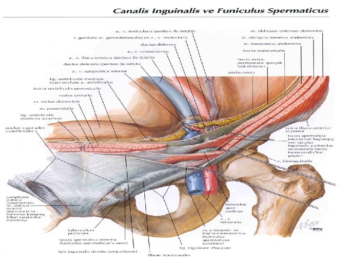

Structures passing through Ingunal Canal • • Spermatic cord in male Round ligament of uterus in female. Ilioinguinal n. . Genital branch of genitofemoral n.

Superficial inguinal ring
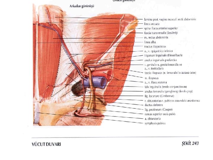

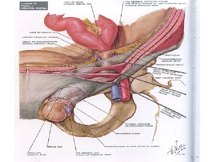
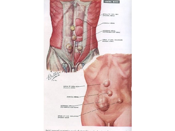
INGUINAL HERNIA • Indirect Inguinal hernia: piece of organ passes through deep ing. ring- courses in inguinal canal – passes through superficial inguinal ring- protrudes outwards. • Direct inguinal hernia: piece of organ pushes directly ant. Abd. wall passes through supeficial inguinal ring – protrudes outwards. • How to differentiate direct and indirect ingunal hernia? reference is the inferior epigastric artery. • If it is lateral to this a. : Indirect inguinal hernia • If it is medial to this a. : Direct inguinal hernia.
")
Femoral canal Femoral hernia (saphenus opening)

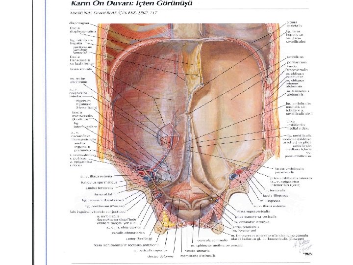
Folds of the ant. Abd. wallperitoneum • When looked from inside the anterior abdominal wall there are some folds of parietal peritoneum : • Median umbilical fold : under which lies the urachus • Medial umbilical fold: under which umbilical artery lies. • Lateral umbilical fold: inferior epigastric vessels lie.

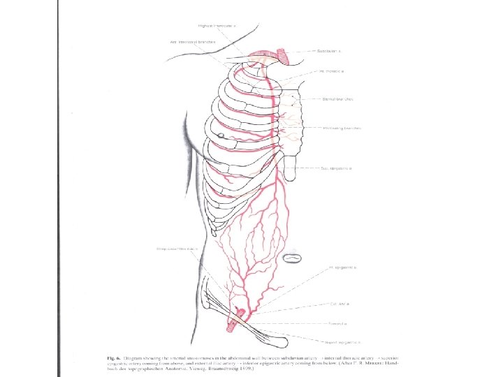
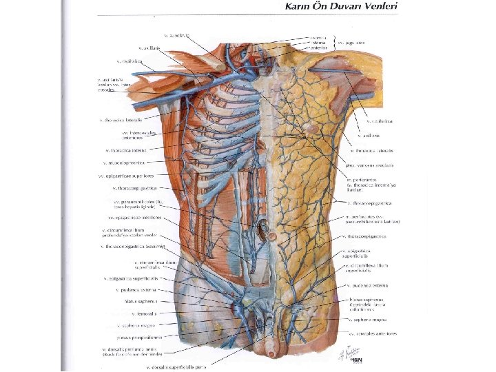
ARTERIES, VEINS and LYMPH OF THE ANT. ABD. WALL • Arteries: Sup. Epigastric. A. + ınferior epigastric a. + superficial circumflex iliac a. + deep circumflex iliac a. + superficial epigastric a. +musculophrenic • Veıns: SAME NAMED. • IMPORTANT ANASTOMOSIS: supeficial epigastric vein- lateral thoracic vein- unite the veins of the superior and inf. Halves of the body. • Lymph: Axillary-above the umbilical region Inguinal- below the umbilical region.

Lymphatics of the abdominal wall

- Slides: 58