Ankle Joint talocruraltalotibialtibiotalartalar mortisetalar Jt RIMT University Department
 RIMT University Department of Physiotherapy Prepared by: Dr.")
Ankle Joint (talocrural/talotibial/tibiotalar/talar mortise/talar Jt. ) RIMT University Department of Physiotherapy Prepared by: Dr. Navkaran Singh

• The term ankle refers specifically to the talocrural joint: that is, the articulation between the distal tibia and fibula proximally and the body of the talus distally. • The ankle is a synovial hinge joint with a joint capsule and associated ligaments.

• It is generally considered to have a single oblique axis with one degree of freedom around which the motions of dorsiflexion/ plantarflexion occur.


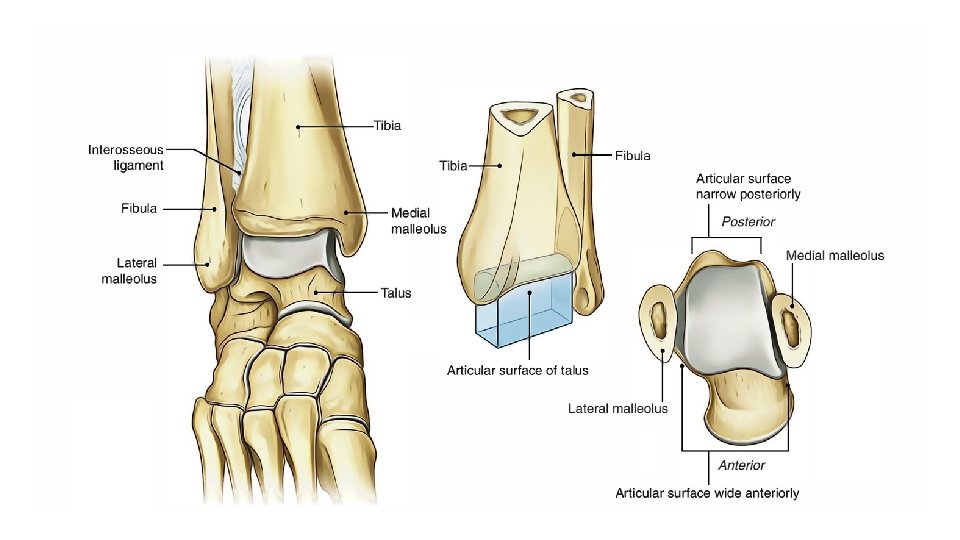
Ankle Joint Structure • The proximal segment of the ankle is composed of : Concave surface of the distal tibia and of the tibial and fibular malleoli. These three facets form an almost continuous concave joint surface that extends more distally on the fibular (lateral) side than on the tibial (medial) side & More distally on the posterior margin of the tibia than on the anterior margin.

The structure of the distal tibia and the malleoli resembles and is referred to as a MORTISE.

• E. g of a mortise is the gripping part of a wrench. • Either the wrench can be fixed (fitting a bolt of only one size) or it can be adjustable (permitting use of the wrench on a variety of bolt sizes). • The adjustable mortise is more complex than a fixed mortise because it combines mobility and stability functions. • The mortise of the ankle is adjustable, relying on the proximal and distal tibiofibular joints to both permit and control the changes in the mortise.

• The proximal and distal tibiofibular joints are anatomically distinct from the ankle joint, but these two linked joints function exclusively to serve the ankle. • Unlike their upper extremity counterparts, the proximal and distal radioulnar joints, the tibiofibular joints do not add any degrees of freedom to the more distal ankle and foot.

However, fusion of the radioulnar joints would have little effect on wrist range of motion (ROM), whereas fusion of the tibiofibular joints may impair normal ankle function by limiting the ability of the talus to move within the ankle mortise.

Proximal Tibiofibular Joint • The proximal tibiofibular joint is a plane synovial joint formed by the articulation of the head of the fibula with the posterolateral aspect of the tibia. • Although the facets of the proximal tibiofibular joint are fairly flat and vary in configuration among individuals, (A slight convexity of the tibial facet and a slight concavity of the fibular facet seem to predominate) • The inclination of the facets may vary from nearly vertical to nearly horizontal in orientation.

• Each proximal tibiofibular joint is surrounded by a joint capsule that is reinforced by anterior and posterior tibiofibular ligaments. • Most typically, the proximal tibiofibular joint is anatomically separate from the knee joint.

• Motion at the proximal tibiofibular joint is variable but consistently small; it has been described as superior and inferior sliding of the fibula and as fibular rotation. ©

Distal Tibiofibular Joint • The distal tibiofibular joint is a syndesmosis, or fibrous union, between the concave facet of the tibia and the convex facet of the fibula. • The distal tibia and fibula do not actually come into contact with each other but are separated by fibroadipose tissue.
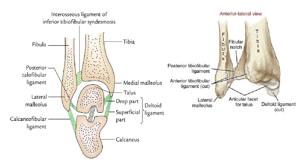

• Although there is no joint capsule, there are several associated ligaments at the distal tibiofibular joint. • Because the proximal and distal joints are linked (the tibia, fibular, and tibiofibular joints are part of a closed chain), all the ligaments that lie between the tibia and fibular contribute to stability at both joints.
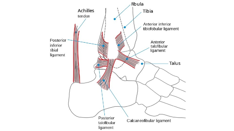

Functions of ligaments • The ligaments of the distal tibiofibular joint are primarily responsible for maintaining a stable mortise. • The ligamentous structures that support the distal tibiofibular joint are the anterior and posterior tibiofibular ligaments and the interosseous membrane. • The interosseous membrane directly supports both proximal and distal tibiofibular articulations.

The distal tibiofibular joint is an extremely strong articulation. Stresses that tend to move the talus excessively in the mortise (e. g. , falling onto the side of the foot) often tear an ankle collateral ligament before the tibiofibular ligaments. • Continued force may fracture the fibula proximal to the distal tibiofibular ligaments before the tibiofibular ligaments will tear.

Why distal tibiofibular joint is an extremely strong articulation? • The function of the ankle (talocrural) joint is dependent on stability of the tibiofibular mortise. • The tibia and fibula would be unable to grasp and hold on to the talus if the tibia and fibular were permitted to separate or if one side of the mortise were missing. • The analogous mortise of a wrench could not perform its function of grasping a bolt if the two pincer segments moved apart every time a force was applied to the wrench. • Conversely, the ankle mortise must have some mobility function to serve; otherwise, a single fused arch would better serve ankle joint function.

The mobility role of the mortise belongs primarily to the fibula. The fibula has, in fact, little weight-bearing function; no more than 10% of the weight that comes through the femur is transmitted through the fibula. Given the relatively small weight-bearing function of the fibula, the hyaline cartilage of the synovial proximal tibiofibular joint appears to be dependent on joint motion (rather than weight-bearing) to maintain nutrition of the cartilage. That is, the proximal tibiofibular joint must be mobile; if the proximal tibiofibular joint is mobile, so too must the distal tibiofibular joint be, because the two joints are mechanically linked.

Distal Articular Surface • The body of the talus forms the distal articulation of the ankle joint. • The body of the talus has three articular surfaces: 1. Large lateral (fibular) facet 2. Smaller medial (tibial) facet 3. Trochlear (superior) facet
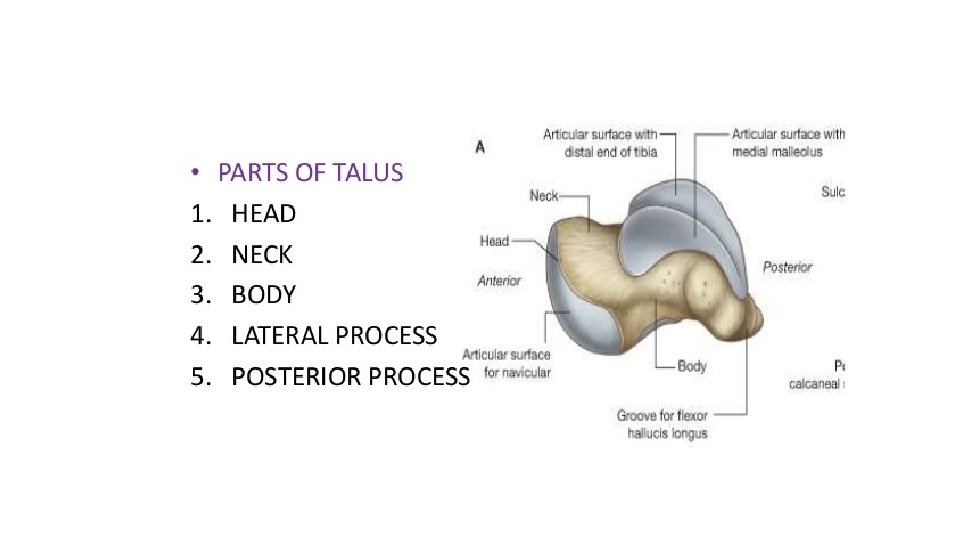

The large, convex trochlear surface has a central groove that runs at a slight angle to the head and neck of the talus. The body of the talus also appears wider anteriorly than posteriorly, which gives it a wedge shape. The degree of wedging may vary among individuals, with no wedging at all in some and a 25% decrease in width anteriorly to posteriorly in others. The articular cartilage covering the trochlea is continuous with the cartilage covering the more extensive lateral facet and the smaller medial facet.

The structural integrity of the ankle joint is maintained throughout the ROM of the joint by a number of important ligaments.

Thank You
- Slides: 26