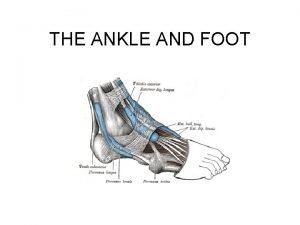
Ankle Foot ANATOMY Mehdi jalali majd Mehdi jalali


- Slides: 79

Ankle & Foot ANATOMY Mehdi jalali majd

Mehdi jalali majd

Mehdi jalali majd

Mehdi jalali majd

Mehdi jalali majd

Talus Mehdi jalali majd

Mehdi jalali majd

Mehdi jalali majd

Mehdi jalali majd

Mehdi jalali majd

Calcaneous Mehdi jalali majd

Mehdi jalali majd

Mehdi jalali majd

Mehdi jalali majd

Mehdi jalali majd

Mehdi jalali majd

Left Navicular Mehdi jalali majd

Ankle & Foot Mehdi jalali majd

Mehdi jalali majd

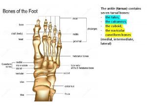
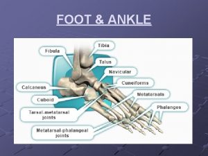
Rearfoot • • Bones – Calcaneus and talus Joint – Subtalar (talocalcaneal) Midfoot • • Bones – Navicular, cuboid, and cuneiform joints – Transverse tarsal: Talonavicular, Calcaneocuboid – Distal intertarsal: Cuneonavicular, Cuboideonavicular, Intercuneiform and cuneocuboid complex Forefoot • • Bones – Metatarsals and phalanges Joints – Tarsometatarsal, Intermetatarsal, Metatarsophalangeal and Interphalangeal Mehdi jalali majd

Fundamental Terminology • Sagittal Plane (med-lat axis) – Dorsiflexion (ext) – Plantar flexion • Frontal Plane (ant-post axis) – Eversion – Inversion • Horizontal Plane (sup-inf axis) – Abduction – Adduction Mehdi jalali majd

Applied Terminology • Pronation – Dorsiflexion – Abduction – Eversion • Supination – Plantar flexion – Adduction – Inversion Mehdi jalali majd

ANKLE Talocrural Joint Structure Mehdi jalali majd

Sup Tibiofibular Joint sup and inf sliding Mehdi jalali majd

Inferior Tibiofibular Joint Mehdi jalali majd

The mortise of the ankle is adjustable the proximal and distal tibiofibular joints permit and control the changes in the mortise Mehdi jalali majd

The three articular surfaces of the talus as part of a cone-shaped surface the larger end facing laterally • trochlea • smaller medial facet • larger lateral facet Mehdi jalali majd

Left Ankle Mehdi jalali majd

Left Ankle (Lat) Mehdi jalali majd

Medial collateral (deltoid) lig Mehdi jalali majd

ANKLE Talocrural Joint Function Mehdi jalali majd

The axis of the ankle joint 14 inclination from the transverse plane & 23 from the frontal plane Mehdi jalali majd

• Ankle dorsiflexion limited by – soft tissue restrictions (calcaneofibular lig & post capsule) – Active or passive tension in the triceps surae • Ankle plantarflexion limited by – soft tissue restrictions (ant talofibular & tibionavicular & ant capsule) – tibialis anterior, extensor hallucis longus, and extensor digitorum longus • side-to-side movement or rotation limited by – The ligaments and assisted by the muscles • medial aspect – The tibialis posterior, flexor hallucis longus, and flexor digitorum longus muscles • lateral aspect – the peroneus longus and peroneus brevis Mehdi jalali majd

Factors that increase the mechanical stability of the fully dorsifexed talocrural joint A. The increased passive tension in connective tissues and muscles B. The trochlear surface of the talus is wider anteriorly than posteriorly Mehdi jalali majd

The compression force on the talocrural joint through the Stance phase of walking Mehdi jalali majd

• Talocrural Joint • Primarily sagittal plane motion • Subtalar Joint • Oblique path motion consisting: Inv – Ever & Abd - Add • Transverse tarsal Joint • More oblique path of motion equally pass through all cardinal planes Mehdi jalali majd

SUBTALAR JOINT (talocalcaneal joint) Mehdi jalali majd

SUBTALAR JOINT (talocalcaneal joint) • The subtalar joint – set of articulations formed by the posterior, middle, and anterior facets of the calcaneus and the talus • Subtalar joint motion – grasp the unloaded calcaneus and twist it in a side-to side and rotary fashion • Mobility at the subtalar joint – allows the foot to assume positions that are independent of the orientation of the superimposed ankle and leg Mehdi jalali majd

Mehdi jalali majd

Articular Structure • The concave posterior articulation occupies about 70% of the total articular surface area • The anterior and middle articulations consist of small, nearly flat joint surfaces. • (see Fig. 14 -8). • The articulation is normally held tightly opposed by – – interlocking shape body weight interosseous ligaments activated muscle Mehdi jalali majd

SUBTALAR JOINT LIGAMENTS • Interosseous: – ant & post interosseous – Cercical • Med – Tibiocalcaneal • Lat – Calcaneofibular • Post: – Talocalcaneal (med & post & lat) Mehdi jalali majd

The ligaments of the subtalar joint posterior cross-sectional view Mehdi jalali majd

Mehdi jalali majd

The superior and inferior extensor retinacula the superior and inferior peroneal retinacula Mehdi jalali majd

osteokinematics at the subtalar joint Mehdi jalali majd

The axis of rotation at the subtalar joint Mehdi jalali majd

Non–weight-bearing motion the right subtalar A. Pronation B. Supination Mehdi jalali majd

Subtalar Joint: ROM • Averaged total inversion exceeds eversion nearly double – Inversion 22. 6 degrees – Eversion 12. 5 degrees • Eversion range of motion is naturally limited by – the distal projecting lateral malleolus – the thick deltoid ligament Mehdi jalali majd

Valgus or calcaneovalgus: increase in the medial angle between the calcaneus and posterior leg Varus or calcaneovarus: decrease in the medial angle between the calcaneus and posterior leg Mehdi jalali majd

• Talocrural Joint • Primarily sagittal plane motion • Subtalar Joint • Oblique path motion consisting: Inv – Ever & Abd - Add • Transverse tarsal Joint • More oblique path of motion equally pass through all cardinal planes Mehdi jalali majd

TRANSVERSE TARSAL or Midtarsal Joint (CHOPART’S) Talonavicular AND Calcaneocuboid Mehdi jalali majd

TRANSVERSE TARSAL JOINTS Lig • Talonavicular (flexibility) – Calcaneonavicular (SPRING) lig 22 • Sustentaculum tali To Plantar surface of Navicular • Calcaneocuboid (rigidity) 23 – Dorsal calcaneocuboid lig – Bifurcat lig • Med (calcaneonavicular) • Lat (calcaneocuboid) – Long plantar lig (calcaneous to 3 rd-4 th lat metatarsal base) – Short plantar lig (plantar calcaneocuboid lig) • plantar ligaments provide excellent structural stability to the lateral side of the foot Mehdi jalali majd

Mehdi jalali majd

Mehdi jalali majd

With the foot fixed internal rotation of the lower limb causes the following associated movements • rearfoot pronation (eversion) • lowering of the medial longitudinal arch, and valgus • stress at the knee • as the rearfoot pronates, the floor "pushes" the forefoot and midfoot into a relatively supinated position Mehdi jalali majd

Control of excessive pronation at the subtalar joint • Controlled pronation of the subtalar joint allowing the foot to accommodate to the shapes and contours of walking surfaces • Improve the "eccentric control" of the muscles (include the supinators of the foot and the external rotators and abductors of the hip), decelerate pronation Mehdi jalali majd

DISTAL INTERTARSAL JOINTS • Cuneonavicular Joints – between the navicular and three cuneiform bones – plantar and dorsal ligaments – help to transfer movements to the forefoot • Cuboideonavicular Joint – – Between navicular and cuboid dorsal, plantar, and interosseous ligaments links the lat and med components of the transverse tarsal joint assisting in transferring movements • Intercuneiform and Cuneocuboid Joint Complex – between the cuneiforms – between lat cuneiform and cuboid – dorsal, plantar, and interosseous ligs Mehdi jalali majd

TARSOMETATARSAL JOINTS • Between the bases of the metatarsals and the distal surfaces of the three cuneiforms and cuboid – first metatarsal with med cuneiform – second metatarsal with the intermediate cuneiform – third metatarsal with the lateral cuneiform – fourth and fifth metatarsal both articulate with the distal surface of the cuboid • Articular surfaces of the tarsometatarsal joints are flat • Held together by dorsal, plantar, and interosseous ligaments • Only the first Joint has a well-developed capsule Mehdi jalali majd

Mehdi jalali majd

METATARSOPHALANGEAL JOINTS • Five metatarsophalangeal joints – – – convex head of metatarsal with proximal phalanx Extensor mechanism poorly defined Four deep transverse metatarsal ligaments plantar plate located on the plantar side of the joint Collateral ligaments spans each MTP joint • courses obliquely from a dorsal-proximal to plantar-distal • a thick cord portion a fanlike accessory portion (attaches to the plantar plate) Mehdi jalali majd

Kinematic • the metatarsophalangeal joints have two degrees of freedom – Extension (dorsiflexion) and Flexion (plantar flexion) in the sagittal plane about a medial-lateral axis – Abduction and Adduction in the horizontal plane about a vertical axis – Both axes of rotation intersect at the center of each metatarsal head • Toes can be hyperextended about 65° • Toes can be Flexed about 30° to 40° • The first toe allows greater hyperextension to near 85° Mehdi jalali majd

INTERPHALANGEAL JOINTS • The IP joint consists of the convex head of the more proximal phalanx with the concave base of the more distal phalanx • Collateral ligaments, plantar plates, and capsules are present, but smaller and less defined • Mobility at the IP joints is limited to flex & ext • Motion tends to be greater at the proximal than the distal joints Mehdi jalali majd

Plantar Arches Mehdi jalali majd

Plantar Arches The longitudinal arch Mehdi jalali majd

Plantar Arches The transverse arch Mehdi jalali majd

ACTION OF THE FOREFOOT DURING THE LATE STANCE PHASE OF GAIT • Depending on the phase of gait, these joints provide an element of flexibility or stability to the forefoot • During the later part of stance midfoot and forefoot must be stable or rigid to accept the stresses with push of – activation of local intrinsic and extrinsic muscles – increased tension in the medial longitudinal arch windlass effect Mehdi jalali majd

Mehdi jalali majd

Extreme pronation at the subtalar joint accompanied by 1) adduction and plantarflexion of the head of the talus 2) eversion of the calcaneus 3) in some instances pronation at the transverse tarsal joint 4) the tarsometatarsal joints undergo supination twist Mehdi jalali majd

Extreme supination at the subtalar joint Is accompanied by • abduction and dorsiflexion of the head of the talus • inversion of the calcaneus • forced supination of the transverse tarsal joint • If the forefoot is to remain on the ground, the tarsometatarsal joints must undergo a counteracting pronation twist. Mehdi jalali majd

Ankle & Foot Muscle Mehdi jalali majd

Mehdi jalali majd

Mehdi jalali majd

Mehdi jalali majd

Mehdi jalali majd

Mehdi jalali majd

Mehdi jalali majd

Mehdi jalali majd

A mechanical model of standing on tiptoes • The force of a contracting gastrocnemius muscle acts with a relatively short internal moment arm from the talocrural joint (A) • Relatively long internal moment arm from the MTJ (B) • Once on tiptoes, the Lo. G due to body weight falls just posterior to the axis of rotation at the MTJ • As a result, body weight acts with a relatively small external moment arm (e) from the MTJ Mehdi jalali majd

The intrinsic muscles of the foot Mehdi jalali majd
 Leila jalali
 Majd alwan
 Metoníma
 Ha egyszer majd megöregszünk
 Ankle foot orthoses
 Ankle foot orthosis project
 Anatomy and physiology of the foot
 Sustentaculum tali
 Cross bridge theory
 Ankle muslces
 You put your right foot in you put your right foot out
 Trigonometry magic triangles
 Iliac nerve
 Foot bone anatomy
 Peronues longus
 Dr. mehdi pain management
 Mehdi nt
 Mehdi namazi
 Mehdi bouguerra
 Mehdi
Mehdi Mehdi sebbane
 R tree java
 Mehdi mojtabavi
 Mehdi khalighi
 Mehdi bouguerra
 Mehdi salek md
 Amir salek md
 General mehdi rahimi
 Mehdi hamadani
 Base deficit
 Inversion et eversion
 Soap injury evaluation
 Cumberland ankle instability tool
 Hammock carry
 Abi normal
 Electronic muscle stimulation ankle sprain
 Calculating abi
 Sugartong splint
 Abdominal reflex response
 How to apply skin traction
 Phong tran surgeon
 Ankle special tests
 Pernous brevis
 Ankle block frca
 Wikimedia
 Ober's test
 Open basket weave taping
 Chapter 19 worksheet the ankle and lower leg
 Prophylactic taping finger
 Popliteal vein
 Ankle eversion
 Rules of mad gab
 Fta ankle
 Twisted ankle
 Homen sign
 Articulatio talo tarsalis
 Rom toes
 Weber classification of ankle fractures
 Carnegie mellon what is rpa robotic process automation
 Sugar tong splint ankle
 Growth plate in ankle
 Advantages and disadvantages of first class levers
 Chapter 15 the ankle and lower leg
 Pab ankle fracture
 End feel pada ankle
 Plantarflexion muscle
 Midtarsal joint
 Ankle joint
 Offred tattoo ankle
 Chaput fragment ankle
 Perpneal tendon
 Regio ankle
 Icd 10 midfoot sprain
 Oficiellt
 Plantaris insertion
 Leg and foot topographical views
 West point ankle sprain grading system
 Lateral dorsum
 Mtp 1 anatomi
 Ankle rom goniometer


