Animal Tissues 1 Histology is the study of


. Adipose cells")

- Slides: 12
Animal Tissues 1
Histology is the study of plants and animal tissues by examining tissues under a light microscope or an electron microscope. A tissue is a group of cells that carry out a particular function. Animals are multicellular organisms with their specialized cells grouped into tissues. In most animals, combinations (group)of various tissues make up functional units called organs, and groups of organs that work together are called systems. For example, the animal digestive system consists of a stomach, small intestine, large intestine, and several other organs, each comprises (cover) of different tissues.
Organs in animals are composed of different types of tissues. Each tissue is composed of cells. Level of organization in animals: Cells Tissues Organs Systems Organism body
Types of Animals Tissues Different types of tissues have different structures and functions. A tissue may be held by a sticky extracellular matrix that bind (link) the cells together in a fabric of fibers (Tissue= cells + extracellular matrix). Types of tissues 1) Epithelial tissue 2) Connective tissue 4) Nervous tissue 3) Muscular tissue
First: Epithelial tissues Characterized by: 1. Cells tightly linked together. 2. The presence of a cell secretion called the basement membrane 3. Covers and lines other tissues and organs. Functions of Epithelium Protection Absorption (skin) (Intestinal villi) Glands can be single epithelial cells, such as the goblet cells that found in the stomach and intestines to secrete mucus. Multicellular glands include the endocrine glands Secretion (glandular cells)
Classification of epithelial tissues Epithelia are classified by the number of cell layers and the shape of cells on the free surface. Classification based on number of cell layers: 1. Simple epithelium is a One layer of cells. 2. Stratified epithelium ﻋﻤﻮﺩﻳﺔ ﻣﻬﺪﺑﺔ ﻃﺒﻘﻴﺔ ﻛﺎﺫﺑﺔ More than one layer of cells. 3. Pseudostratified epithelium One layer of cells but appear to form two layers. ﻣﻜﻌﺒﺔ ﺑﺴﻴﻄﺔ Classification based on cell shape: 1. Cuboidal = cube 2. Columnar = column 3. Squamous = flat ﺣﺮﺷﻔﻴﺔ ﻃﺒﻘﻴﺔ ﺣﺮﺷﻔﻴﺔ ﺑﺴﻴﻄﺔ ﻏﺸﺎﺀ ﻗﺎﻋﺪﻱ ﻋﻤﻮﺩﻳﺔ ﻃﺒﻘﻴﺔ ﻋﻤﻮﺩﻳﺔ ﺑﺴﻴﻄﺔ
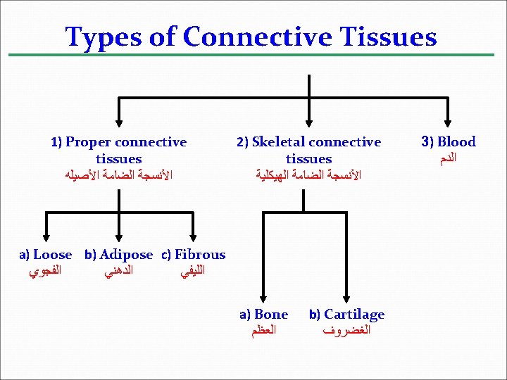
Second: Connective tissues Characterized by 1. Cells widely separated from each other in a matrix that is produced by the cells (cells are scattered ). 2. The matrix generally consists of fibers 3. The matrix may be solid (as in bone), gelatinous, or liquid (as in blood). 4. The functions of connective tissues are to bind and support other tissues.
Types of Fibers in Connective Tissues There are three types of connective tissues fibers, which are all proteins: 1 - Collagenous fibers (white) Are thick and made of collagen and non-elastic 2 - Elastic fibers (Yellow) Are long threads of elastin, which provide a rubbery quality. 3 -Reticular fibers Are very thin and branched, and composed of collagen.
1 - Proper Connective Tissues A- Loose Proper Connective Tissue Binds epithelia to underlying tissues and functions a holding organs in place. It has all three fiber types. It has two cell types: 1 - Fibroblasts Are responsible for making the extracellular fibers. 2 - Macrophages Are amoeboid cells ﺧﻼﻳﺎ ﺃﻤﻴﺒﻴﺔ that swallow and digest microbes, and cellular debris ﺑﻘﺎﻳﺎ ﺍﻟﺨﻼﻳﺎ ﺍﻟﻤﻴﺘﺔ by phagocytosis.
B- Adipose Proper Connective Tissue Function as storage cells for adipose (lipids). Adipose cells contain a large vacuole which in the live cell contains lipids. Cell nucleus and cytoplasm are pushed out to edge ﺣﺎﻓﺔ of cell membrane.
C- Fibrous Proper Connective Tissue It contains large number of collagenous fibers. The functions of this tissue are to attach muscles to bones (tendons )ﺍﻷﻮﺗﺎﺭ , and connect bones to other bones (ligaments )ﺍﻷﺮﺑﻄﺔ at joints ﺍﻟﻤﻔﺎﺻﻞ.