Animal structure and function chap 40 Introduction Anatomy
")

 An amoeba, a single-celled")
 Interstitial fluid")

: Covers all surfaces of the body Epidermis (ectoderm): Outer portion of")

 Epidermis every 2 weeks Stomach")

")


 Duct: Connection from gland to tissue Secrete/absorb chemical")
 Hormones Secreted into blood stream")


 Reticular: Thin,")


, dermis of")

 & collagen Strong, flexible tissue, absorb")


 Cells RBC (erythrocytes) Contain hemoglobin (carries oxygen) WBC (leukocytes) Neutrophils, eosinophils,")


 Sensory,")

 Signaling by hormones STIMULUS (b) Signaling by neurons STIMULUS Endocrine cell Cell body")


 A walrus, an endotherm (b) A lizard, an ectotherm")
 Elevated blood level Islets of Langerhans (sensor,")


- Slides: 68
Animal structure and function (chap 40)
Introduction Anatomy: Biological form Physiology: Biological function Interstitial fluid: Fluid surrounding the cells
Body plan Mouth Gastrovascular cavity Exchange 0. 1 mm (a) An amoeba, a single-celled organism 1 mm (b) A hydra, an animal with two layers of cells
Mouth Food CO 2 Animal body Respiratory system Heart Lung tissue (SEM) Interstitial fluid Nutrients 250 µm Body plan External environment Cells Digestive system Anus Unabsorbed matter (feces) Excretory system Blood vessels in kidney (SEM) Metabolic waste products (nitrogenous waste) 50 µm Lining of small intestine (SEM) 100 µm Circulatory system
Tissues Epithelial Connective Muscle Nervous
Epithelial tissue (epithelium): Covers all surfaces of the body Epidermis (ectoderm): Outer portion of skin Endoderm: Lining of inner surfaces of digestive tract Mesoderm: Inner surface of body cavities
Epithelial tissue Closely packed Tight junctions One/or few cell layers thick Selective absorption in the intestines Rapid gas exchange in lungs Protection from microbes, water loss
Epithelial tissue Regenerative capabilities Liver (gland from epithelial tissues) Epidermis every 2 weeks Stomach lining every 2 -3 days
Epithelial tissue Types Based on cell thickness Shape on exposed surface Simple One layer thick Stratified Multiple layers of cells
Epithelial tissue Shapes of cells Cuboidal: As wide as they are tall (like dice) Columnar: Taller than wide (like bricks on end) Squamous Flat like floor tiles
Epithelial tissues
Epithelial tissue Simple squamous Lining of lungs, capillary walls and blood vessels Simple cuboidal Lining of some glands Simple columnar Lining of stomach, intestines and parts of respiratory tract
Epithelial tissue Stratified squamous Outer layer of skin and mouth Keratin Water resistant protein
Epithelial tissue Exocrine glands (duct system) Duct: Connection from gland to tissue Secrete/absorb chemical solutions Sweat & sebaceous glands Lining of intestines & lungs that secrete mucous
Epithelial tissue Endocrine glands (ductless glands) Hormones Secreted into blood stream
Glands
Connective tissue Holds tissues & organs together Supports, insulates and strengthens Derived from mesoderm Loosely packed cells Scattered in an extracellular matrix
Connective tissue Matrix: Composed of a web of fibers In a foundation of liquid, jellylike or solid Fibers (proteins) are collagen, elastic, or reticular
Connective tissue Collagen: Non-elastic-doesn’t tear easily Elastic: Makes tissue elastic Elastin (protein) Reticular: Thin, branched, joins connective tissue to adjacent tissues
Connective tissue Cells in matrix Fibroblasts: Produce & secrete extracellular matrix Macrophages: Engulf foreign bodies & debris Mast cells & heparin
Connective tissue types 1. Loose connective tissue Beneath skin & between organs Support, insulation, food storage Adipose tissue (fat) Cells become larger when gain weight Shrink with weight loss
Connective tissue 2. Dense connective tissue Tendons, ligaments, sheath around organs (periosteum), dermis of skin Support, strong connections 3. Special connective tissue Cartilage, bone, blood,
Connective tissue
Special connective tissue Cartilage Consists of chondroitin (glycoprotein) & collagen Strong, flexible tissue, absorb stress Joints, ear pinna, nose, intervertebral discs, larynx Chondrocytes: Cartilage cells
Cartilage
Bone Embryos---more cartilage Cartilage is replaced with bone cells or osteocytes Matrix hardens with crystals of calcium phosphate mixed with collagen
Bone Osteoblasts: Lay down new bone Osteoclasts: Dissolve bone Osteons: Unit of bone structure Contains calcified matrix, osteocytes, nerve fibers, blood vessels
Bone Flat bones Long bones Spongy bone: Contains marrow Blood cells formed Compact bone: More dense, gives strength
Bone
Bone
Blood Plasma (matrix) Cells RBC (erythrocytes) Contain hemoglobin (carries oxygen) WBC (leukocytes) Neutrophils, eosinophils, basophils, lymphocytes, monocytes Platelets (thrombocytes)
Blood
Blood Plasma contains Wastes, nourishment Hormones Na+, Ca 2+, other ions Fibrinogen, albumin, antibodies
Connective Tissue Blood Loose connective tissue Plasma White blood cells 55 µm 120 µm Collagenous fiber Cartilage Elastic fiber Fibrous connective tissue Red blood cells 30 µm 100 µm Chondrocytes Chondroitin sulfate 700 µm Adipose tissue Central canal Fat droplets Osteon 150 µm Bone Nuclei
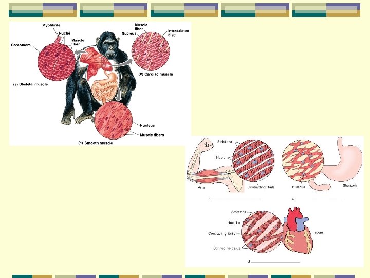
Muscle tissue Movement Organization of actin & myosin filaments Smooth, skeletal & cardiac muscles Striated muscles: skeletal & cardiac Skeletal muscles: voluntary control Smooth & cardiac muscles: involuntary control
Muscle tissue Smooth muscle Walls of blood vessels, stomach, intestines Viscera: Internal organs Made of sheets of cells each with a single nucleus
Muscle tissue Skeletal muscle Attached by tendons to bones Contract move bones
Muscle tissue
Muscle tissue Cardiac muscle Small interconnected cells Linked by gap junctions Openings allow small substances & electrical charges to pass between cells Myocardium Single functioning units
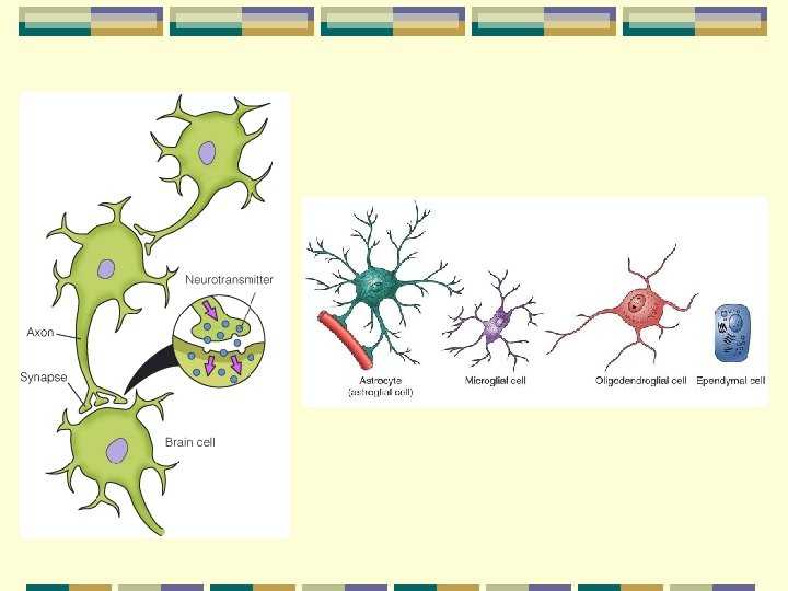
Nerve tissue Neurons Cell body, dendrites, axon Neuroglia Supporting cells Insulate neurons Eliminate foreign bodies
Nerve tissue Dentrites Thin, branched extensions Receive impulses Axons Single extension of cell body Carries impulse away Myelin sheaths, insulating cover
Neurons
Neurons Sensory neurons Eye, ears, surface of skin Motor neurons Brain & spinal cord Interneurons Brain & spinal cord Neurons within the CNS
Neurons
Nervous Tissue Glia Neurons Neuron: Dendrites Cell body Glia Axons of neurons 40 µm Axon (Fluorescent LM) Blood vessel (Confocal LM) 15 µm
Summary Epithelial tissues Simple or stratified Cuboidal, columnar, squamous Connective tissues Loosely packed, tightly packed Special (bone, cartilage, blood) Matrix
Summary Muscle tissues Smooth, cardiac, skeletal Nerve tissues: Neurons (cell body, dentrites, axons) Sensory, motor and interneurons
Coordination Hormones Nervous system Homeostasis
(a) Signaling by hormones STIMULUS (b) Signaling by neurons STIMULUS Endocrine cell Cell body of neuron Axon Nerve impulse Hormone Signal travels everywhere. Signal travels to a specific location. Blood vessel Nerve impulse Axons Response
Homeostasis Dynamic constancy of internal environment Dynamic because conditions fluctuate Narrow range p. H Temp Glucose Oxygen
Regulation 1. Negative feedback loops 2. Positive feedback loops
Negative Feedback
Negative feedback loops Sensors Measure internal environment Integrating center Receives information from sensors Compares to normal range Responds
Negative feedback loops Effectors: Muscles or glands Receive information from center Response
Negative feedback loops Temperature increase Hypothalamus senses deviation Sends signals to relieve heat Sweating & vasodilation Reach baseline Negative feedback stops response
Negative feedback loops Temperature decrease Hypothalamus sends signals Shiver, vasoconstriction Temp to baseline Negative feedback stops response
Thermoregulation
Fig. 40 -9 (a) A walrus, an endotherm (b) A lizard, an ectotherm
Negative feedback loops Glucose (eat a meal) Elevated blood level Islets of Langerhans (sensor, center) Insulin Lowers blood sugar (uptake in muscle, fat & liver cells) Negative feedback stops insulin release
Regulating Blood Sugar
Positive Feedback
Positive feedback loops Uterine contractions Pressure from baby on uterus Causes contractions Causes more stretching More contractions Continues until birth
Positive feedback loop Blood clotting Clotting factors stimulate the formation of more factors Clot forms Maintain blood volume
Bioenergetics Overall flow & transformation of energy in an animal Determines nutritional needs Animal size, activity and environment
Fig. 40 -17 External environment Animal body Organic molecules in food Digestion and absorption Nutrient molecules in body cells Carbon skeletons Cellular respiration Heat Energy lost in feces Energy lost in nitrogenous waste Heat ATP Biosynthesis Cellular work Heat
Metabolic rate Amount of energy an animal uses in a unit of time Torpor: Physiological state of low activity with low metabolism Hibernation: Long term torpor