
Animal Structure and Function An Introduction Chapter 38
- Slides: 78
Animal Structure and Function: An Introduction Chapter 38
KEY CONCEPTS • Structure and function are closely linked at every level of organization
Learning Objective 1 • Compare the structure and function of the four main kinds of animal tissues: epithelial, connective, muscle, and nervous
Tissue • A group of similarly specialized cells • Associated to perform one or more functions
Epithelial Tissue (Epithelium) • A continuous layer (sheet) of cells • • • covering a body surface lining a body cavity Functions in protection, absorption, secretion, or sensation
Connective Tissue 1 • Relatively few cells separated by intercellular substance • • fibers scattered throughout a matrix Intercellular substance fibers • • • collagen fibers elastic fibers reticular fibers
Connective Tissue 2 • Contains specialized cells • • such as fibroblasts and macrophages Functions: • • • joins other body tissues supports body and organs protects underlying organs
Muscle Tissue • Consists of cells specialized to contract • Each cell is an elongated muscle fiber • many contractile units (myofibrils)
Nervous Tissue • Neurons • • • elongated cells specialized for transmitting impulses Glial cells • support and nourish neurons
Learning Objective 2 • Compare the structure and function of the main types of epithelial tissue
Epithelial Tissue • Epithelial cell shapes • • squamous, cuboidal, columnar Epithelial tissue structure • • simple, stratified, pseudostratified (See Table 38 -1)
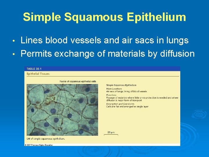
Simple Squamous Epithelium Lines blood vessels and air sacs in lungs • Permits exchange of materials by diffusion •
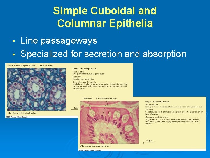
Simple Cuboidal and Columnar Epithelia Line passageways • Specialized for secretion and absorption •
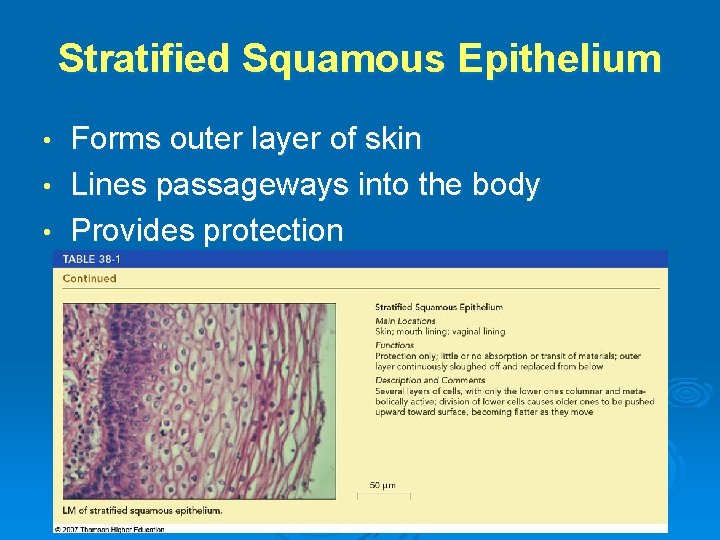
Stratified Squamous Epithelium Forms outer layer of skin • Lines passageways into the body • Provides protection •
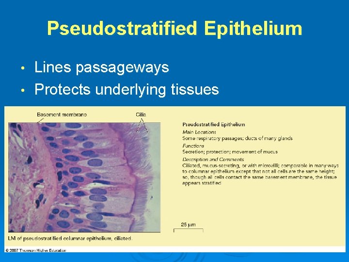
Pseudostratified Epithelium Lines passageways • Protects underlying tissues •
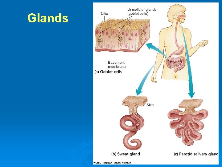
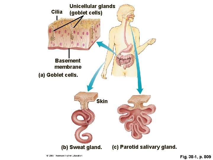
Glands 1 • Specialized epithelial tissue • Goblet cells • unicellular exocrine glands that secrete mucus
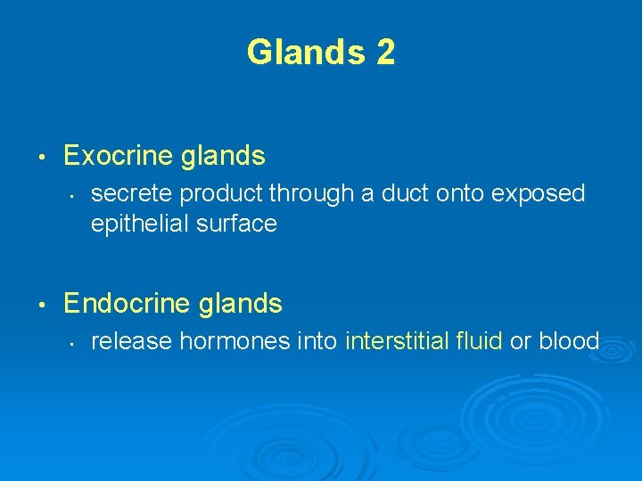
Glands 2 • Exocrine glands • • secrete product through a duct onto exposed epithelial surface Endocrine glands • release hormones into interstitial fluid or blood
Glands
Cilia Unicellular glands (goblet cells) Basement membrane (a) Goblet cells. Skin (b) Sweat gland. (c) Parotid salivary gland. Fig. 38 -1, p. 809
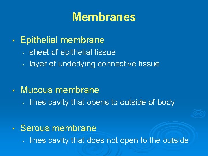
Membranes • Epithelial membrane • • • Mucous membrane • • sheet of epithelial tissue layer of underlying connective tissue lines cavity that opens to outside of body Serous membrane • lines cavity that does not open to the outside
Learning Objective 3 • Compare the main types of connective tissue • Summarize their functions
Connective Tissues • Cells embedded in intercellular substance • • microscopic collagen fibers, elastic fibers, reticular fibers (thin branched fibers) scattered through a matrix (thin gel of polysaccharides)
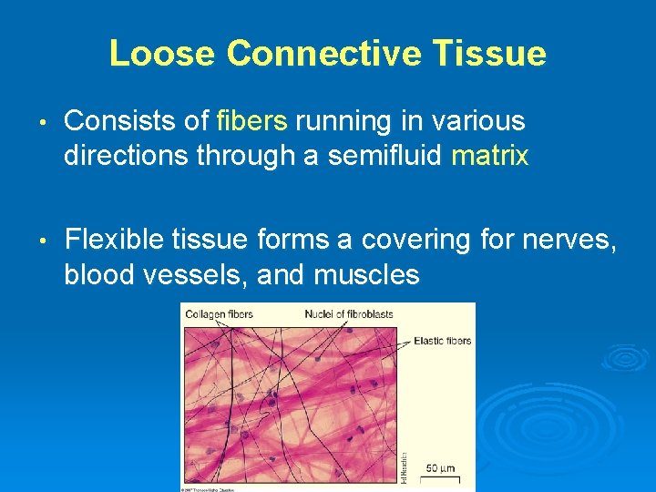
Loose Connective Tissue • Consists of fibers running in various directions through a semifluid matrix • Flexible tissue forms a covering for nerves, blood vessels, and muscles
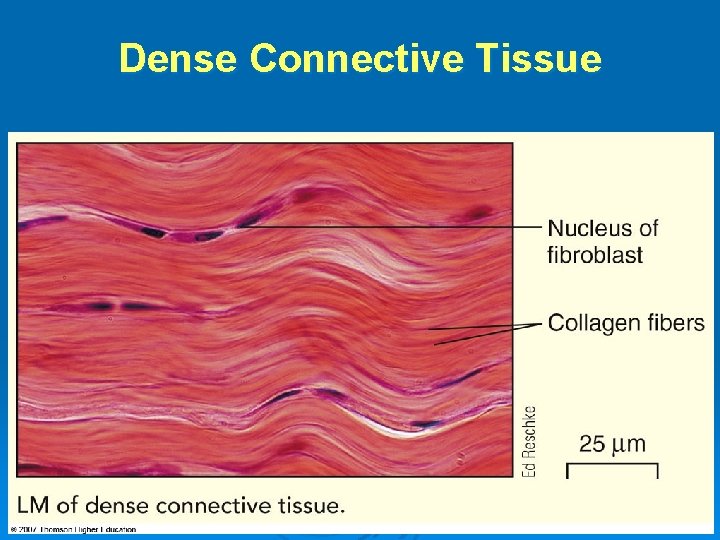
Dense Connective Tissue • Stronger, less flexible than loose connective tissue • Collagen fibers arranged in definite pattern • Forms • • tendons (connect muscles to bones) ligaments (connect bones to bones)
Dense Connective Tissue
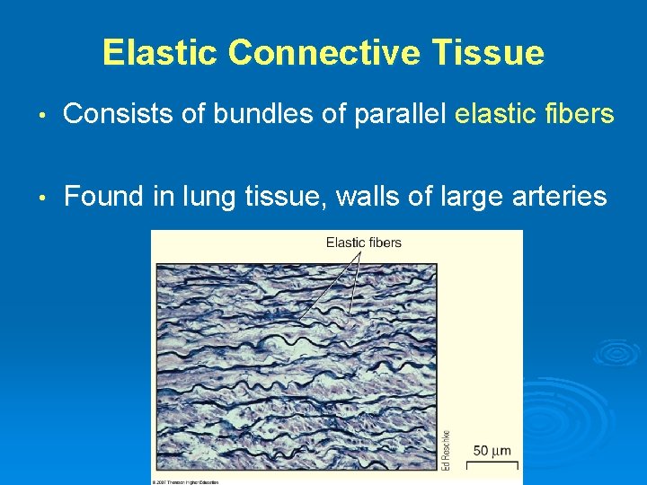
Elastic Connective Tissue • Consists of bundles of parallel elastic fibers • Found in lung tissue, walls of large arteries
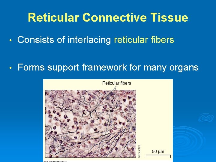
Reticular Connective Tissue • Consists of interlacing reticular fibers • Forms support framework for many organs
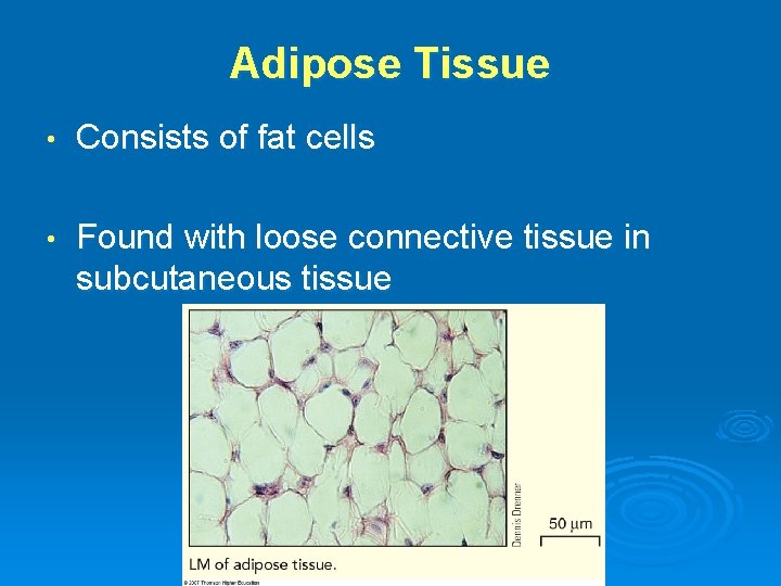
Adipose Tissue • Consists of fat cells • Found with loose connective tissue in subcutaneous tissue
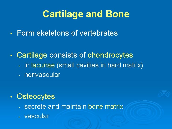
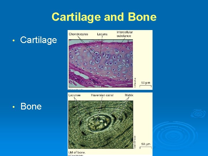
Cartilage and Bone • Form skeletons of vertebrates • Cartilage consists of chondrocytes • • • in lacunae (small cavities in hard matrix) nonvascular Osteocytes • • secrete and maintain bone matrix vascular
Cartilage and Bone • Cartilage • Bone
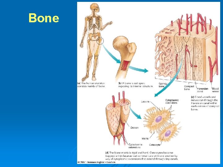
Bone
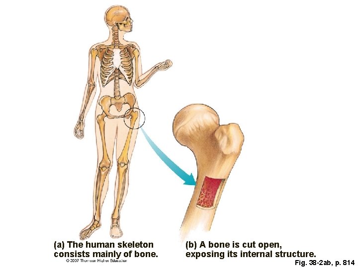
(a) The human skeleton consists mainly of bone. (b) A bone is cut open, exposing its internal structure. Fig. 38 -2 ab, p. 814
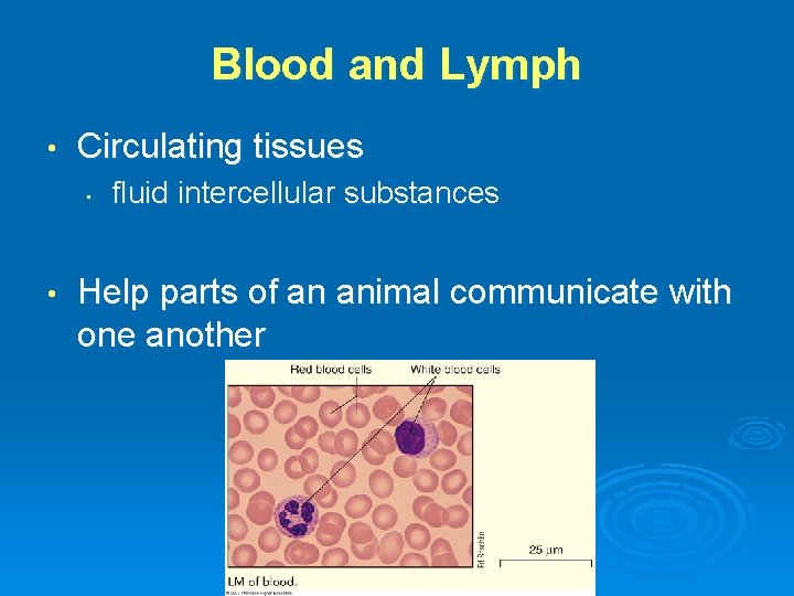
Blood and Lymph • Circulating tissues • • fluid intercellular substances Help parts of an animal communicate with one another
Learning Objective 4 • Contrast the three types of muscle tissue and their functions
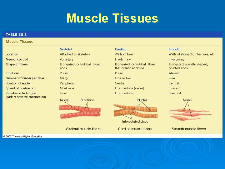
Skeletal Muscle • Striated and under voluntary control • Elongated, cylindrical fibers with several nuclei • Skeletal muscles contract, move parts of the body
Cardiac Muscle • Striated, contractions are involuntary • Elongated, cylindrical fibers branch and fuse; one or two central nuclei • Muscle contracts, heart pumps blood
Smooth Muscle • No striations, contractions involuntary • Elongated, spindle-shaped fibers with a single central nucleus • Smooth muscle moves body organs (example: pushes food through digestive tract)
Muscle Tissues
Learning Objective 5 • How does the structure of the neuron relate to its function?
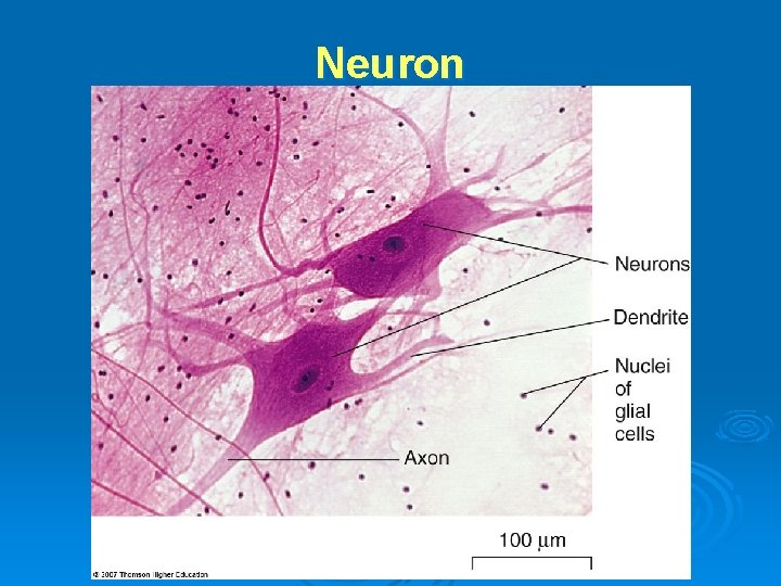
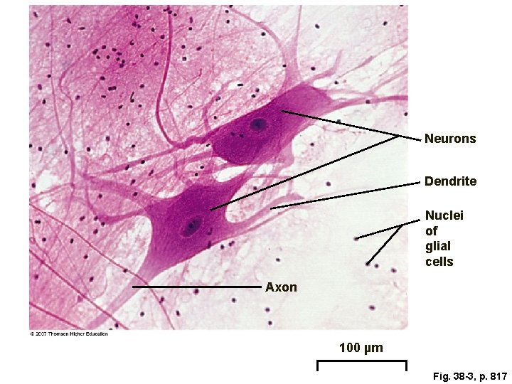
Neuron • Elongated cell • Receives and transmits information • Synapse • a junction between neurons
Neuron • Dendrites • • • receive signals transmit signals to cell body Axon • • transmits signals away from cell body to other neurons, muscles, glands
Neuron
Neurons Dendrite Nuclei of glial cells Axon 100 µm Fig. 38 -3, p. 817
KEY CONCEPTS • The main types of tissues in a complex animal are epithelial, connective, muscle, and nervous
Learning Objective 6 • Describe the organ systems of a mammal • Summarize the homeostatic actions of each organ system
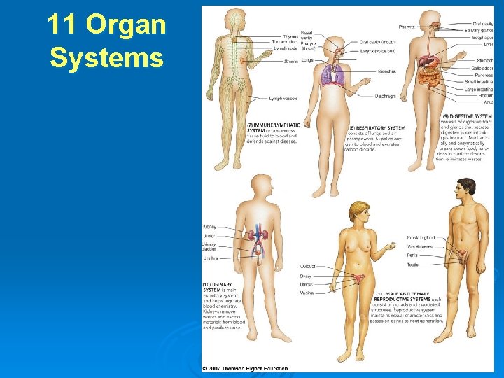
Organ Systems • Tissues and organs working together • In mammals, 11 organ systems work together in the organism • Each organ system functions to maintain homeostasis
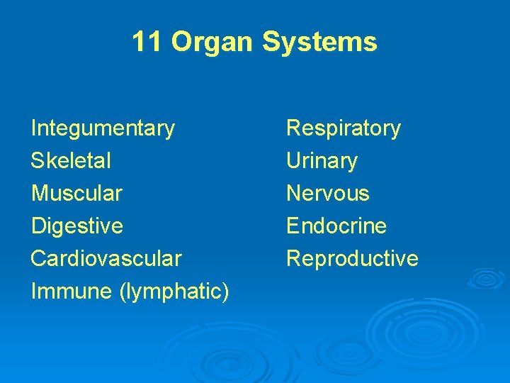
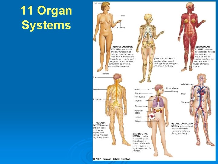
11 Organ Systems Integumentary Skeletal Muscular Digestive Cardiovascular Immune (lymphatic) Respiratory Urinary Nervous Endocrine Reproductive
11 Organ Systems
11 Organ Systems
Insert “Human organ systems” organ_systems_v 2. swf
Watch body systems work together by clicking on the figure in Thomson. NOW.
KEY CONCEPTS • Tissues and organs form the 11 main organ systems of a complex animal
Learning Objective 7 • Define homeostasis • Contrast negative and positive feedback systems
Homeostasis • Balanced internal environment (steady state) • Homeostatic mechanisms • control processes that maintain conditions
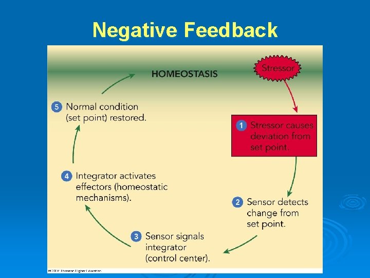
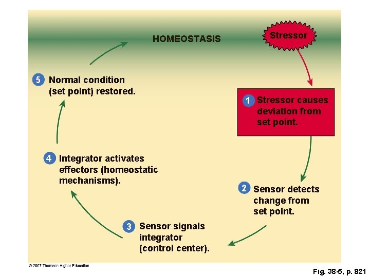
Negative Feedback Systems 1 • Maintain dynamic equilibrium (homeostasis) 1. Stressor • causes change in some steady state 2. Triggers a response • that opposes the change
Negative Feedback Systems 2 3. Sensor detects change • a deviation from desired condition (set point) 4. Sensor signals an integrator (control center) 5. Integrator activates effectors • organs or processes that restore steady state
Negative Feedback
HOMEOSTASIS 5 Normal condition (set point) restored. 4 Integrator activates effectors (homeostatic mechanisms). Stressor 1 Stressor causes deviation from set point. 2 Sensor detects change from set point. 3 Sensor signals integrator (control center). Fig. 38 -5, p. 821
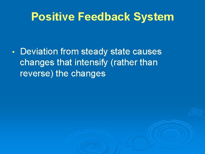
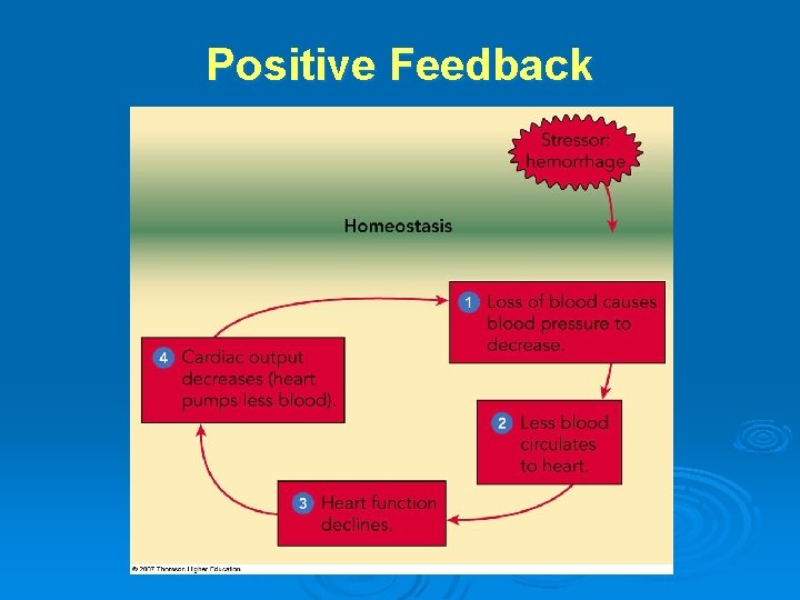
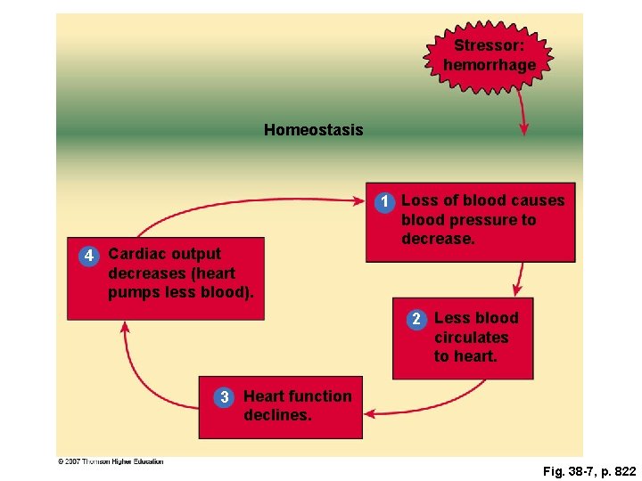
Positive Feedback System • Deviation from steady state causes changes that intensify (rather than reverse) the changes
Positive Feedback
Stressor: hemorrhage Homeostasis 4 Cardiac output decreases (heart pumps less blood). 1 Loss of blood causes blood pressure to decrease. 2 Less blood circulates to heart. 3 Heart function declines. Fig. 38 -7, p. 822
KEY CONCEPTS • Homeostatic mechanisms are responsible for the body’s automatic tendency to maintain a relatively stable internal environment
Learning Objective 8 • Compare the costs and benefits of ectothermy
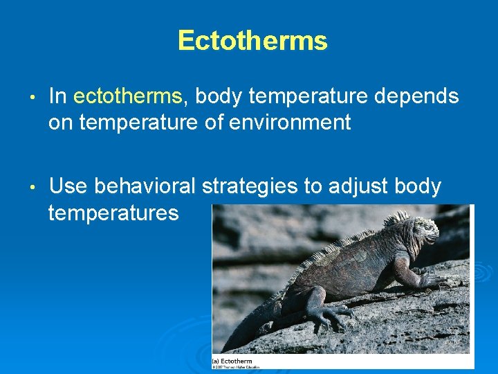
Thermoregulation • Process of maintaining body temperature within certain limits • • despite changes in surrounding temperature Animals have different structural, behavioral, and physiological strategies
Ectotherms • In ectotherms, body temperature depends on temperature of environment • Use behavioral strategies to adjust body temperatures
Costs and Benefits • Benefits of ectothermy • • • very little energy used to maintain the metabolic rate ectotherms can survive on less food Disadvantage of ectothermy • activity limited by daily and seasonal temperature conditions
Learning Objective 9 • Compare the costs and benefits of endothermy • Describe strategies animals use to adjust to challenging temperature changes
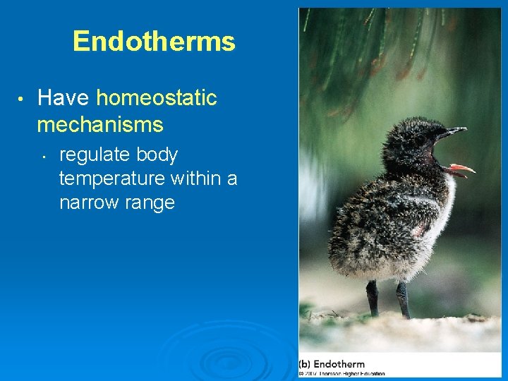
Endotherms • Have homeostatic mechanisms • regulate body temperature within a narrow range
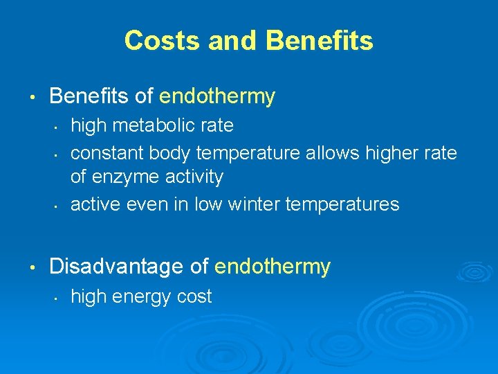
Costs and Benefits • Benefits of endothermy • • high metabolic rate constant body temperature allows higher rate of enzyme activity active even in low winter temperatures Disadvantage of endothermy • high energy cost
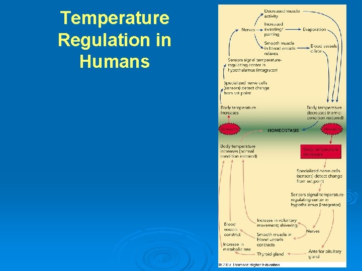
Temperature Regulation in Humans
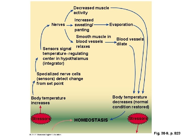
Decreased muscle activity Nerves Increased sweating/ panting Evaporation Smooth muscle in blood vessels relaxes Blood vessels dilate Sensors signal temperature- regulating center in hypothalamus (integrator) Specialized nerve cells (sensors) detect change from set point Body temperature decreases (normal condition restored) Body temperature increases Stressors HOMEOSTASIS Stressors Fig. 38 -9, p. 823
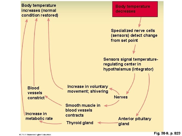
Body temperature increases (normal condition restored) Body temperature decreases Specialized nerve cells (sensors) detect change from set point Sensors signal temperatureregulating center in hypothalamus (integrator) Blood vessels constrict Increase in metabolic rate Increase in voluntary movement; shivering Nerves Smooth muscle in blood vessels contracts Thyroid gland Anterior pituitary gland Fig. 38 -9, p. 823
Insert “Control of human body temperature” hot_guy_m. swf
Acclimatization • Process of adjustment to seasonal changes
Torpor • • Hibernation • • adaptive hypothermia (in small endotherms when surrounding temperature drops) long-term torpor in response to winter cold Estivation • torpor due to lack of food or water during summer heat
KEY CONCEPTS • Thermoregulation contributes to homeostasis
Insert “Human thermoregulation” temp_regulation. swf
Explore negative feedback and temperature regulation in humans and other animals by clicking on the figures in Thomson. NOW.