Animal Physiology Respiration Hierarchy within living organisms Biological

Animal Physiology: Respiration

Hierarchy within living organisms….

Biological Systems in Animals n n n A system is a group of organs that work together and provide an organism with an advantage for survival A system is the most complex organization in your body and the final level of the progression from cells to tissues to organ systems Systems work alone and with other systems to allow your body to maintain homeostasis ¡ HOMEOSTASIS is a stable/balanced internal environment that allows you (and your cells) to survive.
")
Respiration n Respiration includes all processes involved in the exchange of oxygen (O 2) and carbon dioxide (CO 2) between cells and their environment.
. Gas exchange")
This includes: n n n Breathing (exchange between internal and external environment). Gas exchange across a respiratory surface (Exchange of gases at alveoli in lungs and between circulatory system and body cells) Cellular respiration (use of oxygen to burn glucose for energy! … That’s WHY we need the oxygen in the first place)
 Respiratory System n n Consists of: ¡ passages that transport air to")
Human (Mammalian) Respiratory System n n Consists of: ¡ passages that transport air to and from the lungs ¡ The air sacs of the lungs in which gas exchange takes place Divided into upper respiratory tract and lower respiratory tract: Upper – consists of parts outside thoracic cavity – nose, nasal cavity, pharynx, larynx, and upper trachea Lower – consists of parts inside thoracic cavity – lower trachea, bronchi and bronchioles, and lungs.

Upper and Lower Respiratory Tract

The Task of Respiration n n Whether an organism is single celled or multicellular each cell must be able to uptake O 2 and get rid of CO 2. This is GAS EXCHANGE. Every respiratory system has these requirements: ¡ Thin permeable membrane for diffusion of molecules ¡ Large surface area ¡ Blood supply ¡ Breathing system to obtain O 2 nhttp: //www. youtube. com/watch? v=Mr. Dbi. KQOtl. U

Mammalian Respiratory System Requirements 1. 2. 3. 4. Thin permeable membrane for diffusion of molecules Large surface area Blood supply Breathing system to obtain O 2

Your Task n n Fill in the diagram of the respiratory system and the table of structures. Be able to trace the path of an oxygen molecule from the air around to until it reaches the cells of your body.

Parts of Our Respiratory System n Air enters through our nasal cavities or the mouth (most breathing is through the nose)

Nasal Passage: n n n Filters air – traps foreign particles with nostril hairs and mucus. Warms air – blood vessels lining the nasal passages warm the air which allows diffusion of oxygen to occur faster. Moistens air – mucus also moistens air. Air needs to be moist so oxygen will dissolve at the alveolar surface.

Pharynx n n n Common passage for both food and air It continues to filter, warm and moisten the air At the end of the pharynx it branches into the esophagus (carries food) and the trachea (carries air)

Trachea n n Hollow tube with cartilage rings that provide strength to keep the tube open. The rings also provide flexibility so you can twist your need without cutting off flow. Trachea is lined with: ¡ ¡ Mucus – trap particles and moisten air Cilia - are microscopic hair-like structures. They beat up to 16 X per second, pushing trapped particles up into the pharynx to be either swallowed or coughed out. Polluted air can clog or paralyze cilia.

Trachea n At the top of the trachea, are the ¡ Epiglottis – a flap of tissue that covers over the top of the trachea during swallowing. ¡ Larynx – voice box consisting of two bands of tissue (called vocal cords) stretched across the top of the trachea. Air forced between vocal cords causes them to vibrate, producing sound.

Bronchi n n n The trachea branches into the bronchi which are extensions of muscle and cartilage that are lined with cilia and mucus. Each bronchus (singular) takes air to each lung. Bronchi branch into smaller and smaller bronchioles reaching all parts of the lungs.

Lungs n n Lungs are a pair of large soft bag-like organs consisting of a right lung (three lobes) and a left lung (2 lobes). The left side is smaller to make room for the heart. Lungs are primarily made of alveoli which are microscopic air sacs.

Alveoli n n The alveoli increase surface area for maximum gas exchange. Each is surrounded by capillaries so that gases can diffuse in and out of the blood easily. Alveolar walls consist of flat epithelial cells, one cell thick and are moist to allow diffusion of oxygen in and carbon dioxide out. Alveoli are arranged in clusters at the end of the bronchioles.

Alveoli

Cellular Respiration n n C 6 H 12 O 6 + 6 O 2 + 36 ADP + 36 Pi 6 CO 2 + 6 H 2 O + 36 ATP + thermal energy ATP powers all energy-requiring processes; growth, movement, building molecules
 nose: opening supported by bone and cartilage provides entrance for air nasal")
Structure Function(s) nose: opening supported by bone and cartilage provides entrance for air nasal cavity: cavity divided by the nasal septum mucous membrane filters, warms, and moistens air particles trapped and removed from air sinuses: spaces in bones of the skull lined with mucous membranes continuous with nasal cavity – also cleans air pharynx: back of mouth before entrance to trachea common passageway for air and food larynx: enlargement at top of trachea, composed of muscle and cartilage passageway for air and helps prevent foreign objects from entering trachea contains vocal chords: sounds produced by vibration as air passes over them trachea: pathway supported by cartilaginous rings mucous lining filters air pathway forks into right and left bronchi bonchi/bronchioles: branching tubes that becomes smaller and smaller, carrying air to sacs within lungs called alveoli mucous lining branching increases surface area for air carried deep into lungs mucous filters *bronchus = main tubes branching away from trachea *bronchioles = smaller branches alveoli: airs sacs - one cell thick, surrounded by capillaries gas exchange

More on gas exchange: • At LUNGS: – high alveolar O 2 means oxygen diffuses from air sac into bloodstream (at capillary) – high capillary CO 2 means carbon dioxide diffuses from blood into air sac (alveoli) • At BODY CELLS: – Oxygen diffuses from blood into cells, and carbon dioxide diffuses from cells into blood (blood travels back to lungs to get rid of this waste).

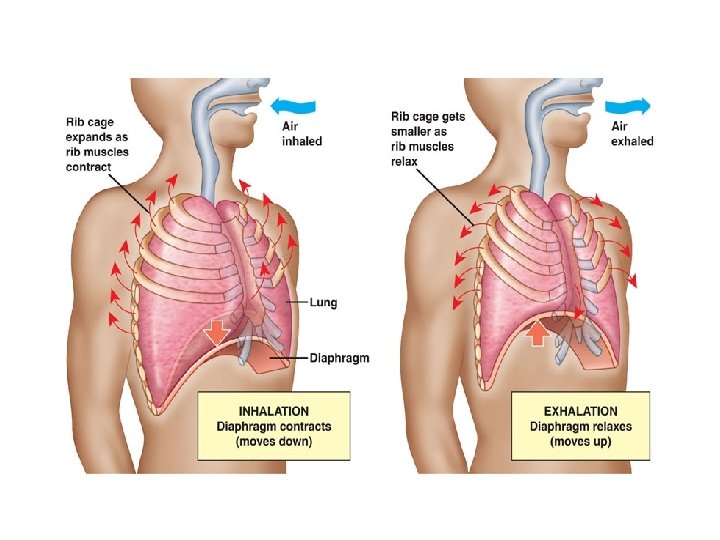
How we Breathe • Air within the lungs must be exchanged regularly in order to provide a fresh supply of oxygen and to carry away waste and carbon dioxide gas. • This involves two main stages of breathing: inspiration (inhaling), in which air is moved into the lungs, and exhalation (exhaling) where air is forced out of the lungs Understanding Air Pressure • Air will always move from an area of high pressure to an area of low pressure • If the air pressure inside the lungs is less than outside of the lungs, then the air will move into the lungs • Similarly, if the air pressure is greater inside the lungs than outside, the air will move out of the lungs

• The lungs are located within the thoracic cavity surrounded by the ribs and a thin sheet of muscle called the diaphragm. • During inspiration, the diaphragm contracts and moves downward, while the ribcage moves upward and out by the actions of the external intercostal muscles. This results in an increase in volume of the chest cavity; this lowers air pressure inside the chest. • Air then moves down through the trachea and fills the lungs • During expiration, the diaphragm relaxes and moves upward while the ribcage moves inward and down. This decreases the volume and increases the air pressure, forcing air out of the lungs. • Lungs continuously expand contract. This constant motion could result in damage to the delicate surface of the lungs. To prevent such damage, special pleural membranes cover the lungs and the inner walls of the chest cavity.

Video • http: //www. youtube. com/watch? v=Mr. Dbi. K QOtl. U

Control of Breathing Rate • The pattern of alternating inspiration and expiration is controlled by the autonomic nervous system, by nerve impulses form a breathing centre in your brain • The brain does not monitor oxygen levels to control breathing rate, instead it measures carbon dioxide levels in the blood as an indicator of the level of activity (medulla oblongata -between your brain and spine. • the medulla oblongata sends out nerve impulses to direct the action of the intercostals muscles and diaphragm • As you exercise vigorously, you consume oxygen quickly and produce a lot of carbon dioxide. • chemoreceptors in the aorta and carotid artery send messages to the MO to increase breathing rate if CO 2 levels are too high • The greater the level of carbon dioxide in your blood, the faster the breathing rate will be.

Breathing at Extremes • At high altitudes, there is less oxygen in the air, and our bodies compensate both by increasing our breathing rate and, over a period of days and weeks, by gradually increasing our number of red blood cells. • Scuba divers always carry their air supply in tanks and use special devices called regulators to compensate for the changes in air pressure at different depths. • Professional divers may use mixtures of gases, for safety and to extend their dive times. • Pure oxygen is often deadly when breathed at depths below approximately 7 m.

Your Task n n n Read pp. 438 -445 # 1 -5, 7 -9, 11 Learn about Disorders of the respiratory System (Know at least 2 well for the test. ) Read pp. 452 – 458
- Slides: 29