ANIMAL HISTOLOGY BIOL 241 Topic 12 II Female

ANIMAL HISTOLOGY BIOL 241 Topic 12 -II: Female Reproductive System Dr. Issa Al-Amri Department of Biological Sciences & Chemistry College of Arts & Sciences

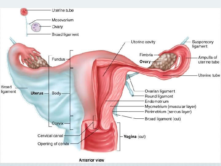
Introduction A. Female Reproductive System consists of the paired ovaries and oviducts; uterus, vagina, and external genitalia; and paired mammary glands B. It undergoes marked changes at the onset of puberty, which is initiated by menarche. C. It exhibits monthly menstrual cycles and menses from puberty until end of reproductive years, which terminate at menopause.

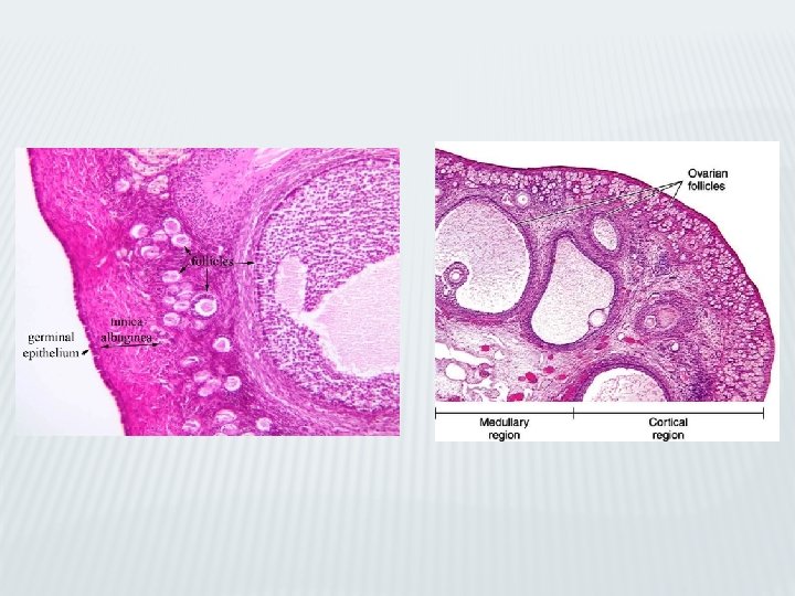
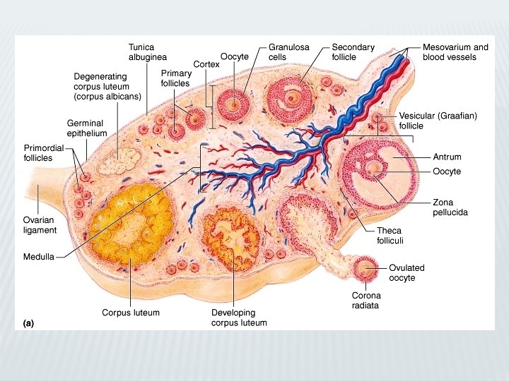
Ovaries I. Ovaries: 1. Ovaries covered by a simple cuboidal epithelium called germinal epithelium. 2. Deep to germinal epithelium, ovaries possess a capsule, tunica albuginea, composed of dense, irregular collagenous connective tissue. 3. Each ovary subdivided into cortex and medulla.

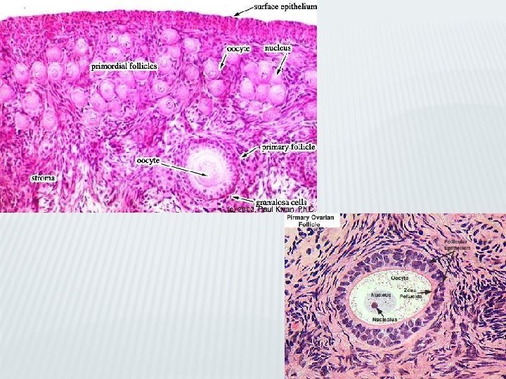
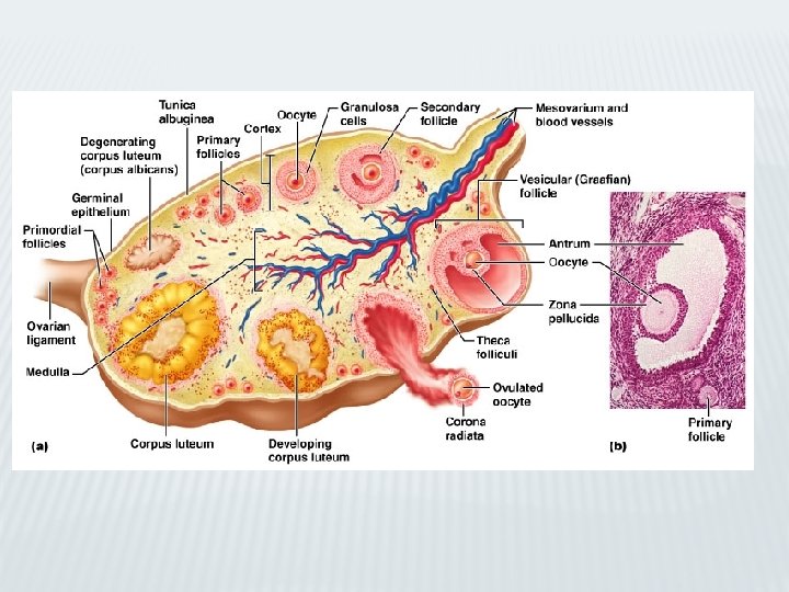
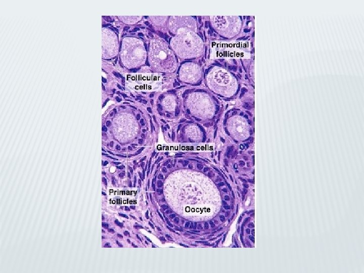
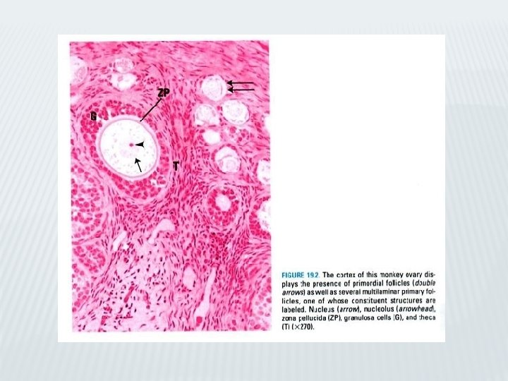
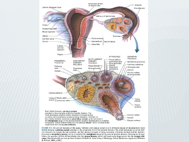
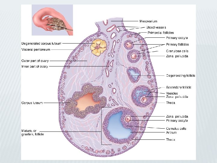
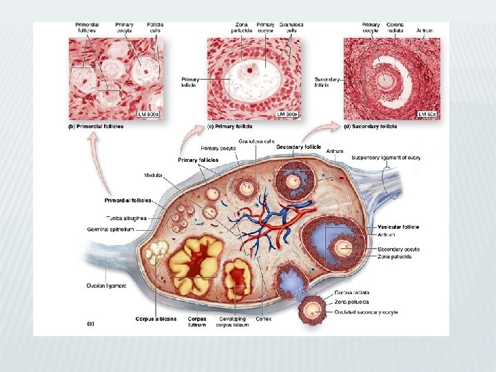
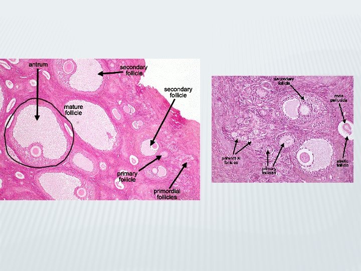
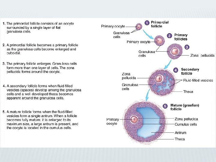
Ovaries: follicles: primary oocytes A. Ovarian cortex: • Consists of ovarian follicles (various stages of development) and connective tissue stroma (contain cells respond to hormonal stimuli). 1. Ovarian follicles: a. Primordial follicles: composed of primary oocyte enveloped by layer of flat follicular cells. (1) Primary oocytes: (a) Have prominent, acentric, vesicular-appearing nucleus (germinal vesicle) with single nucleolus. (b) Many Golgi complexes, mitochondria, RER, well-developed annulate lamellae. (c) become arrested in prophase of meiosis I by paracrine factors produced by follicular cells during fetal life and remain in this stage until ovulation.
 Follicular cells: (a) Attached to one another by desmosomes. (b)")
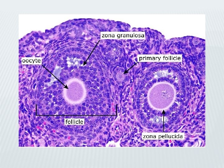
Ovaries: primary follicles (2) Follicular cells: (a) Attached to one another by desmosomes. (b) Separated from surrounding stroma by basal lamina. b. Growing follicles: (1) Primary follicles: are not dependent on follicle-stimulating hormone (FSH) for their development. • Contain amorphous layer (zona pellucida) surrounding and produced by primary oocyte; basal lamina present at interface of follicular cells with stroma.

TEM: primary follicle
 Secondary (antral) follicles: (a) Established when fluid (liquor folliculi; containing")
Ovaries: secondary follicles (2) Secondary (antral) follicles: (a) Established when fluid (liquor folliculi; containing hormones such as activin, estradiol, follistatin, inhibin, and progesterone) begins to accumulate in spaces between granulosa cells. • The fluid-filled spaces begin to coalesce, to form single large cavity called antrum. (a) They are dependent on FSH. (b) Narrow processes extend from granulosa cells into zona pellucida. (c) Granulosa cells contact each other via gap junctions, also form gap junctions with cell membrane of primary oocyte.
 and cortex(Co). -Medulla: large blood vessels(BV)")
Ovary Figure 1 : Ovary -Subdivided into medulla(Me) and cortex(Co). -Medulla: large blood vessels(BV) cortical vascular supply. -Cortex: many ovarian follicles, most very small (arrows). -Few maturing follicles reached Graafian follicle(GF) stage. -Thick, fibrous connective tissue capsule: tunica albuginea(TA). -Germinal epithelium(GE), mesovarium(Mo) not only suspends ovary but also conveys vascular supply to medulla. Figure 2: Ovary Figure 3: Primary follicles Figure 4: Secondary follicles -Higher mag. of Figure 1. -Germinal epithelium(GE) covers tunica albuginea(TA). This region of cortex(Co) has many primordial follicles(PF). -Cconnective tissue of ovary highly cellular: stroma(St). -Box: primary oocyte(PO), nucleus(N), nucleolus (arrow). -Single layer of flat follicular cells(FC) surrounding oocyte. -Tunica albuginea(TA), germinalepithelium(GE). -Single layer of cuboidal follicular cells(FC) surround small primary oocyte(PO), nucleus(N) -Multilaminar primary follicle displays primary oocyte(PO) -Follicular cells(FC) form stratified layer around oocyte, separated from it by zona pellucida(ZP). -Stroma(St) re-organized around follicle to form theca interna(TI). Basal membrane(BM) between follicular cells and theca interna. -Stratification of follicular cells(FC), follicular fluid(FF) appear in intercellular spaces, coalesces into several Call. Exner bodies. -Stroma surrounding follicular cells rearranged to form cellular theca interna(TI) and fibrous theca externa(TE).
 follicle: (1) Dominant graafian follicle is one follicle")
Ovaries: graffian follicle c. Graafian (mature) follicle: (1) Dominant graafian follicle is one follicle among secondary follicles that will ovulate. • FSH independent, manufactures hormone inhibin (shuts off FSH release by basophils of anterior pituitary) causing atresia of other developing follicles (secondary and nondominant graafian follicles). (2) Measures 2. 5 cm in diameter, appear as large bulge on surface of ovary.
 Primary oocyte positioned off center on small mound of granulosa")
Ovaries: graffian follicle (3) Primary oocyte positioned off center on small mound of granulosa cells (cumulus oophorus) projects into liquor folliculi-containing antrum of follicle. • Granulosa cells surround zona pellucida (corona radiata). Other granulosa cells line antrum, forming membrana granulosa. (4) Theca interna cells manufacture androgens (transferred to granulosa cells) which are converted into estrogens. (5) Theca externa is mostly collagenous. It contains a few smooth muscle cells and many blood vessels, which provide nourishment to theca interna.
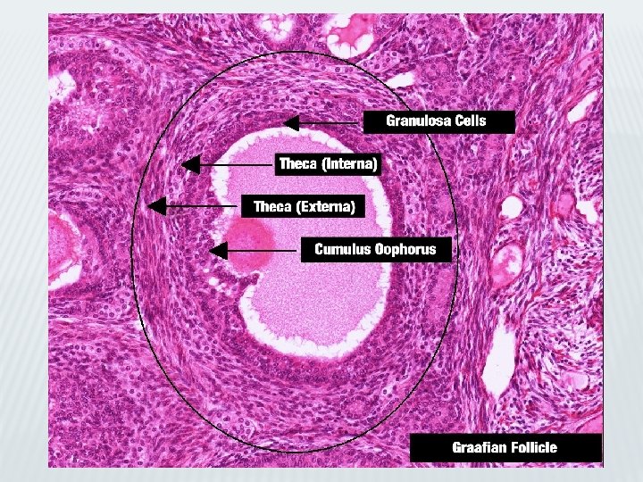
 fills single chamber, :")
Ovary, Corpus Luteum Figure 1 : Graafian follicle -Follicular fluid(FL) fills single chamber, : antrum, surrounded by wall of granulosa (follicular) cells known as membrana granulosa(MG). -Granulosa cells, surround primary oocyte(PO), jut into antrum as cumulus oophorus(CO). -Basal membrane(BM), separates granulosa cells from theca interna(TI). -Theca externa(TE) merges with surrounding stroma. Figure 2: Graafian follicle Figure 3: Corpus luteum Figure 4: Corpus luteum -Higher mag. of Figure 1. -Primary oocyte(PO), nucleus(N), zona pellucida(ZP). -Processes (arrows) of surrounding follicular cells extend into this acellular region. -Single layer of follicular cells radiate as a crown at periphery of primary oocyte: corona radiata(CR). -Basal membrane(BM), theca interna(TI), theca externa(TE). -Cells of membrana granulosa enlarge, become vesicular in appearance and folded: granulosa lutein cells(GL). -Spaces between folds occupied by connective tissue elements, blood vessels, cells of theca interna (arrows). -Theca interna cells also enlarge, become glandular: theca lutein cells. -Remnants of antrum filled with fibrin serous exudate will be replaced by connective tissue elements. -Higher mag. of Figure 3. -Granulosa lutein cells(GL), connective tissue(CT) elements. -GL display round nuclei(N), mostly in center of large round cells (arrowheads). -Center of field occupied by fold, housing theca lutein cells(TL), connective tissue(CT) and vascular(BV) elements.

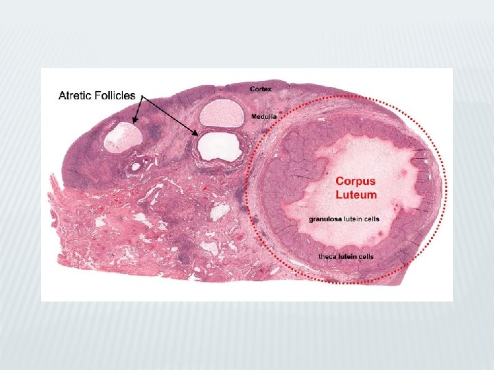
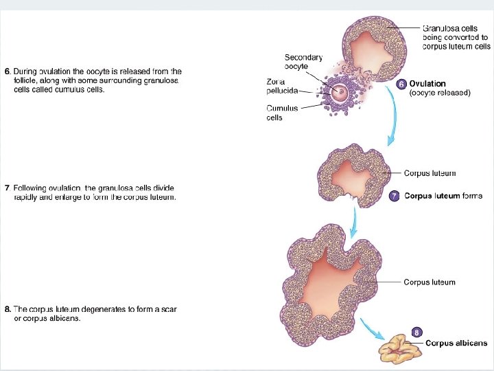
Ovaries: corpus luteum 2. Corpus hemorrhagicus: formed from remnants of graafian follicle. 3. Corpus luteum: (1) Formed from corpus hemorrhagicus. (2) Composed of granulosa lutein cells (modified granulosa cells) and theca lutein cells (modified theca interna cells). (3) Formation of this richly vascularized temporary endocrine gland is dependent on LH.
, pale")
Ovaries: corpus luteum a. Granulosa lutein cells: b. Large (30 µm in diameter), pale cells contain abundant SER, RER, many mitochondria, well-developed Golgi complex, and lipid droplets. (1) Derived from cells of membrana granulosa. (2) Function: manufacture most of body’s progesterone and convert androgens formed by theca lutein cells to estrogens. b. Theca lutein cells: (1) Small (15 µm in diameter) cells concentrated mainly along periphery of corpus luteum. (2) Derived from cells of theca interna. (3) Function. manufacture progesterone androgens and small amounts of estrogen.

Ovaries: corpus luteum 4. Corpus albicans: • Remnant of degenerated corpus luteum. • Its formation due to hypoxic conditions present in corpus luteum as fibroblasts manufacture an overabundance of collagen. • The fibrotic event elicits the arrival of T cells that release interferon-γ, a chemoattractant for macrophages. • These cells release tumor necrosis factor α, a cytokine that drives both granulosa lutein and theca lutein cells into apoptosis. • As cell death and fibrosis progresses, corpus albicans contracts and becomes a small scar on surface of ovary.
; cytoplasm appears")
Ovary and Oviduct Figure 1 : Corpus luteum -Large granulosa lutein cells(GL); cytoplasm appears vesicular. -Nuclei(N) of these cells farther away from each other than the nuclei of smaller theca lutein cells(TL), which also appear darker staining (arrow-heads). -Flattened nuclei (arrows) belong to various connective tissue cells. Figure 2: Corpus albicans Figure 3: Oviduct (C. S) Figure 4: Oviduct (C. S) -c. Crpus luteum invaded by macrophages phagocytose dead cells, leaving behind acellular fibrous tissue(FT). -Vascular supply(BV) regressed, and entire corpus albicans appears pale in comparison to dark staining of surrounding ovarian stroma(St). -Corpus albican will regress until it becomes small scar on surface of ovary. -Referred to fallopian or uterine tube, extends from ovary to uterine cavity. -Suspended from body wall by broad ligament(BL), which conveys vascular supply(BV) to serosa (S) of oviduct. -Thick muscularis(M): inner circular and outer longitudinal muscle layers. -Mmucosa(Mu): longitudinal folds in infundibulum and ampulla: they subdivide lumen(L) into labyrinthine spaces. -Higher mag. of Figure 3. -Thickness of oviduct wall with: vascular(BV)serosa(S); envelops thick muscularis, whose outer longitudinal(OL) and inner circular(IC) layers not well delineated. -Mucosa(Mu) highly folded and lined by simple columnar epithelium(Ep). -Loose connective tissue of lamina propria(LP) richly vascularized (arrows).

Ovaries: atretic follicles – ovarian medulla 5. Atretic follicles: a. Follicles (in various stages of maturation) undergoing degeneration. b. Present in ovary; after graafian follicle ovulates, the remaining graafian and secondary follicles degenerate. c. Show pyknotic changes in nuclei of granulosa cells and other degenerative changes. C. Ovarian medulla: • Contains large blood vessels, lymphatic vessels, and nerve fibers in a loose connective tissue stroma. • Possess small number of estrogen-secreting interstitial cells and few androgen-secreting hilus cells.
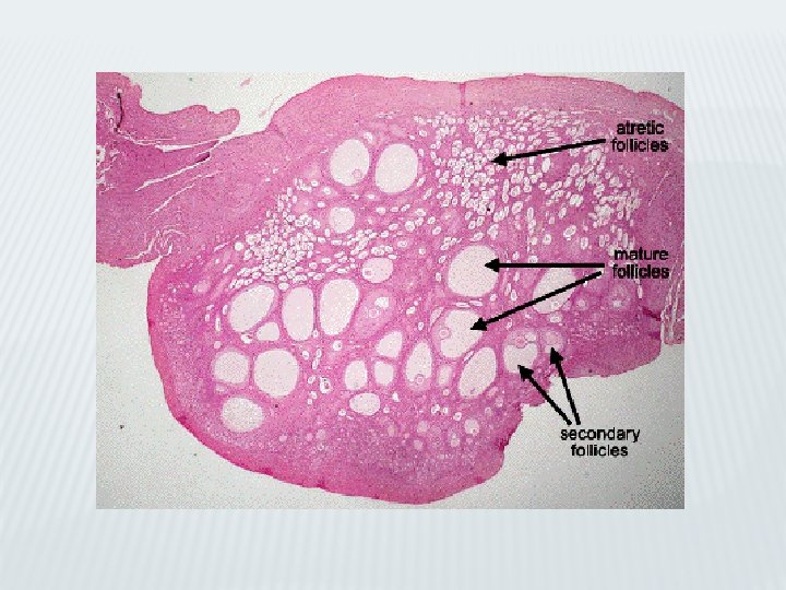

H&E Ovary: cortex, medulla and follicles at various stages of development
 • Each of two oviducts is subdivided into four")
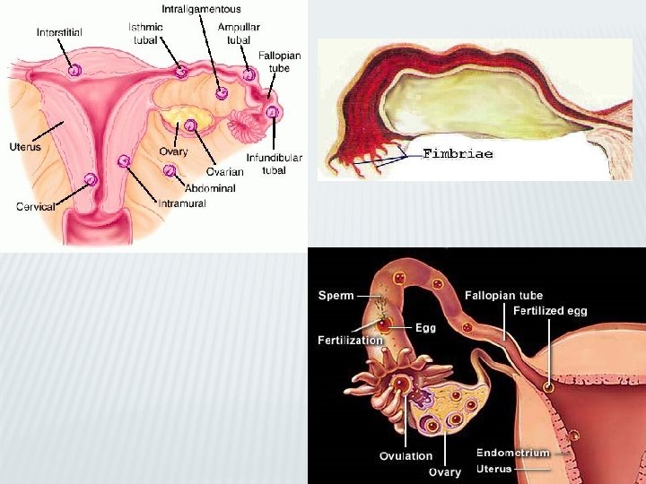
Oviducts II. Oviducts (Fallopian Tubes) • Each of two oviducts is subdivided into four regions: 1. Infundibulum: has a fimbriated end. 2. Ampulla: most common site of fertilization; 3. Isthmus, and 4. Intramural portion: parallel to the wall of uterus. • Wall of each oviduct consists of: mucosa, muscularis, and serosa.

H&E: Wall of oviduct
 Figure 1 : Oviduct (C. S) -Higher mag. of Figure 4")
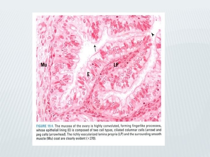
Oviduct (LM, EM) Figure 1 : Oviduct (C. S) -Higher mag. of Figure 4 of previous slide. -Inner circular muscle (IC) layer of the muscularis. -Lamina propria(LP) narrow in this region (arrows)but presents longitudinal epithelially lined folds. -Core of these folds made of vascular(BV), loose, highly cellular connective tissue(CT). -Simple columnar epithelium(Ep) lines labyrinthine lumen(L) of oviduct. Figure 2: Oviduct (C. S) Figure 3: Oviduct epithelium -Higher mag. of Figure 1. -Lamina propria(LP) highly cellular, loose connective tissue richly vascularized. -Basal membrane(BM) separating connective tissue from epithelial lining. -Epithelium made of two different cell types, a thinner peg cell(PC), no cilia but whose apical extent bulges above the ciliated cells. -These bulges (arrowheads) contain nutritive materials that nourish gametes. -Second cell type is ciliated cell(CL), whose cilia move in unison with those of neighboring cells, propelling nutrient material toward uterine lumen. -Human oviduct at midcycle (day 14) presents two types of epithelial cells, the peg cell(PC) and the ciliatedcell(CC). -Former are secretory cells, as indicated by their extensive Golgi apparatus(GA) situated in the region of cell apical to nucleus(N). -Electron-dense secretory products (arrows) in expanded, apical free ends of these cells. -Some ciliated cells display large accumulations of glycogen(Gl) at either pole of the nucleus.

Oviducts: mucosa A. Mucosa: • • Has extensive longitudinal folds in infundibulum. Degree of folding progressively decreases in remaining three regions of the oviduct. 1. Epithelium: simple columnar, consists of peg cells and ciliated cells. • Ciliated cells: (1) Ciliated cells: contain many cilia, which beat mostly toward lumen of uterus. (2) Function: aid in transport of developing embryo to uterus. 2. Lamina propria: consists of loose connective tissue containing reticularfibers, fibroblasts, mast cells, and lymphoid cells.

Mucosa of oviduct

Oviducts: muscalaris , serosa B. Muscularis: 1. Composed of an ill-defined inner circular and an outer longitudinal layer of smooth muscle. 2. Function: By contracting rhythmically; muscularis assists in moving embryo toward uterus. C. Serosa: • Composed of simple squamous epithelium overlying a thin connective tissue layer, covers outer surface of oviduct.

Diagram of cross section of oviduct
, and cervix.")
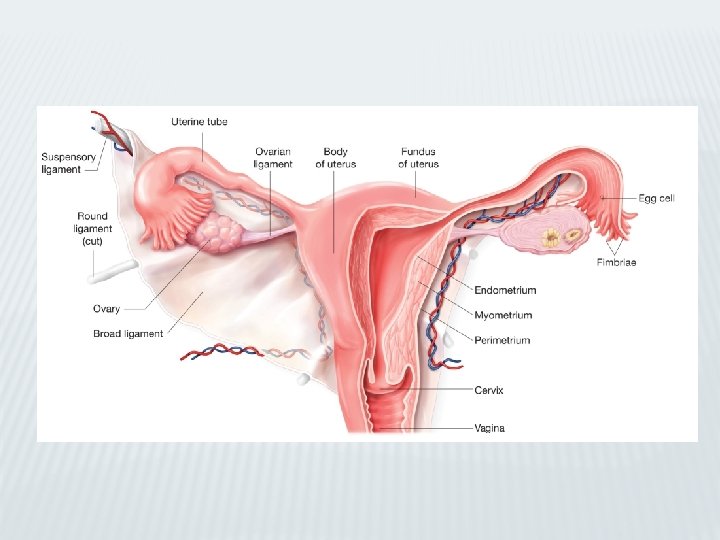
Uterus III. Uterus • Uterus has three regions; the fundus, body (corpus), and cervix. A. Uterine wall: • Consists of the endometrium, myometrium, and adventitia (or serosa).

Uterus: uterine wall: endometrium 1. Endometrium: • Composed of: epithelial lining and gland-rich connective tissue stroma; undergoes hormone-modulated cyclic alterations during menstrual cycle. • Lined by simple columnar epithelium containing secretory and ciliated cells. • Its stroma similar to mesenchymal connective tissue, with stellate cells and abundance of reticular fibers. • Stroma contain macrophages and leukocytes. • Stroma houses simple tubular glands of endometrium.
 Functional layer (functionalis): • Thick")
Uterus: uterine wall: endometrium b. Layers of endometrium: (1) Functional layer (functionalis): • Thick superficial layer of endometrium that is sloughed and reestablished monthly as result of hormonal changes during menstrual cycle. (2) Basal layer (basalis): • Deeper layer of endometrium that is preserved during menstruation. • Has endometrial glands, which have basal cells that provide a source for reepithelialization of endometrium after functional layer is shed.

Uterus: uterine wall: endometrium c. Endometrial vascular supply: • Consists of two types of arteries derived from vessels in stratum vasculare of myometrium. (1) Coiled arteries: extend into functional layer and undergo pronounced changes during various stages of menstrual cycle. (2) Straight arteries: do not undergo cyclic changes and terminate in basal layer.

Uterus: uterine wall: myometrium 2. Myometrium: a. thick smooth muscle tunic of uterus. b. composed of inner and outer longitudinal layers and a thick middle circular layer (stratum vasculare). c. thickens during pregnancy. d. Near end of pregnancy; develops many gap junctions between its smooth muscle cells (It coordinate contraction of muscle cells during parturition).

Uterus: uterine wall: external covering 3. External covering: a. Serosa: present over surfaces of uterus bulging into peritoneal cavity. b. Adventitia: present along retroperitoneal surfaces of uterus.

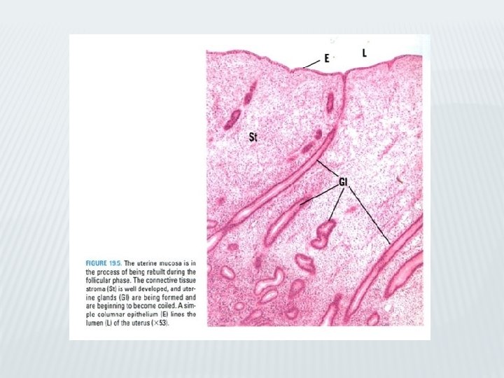
Uterus Figure 1 : Uterus, follicular phase -consists of three layers: external serosa not clear here. -Myometrium(My) made of smooth muscle: outer longitudinal(OL), middle circular(MC), and inner longitudinal(IL). -Endometrium(En): basal layer(B) and functional layer(F). -Functional layer is in process of being built up and forming glands(GL) are straight. -Deeper aspects of some of these glands display branching (arrow). Figure 2: Uterus, follicular phase Figure 3: Uterus, luteal phase Figure 4: Uterus, early luteal phase -Higher mag. of Figure 1. -Functional layer(F) lined by simple columnar epithelium(Ep) displaying mitotic activity (arrows). -Forming glands(GL) consist of simple columnar epithelium(Ep) whose cells actively dividing. -Stroma(St) highly cellular, vascular supply(BV). -Myometrium(My) of uterus remains constant during various endometrial phases. -Its three layers, richly vascularized: stratum vasculare(SV). -Endometrium(En) richly endowed with glands(GL). -Higher mag. of Figure 3. -Functional layer of endometrium covered by simple columnarepithelium(Ep), -Separating endometrial stroma(St)from uterine lumen(L). -Glands(GL), also composed of simple columnar epithelium, -These glands dilated and their lumina contain slight amount of secretory product (arrow).
 become quite tortuous and corkscrew-shaped,")
Uterus Figure 1 : Uterus, midluteal phase -Endometrial glands(GL) become quite tortuous and corkscrew-shaped, and simple columnar cells(CC) accumulate glycogen (arrow). -During this phase of endometrium, glycogen basally located, displacing nucleus(N) toward center of cell. -Stroma(St) undergoing decidual reaction in that some of the con-nective tissue cells enlarge as they become engorged with lipid and glycogen. Helical artery(HA) Figure 2: Uterus. Late luteal phase -endometrium glands assume a characteristic ladder shape(arrows). -Simple columnar epithelial cells(CC) are pale, position of glycogen now apical (arrowheads) rather than basal. -Apical location of glycogen imparts a ragged, torn appearance to free surface of these cells. -Lumina(L)of glands filled with glycogen-rich, viscous fluid. -Stroma(St) infiltrated by many leukocytes(Le). Figure 3: Uterus, menstrual phase -Of endometrium : periodic constriction and sequential opening of helical arteries(HA), result in ischemia with necrosis of superficial aspect of functional layer. -Due to contractions, sudden spurts of arterial blood detach necrotic fragments(NF) of superficial layers of endometrium that then dischargedas menstrual flow. -Endometrial stroma becomes engorged with blood, increasing degree of ischemia, entire functional layer desquamated. Lumen(L) no longer have complete epithelial lining (arrowheads). Figure 4: Uterus, menstrual phase -Higher mag. of Fig. 4: endometrial glands(GL) torn and necrotic fragment(NF) detached from functional layer(F) of endometrium. Stroma(St) infiltrated by leukocytes , whose dense nuclei(N) mask most of endometrialcells.
- Slides: 53