ANIMAL HISTOLOGY BIOL 241 Topic 11 Urinary System

ANIMAL HISTOLOGY BIOL 241 Topic 11: Urinary System Dr. Issa Al-Amri Department of Biological Sciences & Chemistry College of Arts & Sciences

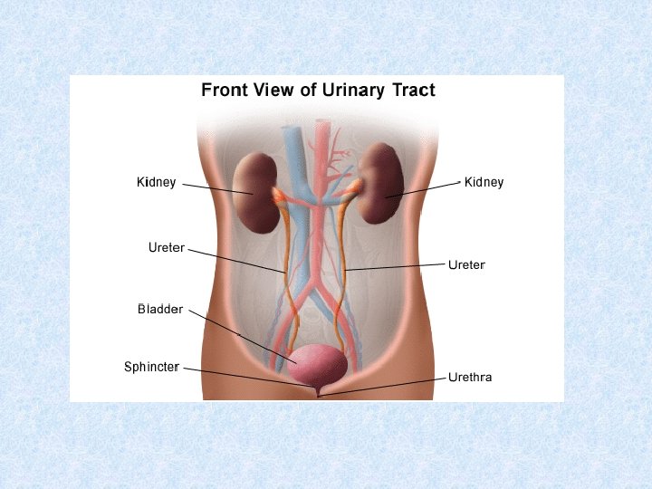
Introduction A. Urinary system composed of paired kidneys and ureters and bladder and urethra. B. Function: urinary system produces and excretes urine, thereby clearing blood of waste products. • Kidneys also regulate electrolyte levels in the extracellular fluid and synthesize renin and erythropoietin.
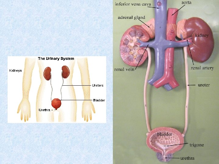
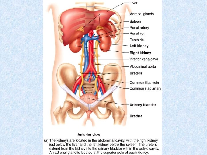

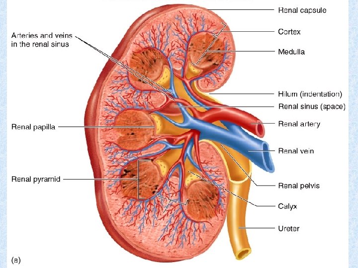
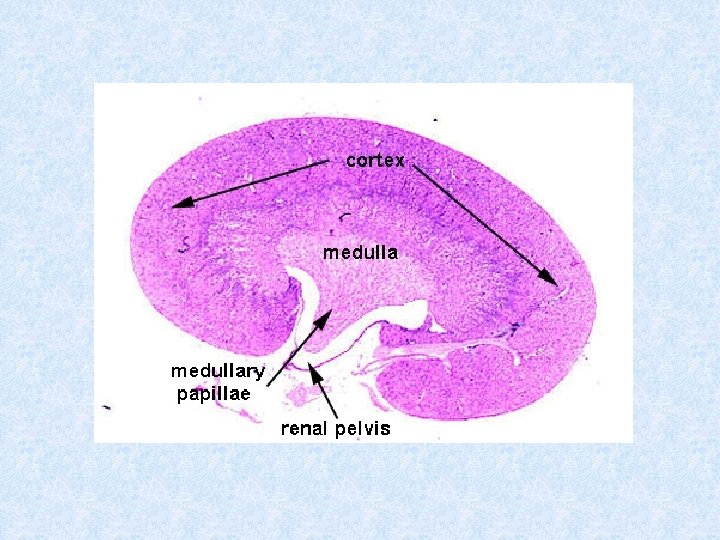
Kidneys I. Kidneys A. General structure: 1. Kidneys are paired bean-shaped organs enveloped by a thin capsule of connective tissue. 2. Each kidney divided into outer cortex and inner medulla. 3. Each kidney contains 2 million nephrons. A nephron and collecting tubule into which it drains form a uriniferous tubule. B. Renal hilum: a concavity on medial border of kidney. It houses arteries, veins, lymphatic vessels, nerves, and the renal pelvis.
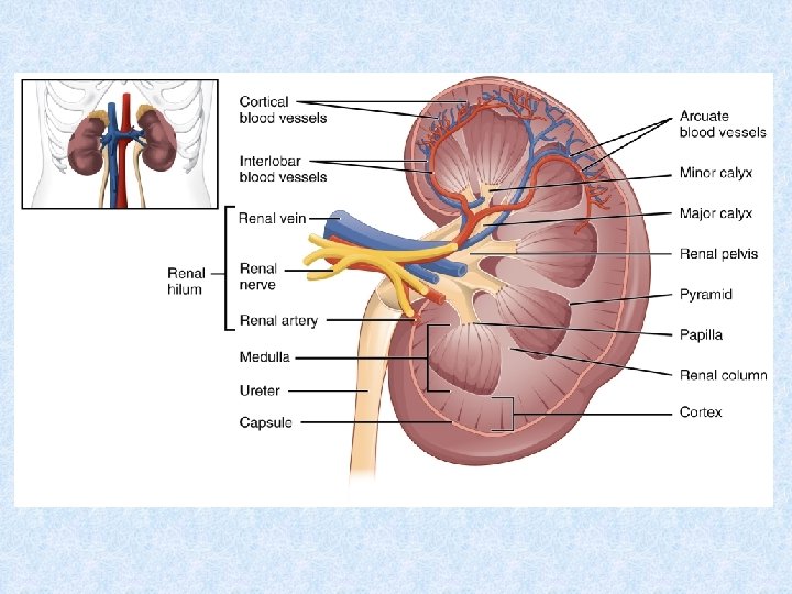

Kidneys C. Renal pelvis: funnel-shaped expansion of upper end of ureter. It is continuous with major renal calyces, which in turn have several small branches, the minor calyces. D. Renal medulla: lies deep to the cortex but sends extensions (medullary rays) into cortex. 1. Renal (medullary) pyramids are conical or pyramidal structures that compose the bulk of renal medulla. a. Each kidney contains 10 - 18 renal pyramids. b. Each pyramid consists of thin limbs of loops of Henle, blood vessels, and collecting tubules. 2. Renal papilla is located at the apex of each renal pyramid. It has a perforated tip (area cribrosa) that projects into the lumen of a minor calyx.

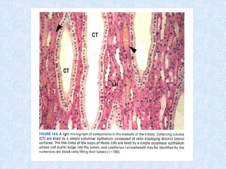
Renal Medulla Figure 1 : Renal medulla -Connective tissue elements among the tubules and vessels are very sparse and constitute mainly fibroblasts, macrophages, and fibers (asterisks). -Collecting tubules(CT). -Thick limbs of Henle’s loop(TH), thin limbs of Henle’s loop(TL). -Arteriolae rectae spuriae(AR), venulaerectae spuriae(VR). Figure 2: Renal papilla (C. S) Figure 3: Renal papilla (C. S) Figure 4: Renal medulla (L. S) -Collecting tubules(CT) with cuboidal cells. -Thin limbs of Henle’s loop(TL). -Arteriolae rectae spuriae(AR) venulae rectae spuriae(VR) -Formed connective tissue elements (asterisks). -Thick limb of Henle’s loop(TH). -Deeper medulla, collecting tubules merge with each other, forming larger structures. T -The largest ducts known as papillaryducts (PD), or ducts of Bellini, with tall, pale columnar cells and lateral plasma membranes (arrows). -Thin limbs of Henle’s loop(TL). -Arteriolae rectae spuriae(AR), thevenulae rectae spuriae(VR) -Connective tissue elements marked by asterisks. -Collecting tubule(CT), withtall cuboidal cells -Collecting tubule flanked by thick limbs of Henle’s loop(TH). -Vasa recta filled with blood, and thickness of their walls identifies whether they are arteriolae rectae spuriae(AR) or venulae rectae spuriae(VR). -Thin limbof Henle’s loop(TL).

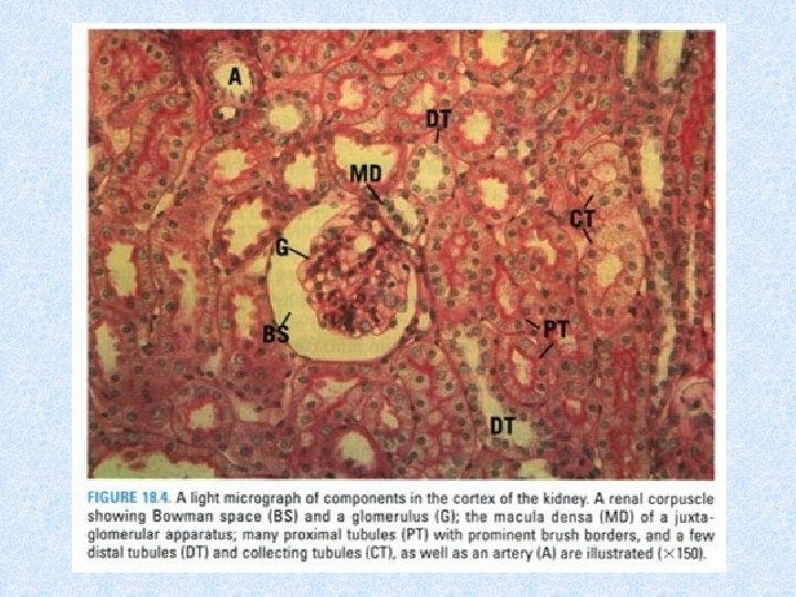
Kidneys E. Renal cortex: superficial layer of kidney beneath capsule. It consists of renal corpuscles and convoluted tubules. 1. Renal columns of Bertin: extensions of cortical tissue between adjacent renal pyramids. 2. Medullary rays: groups of straight tubules that extend from the base of each renal (medullary) pyramid into the cortex. F. Renal lobe: consists of a renal pyramid and its closely associated cortical tissue.
, cortex(C), medulla(M), medullary rays(MR). -cortical")
Kidney Figure 1 : Kidney cortex and mudella -Capsule(Ca), cortex(C), medulla(M), medullary rays(MR). -cortical labyrinth(CL). renal corpuscles(RC). , -These are the first part of nephrons, and their location in cortex indicative of their time of development as well as of their function. -They referred to: superficia l(1), midcortical (2), or juxtamedullary nephrons(3). -Lobule extends into medulla, but its borders are undefinable histologically (approx. vertical lines). -Arcuate vessels(AV), in cortical labyrinth; interlobular vessels(IV). Figure 2: Kidney capsule Figure 3: Kidney cortex Figure 4: Colored colloidin-injected kidney -Capsule(Ca), fibroblasts (Fb), capsular vessels(CV). – Capillary network (CN), proximal convoluted tubules(PT). -Two renal corpuscles(RC), Bowman’s space(BS). -Proximal convoluted tubules(PT), distal convoluted tubules(DT), macula densa(MD). -Medullary rays contain pars recta(PR) of proximal tubule. -Ascending thick limbs of Henle’s loop(AT), and collecting tubules(CT). -Each renal corpuscle has tufts of capillaries, glomerulus(G), supplied by afferent glomerular arteriole(AA), drained by efferent glomerular arteriole(EA). -Large vessel on lower right is interlobular artery(IA)

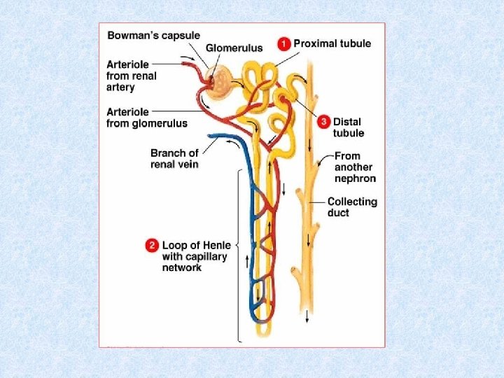
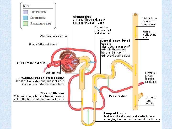
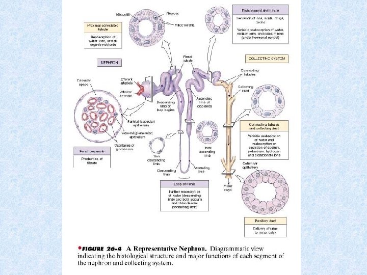
Uriniferous tubules III. Uriniferous Tubules A. Nephrons: • Consist of renal corpuscle, proximal convoluted tubule, loop of Henle, and distal convoluted tubule. 1. Classification: • Nephrons classified as cortical or juxtamedullary, depending on location of renal corpuscle. • Juxtamedullary nephrons possess longer loops of Henle than cortical nephrons and are responsible for establishing the interstitial concentration gradient in the medulla.
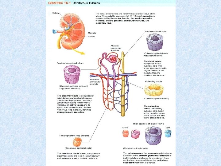
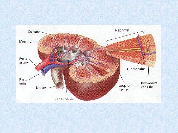

Diagram of a nephron
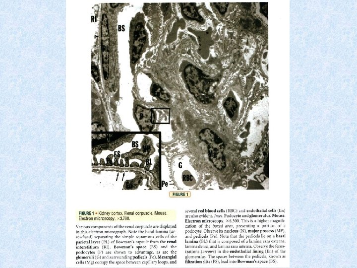

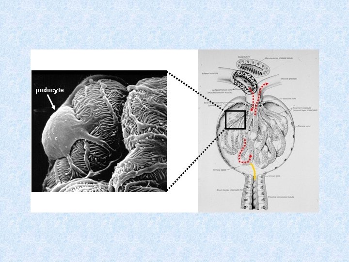
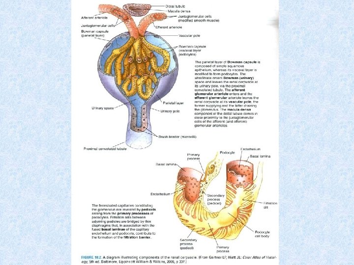
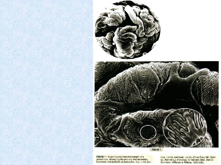
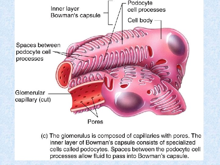
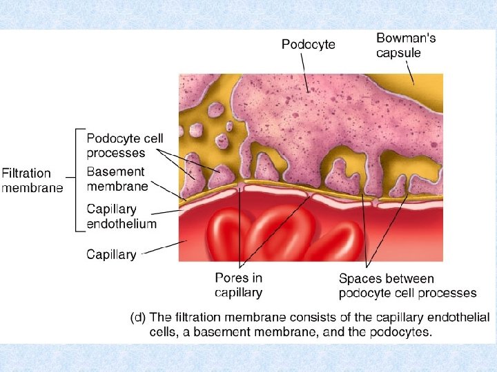
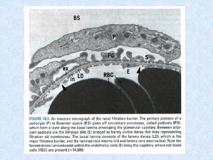
Uriniferous tubules b. Podocytes: modified epithelial cells that form visceral layer of Bowman capsule. • Contain several primary processes that give rise to many secondary processes called pedicels. (1) Pedicels: a. Pedicels embrace the glomerular capillaries and interdigitate with pedicels arising from other primary processes. b. Their surfaces facing the Bowman space are coated with podocalyxin, a protein that is thought to maintain their organization and shape. (2) Filtration slits: elongated spaces between adjacent pedicels. Diaphragms, composed of a layer of filamentous material, bridge each filtration slit.
")
Uriniferous tubules c. Renal glomerulus: tuft of capillaries that extends into Bowman capsule. (1) Glomerular endothelial cells: (a) Form inner layer of capillary walls. (b) Have thin cytoplasm that is thicker around nucleus, where most organelles are located. (c) Possess large fenestrae (60– 90 nm in diameter) but lack thin diaphragms that typically span the openings in other fenestrated capillaries.
. -Glomerulus(G),")
Renal Cortex Figure 1 : Kidney cortical labyrinth -Cconvoluted portion of the proximaltubule(PT). -Glomerulus(G), Bowman’s capsule (podocytes) associated with glomerulus, Bowman’s space(BS), -into which ultrafiltrate expressed from capillaries -Parietal layer(PL) of Bowman’s capsule made of simple squamous epithelium. -Mesangial cells present in renal corpuscle. -Darker-staining proximal tubules(PT). - Lighter-staining distal tubules(DT). Figure 2: Kidney cortical labyrinth Figure 3: Kidney cortical labyrinth Figure 4: Juxtaglomerular apparatus -Vascular pole(VP): region where afferent and efferent glomerular arterioles enter and leave renal corpuscle. -Modified smooth muscle cells of afferent glomerular arterioles known as juxtaglomerular cells (JC). -macula densa (MD) region of distal tubule. -Proximal tubules(PT), rich vascularity (BV) of renal cortex. -Scant amount of connective tissue elements (arrows). -Afferent glomerular arteriole (AA). -Eefferent glomerular arteriole(EA) -Mesangial cells(Mg), distal(DT), proximal(PT) tubules. -Endothelial cell(En), podocytes(P), visceral cell layer of Bowman’s capsule, by a thick basal lamina (arrows). -Mesangial cells(Mg) -Major processes (asterisks) of podocytes -Macula densa(MD) region of distal tubule and apparent. juxtaglomerular cells(JC). -Modified smooth muscle cells of afferent glomerular arteriole(AA). granules (arrowheads) in JC. -Nuclei (asterisks) of endothelial cells of afferent

LM of glomerulus and tubules

TEM of glomerulus and tubules
 and podocytes with fenestrated processes (FPs)")
TEM of glomerulus basement memebrane (GBM) and podocytes with fenestrated processes (FPs)

Uriniferous tubules d. Renal filtration barrier: 1. Structure: composed of fenestrated endothelium of glomerular capillaries, basal lamina, and filtration slits with diaphragms between pedicels. 2. Function: permits passage of water, ions, small molecules from bloodstream to capsular space but prevents passage of large and -vely charged proteins, forming ultrafiltrate of blood plasma in Bowman space: (a) Laminae rarae: contain heparan sulfate, assists in restricting passage of -vely charged proteins into Bowman space. (b)Lamina densa: contains type IV collagen, acts as selective macromolecular filter preventing passage of large protein molecules (MW < 69, 000 daltons) into Bowman space.
: a. Lined by a layer of irregularly")
Uriniferous tubules 3. Proximal convoluted tubule (PCT): a. Lined by a layer of irregularly shaped (cuboidal to columnar) epithelial cells with microvilli (forming brush border). cells exhibit following structures: (1) Apical canaliculi, vesicles, and vacuoles (endocytic complex), function in protein absorption. (2) Prominent interdigitations along their lateral borders, which interlock adjacent cells with one another. (3) Numerous mitochondria compartmentalized in basal region by extensive infoldings of basal plasma membrane, which supply energy for active transport of Na+ out of tubule.
 (2) (3) Descending thick limb of")
Uriniferous tubules 4. Loop of Henle: a. (1) (2) (3) Descending thick limb of Henle loop: Descending limb of Henle loop (pars recta) of proximal tubule. Lined by simple cuboidal epithelium; has brush border. Function: resorb, exchange, and secrete similar to that of proximal convoluted tubule. b. Thin limb of Henle loop: (1) Composed of descending segment (loop) and ascending segment, lined by simple squamous epithelial cells (few short microvilli). Nuclei bulge into lumen. (2) In juxtamedullary nephrons, thin limb divided into three portions on basis of shape of epithelial cells, organelle content, depth of tight junctions, and permeability to water.
 Ascending thick limb of")
Uriniferous tubules c. Ascending thick limb of Henle loop: (1) Ascending thick limb of Henle loop (pars recta) of distal tubule. (2) Lined by cuboidal epithelial cells contain few microvilli, apical nucleus, and mitochondria compartmentalized within basal plasma membrane infoldings. (3) Establishes a gradient of osmolarity in medulla. (4) Ascending thick limb returns to renal corpuscle of origin, where it is in close association with afferent and efferent glomerular arterioles. • In this region, the wall of tubule is modified, forming macula densa, which is part of juxtaglomerular (JG) apparatus.
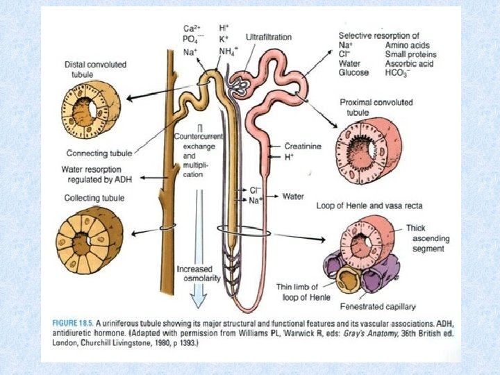

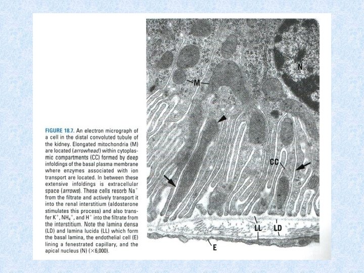
Uriniferous tubules 6. Distal convoluted tubule: a. Continuous with macula densa, similar to ascending thick limb of Henle loop. b. Shorter, wider lumen than proximal tubule (no brush border). c. Function: resorbs Na+ from filtrate and transports it to renal interstitium (stimulated by aldosterone). Also transfers K+, NH 4+, and H+ into filtrate from interstitium. 7. Connecting tubule: short segment between distal convoluted tubule and collecting tubule into which it drains. Lined by two types of epithelial cells: a. Principal cells: have many infoldings of basal plasma membrane. Remove Na+ from filtrate and secrete K+ into it. b. Intercalated cells: have many apical vesicles and mitochondria. Remove K+ from filtrate and secrete H+ into it.
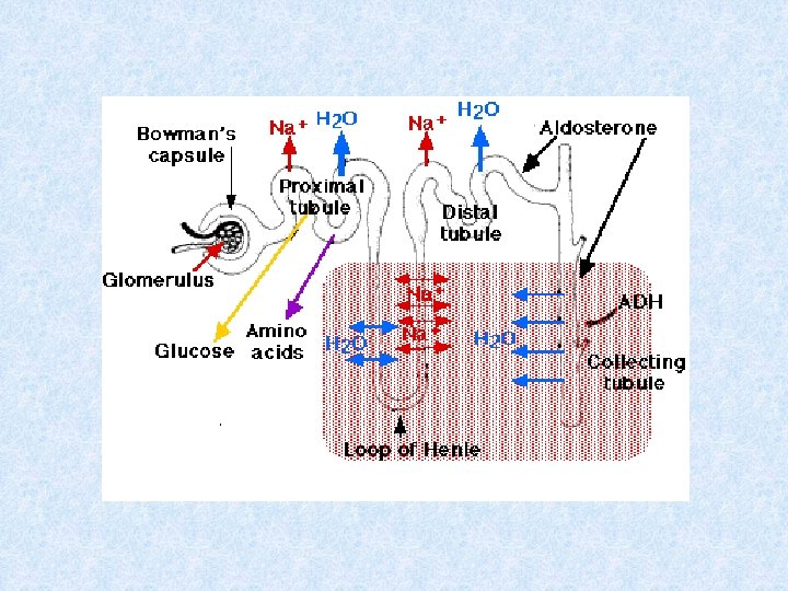

Uriniferous tubules 8. Collecting tubules: • Have segments in both cortex and medulla and converge to form larger and larger tubules. 1. Cortical collecting tubules: • Located within medullary rays, few in cortex (cortical labyrinth). Lined by simple epithelium containing two types of cuboidal cells. a. Principal (light) cells: contain round central nucleus and single central cilium. b. Intercalated (dark) cells: less numerous than principal cells and contain microplicae (folds) on their apical surface and numerous apical cytoplasmic vesicles.

Uriniferous tubules 2. Medullary collecting tubules. a. In outer medulla: medullary collecting tubules contain both principal and intercalated cells in their lining epithelium. b. In inner medulla: collecting tubules lined by principal cells. 3. Papillary collecting tubules (ducts of Bellini): a. Large collecting tubules (200– 300 µm diameter) formed from converging smaller tubules. b. Lined by a simple epithelium composed of columnar cells that have single central cilium. c. Empty at area cribrosa, a region at apex of each renal pyramid has 10 - 25 openings through which urine exits into a minor calyx.

Renal blood circulation IV. Renal Blood Circulation: • The renal blood circulation is extensive, with total blood flow through both kidneys of about 1200 m. L per minute. • At this rate, all of the circulating blood in the body passes through the kidneys every 4 to 5 minutes.

Excretory passages VI. Excretory Passages 1. Excretory passages include minor and major calyces and renal pelvis, located within each kidney, and ureters, urinary bladder, and urethra, located outside kidneys. 2. These structures contain three-layer wall composed of: - Mucosa of transitional epithelium (except in the urethra) Lamina propria of connective tissue Muscularis (smooth muscle) Adventitia

Excretory passages A. Ureter 1. Ureter conveys urine from renal pelvis of each kidney to urinary bladder. 2. It has transitional epithelium that is thicker and contains more cell layers than renal calyces. 3. Contains two-layer muscularis (inner longitudinal and outer circular layer of smooth muscle) in its upper two-thirds. • Lowest third contains additional outer longitudinal layer of smooth muscle. 1. Contracts its muscle layers, producing peristaltic waves that propel urine so it enters bladder in spurts.
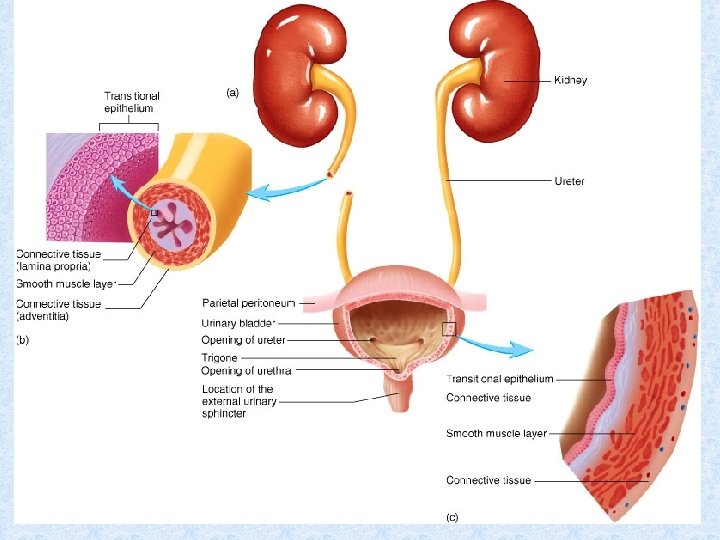

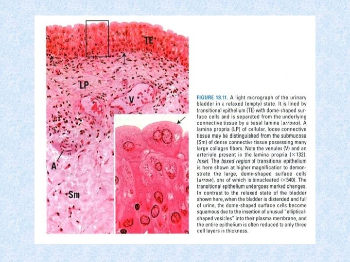
Excretory passages B. Urinary bladder. • Contains transitional epithelium, thin lamina propria of fibroelastic connective tissue, and three-layer muscularis: 1. Epithelium of relaxed bladder: a. 5 -6 cell layers thick, rounded dome-shaped cells bulge into lumen. b. Contain unique plaques in thick luminal plasma membrane, and flattened elliptical vesicles in cytoplasm. 1. a. b. c. Epithelium of distended bladder: Epithelium is only three to four cell layers thick. Has squamous superficial cells. Much thinner and has larger luminal surface than relaxed bladder; this results from insertion of elliptical vesicles into luminal plasma membrane of surface cells.

Excretory passages C. Urethra 1. Urethra conveys urine from bladder outside the body. In males, the urethra also carries semen during ejaculation. 2. Has two-layer muscularis consisting of inner longitudinal and outer circular layer of smooth muscle. 3. Surrounded by external sphincter of skeletal muscle, which permits voluntary closure.
 -Lumen(L) and thick lining")
Ureter and Urinary Bladder Figure 1 : Ureter (C. S) -Lumen(L) and thick lining epithelium(E). -Interface between subepithelial connective tissue(SCT), smooth muscle coat(SM indicated by arrows. -Muscle coat surrounded by fibrous adventitia(Ad), -Wall of the ureter consists of: mucosa (epithelium and underlying connective tissue), muscularis, and adventitia. Figure 2: Ureter (C. S) Figure 3: Urinary bladder Figure 4: Urinary bladder -Thick, transitional epithelium whose free surface possesses characteristic dome-shaped cells(D). . -Muscularis consists of three layers of smooth muscle: inner longitudinal (IL), middle circular (MC), and outer longitudinal (OL). -Adventitia (Ad) composed of fibrous connective tissue -Mucosa may or may notdisplay folds. (arrows). -Transitional epithelium(TE). -Muscularisis layers : inner longitudinal(IL), middle circular(MC), and outer longitudinal(OL). -Transitional epithelium(TE), -Lamina pro-pria(LP), submucosa(Sm). -Vascularity of this region demonstrated by numerous venules (V) and arterioles(A). -Transitional epithelium demonstrate large, dome-shaped cells (arrow) at free surface.
- Slides: 53