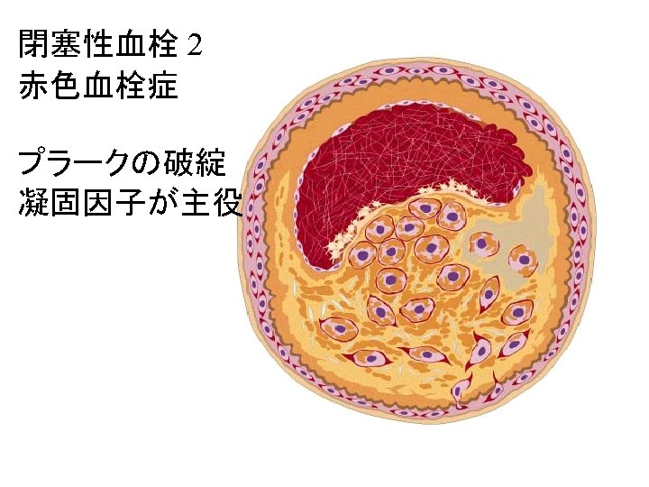
Angina pectoris a syndrome characterized by paroxysmal constricting

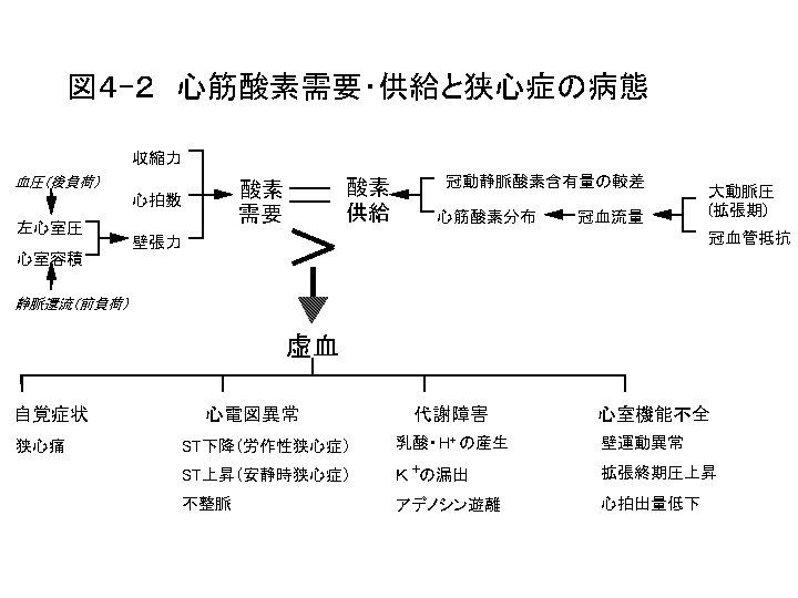
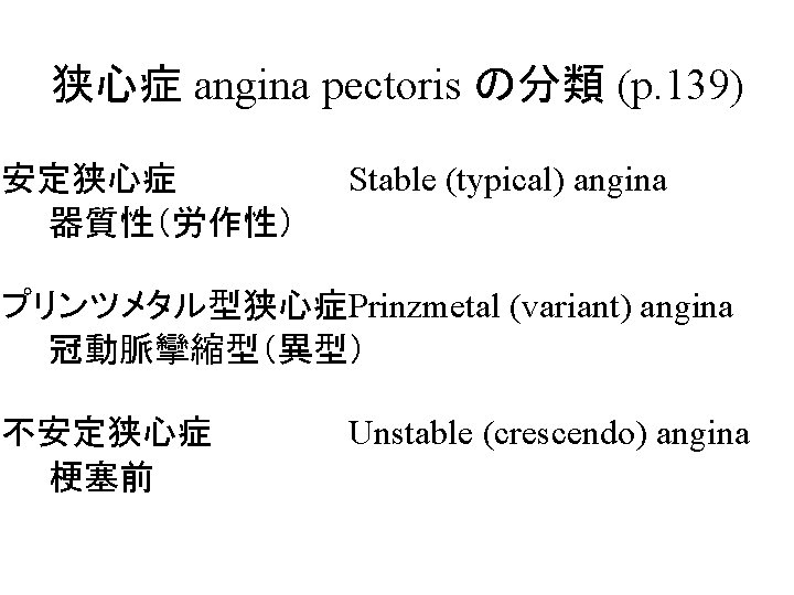
Angina pectoris 狭心症 a syndrome characterized by paroxysmal, constricting pain below the sternum, most easily precipitated by exertion or excitement and caused by ischemia of the heart muscle, usually due to a coronary artery disease, as arteriosclerosis. Angina: (語源) any attack of painful spasms characterized by sensations of choking or suffocating. < L angina quinsey, for * ancina < Gk strangulation, hanging 病態 治療原則
 and after 4. 5 min of exercise (bottom).")
Lead V 4 at rest (top) and after 4. 5 min of exercise (bottom). There is 0. 3 m. V of horizontal ST-segment depression, indicating a positive test for ischemia.

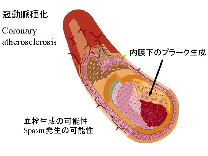
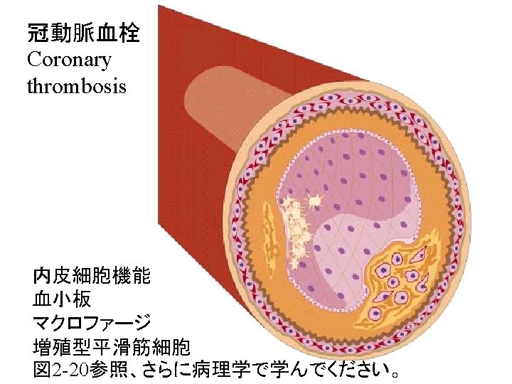
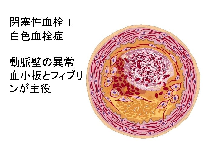
Coronary Angiography Stenosis: narrowing of the artery, due most likely to atherosclerosis. Damage to the intima due to atherosclerosis is a major cause of thrombosis in the arteries. The resultant ischemia of the myocardium can lead to ischemic necrosis, i. e. , an infarct [Myocardial Infarction]. 1. What therapeutic agent can be used to lyse the clots in coronary vessels?

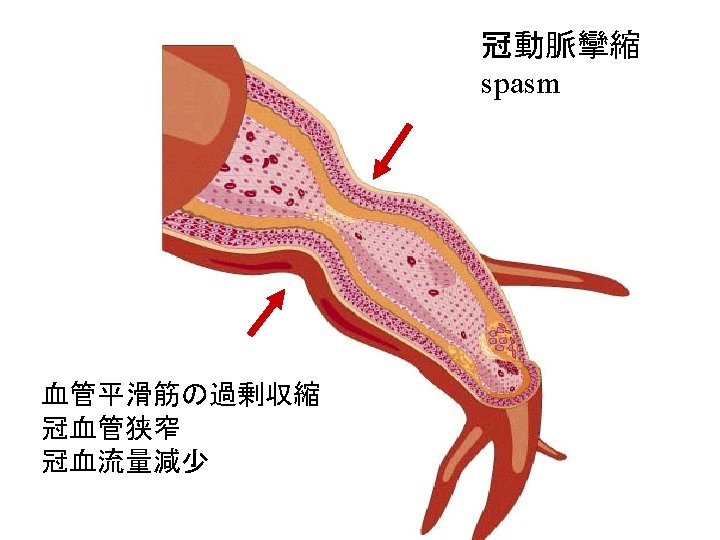
Dynamic coronary obstruction Spasm Rest angina tone

Dynamic coronary obstruction • This figure shows how the caliber of eccentric coronary artery stenoses may change, with considerable variation in the degree of stenosis resistance and the propensity to produce angina. • Both increased vascular tone (first two examples, spasm & tone ) and decreased vascular tone (third example) are depicted.

 nitrates (nitroglycerin) vasodilators (Ca")
Correction of the imbalance increasing delivery (by increasing coronary flow) nitrates (nitroglycerin) vasodilators (Ca antagonists, KCOs) coronary bypass grafts or angioplasty (PTCA) percutaneous transcatheter coronary angioplasty decreasing oxygen demand (by decreasing cardiac work) b-blockes, nitrates (nitroglycerin) vasodilators (Ca antagonists, KCOs) *against atherosclerosis and thrombus formation
")
Artery blocked by thrombus on fissured atherosclerotic plaque Zone of perfusion (area at risk) next

Myocardium 0 hr 24 hr Obstructed coronary art. Endocardium area at risk Zone of necrosis Epicardium Progression of myocardial necrosis after coronary artery occlusion. Necrosis begins in a small zone of the myocardium beneath the endocardial surface in the center of the ischemic zone. This entire region of myocardium (dashed outline) depends on the occluded vessel for perfusion and is the area at risk. Note that a very narrow zone of myocardium immediately beneath the endocardium is spared from necrosis because it can be oxygenated by
 a")
Principle of stress radionuclide imaging for the detection of coronary artery disease. (1) a normal coronary artery and an artery with significant stenosis (top), (2)(2) the myocardial territory (perfusion image), and (3)(3) the ventricular wall (wall motion image) supplied by each artery.

Distribution of regional myocardial blood flow • The distribution of regional myocardial blood flow in the myocardium is heterogeneous, i. e. , there is less blood flowing in the ischemic myocardial bed (blue area) than in the normal myocardial bed.
 最大運動時")
Coronary blood flow at rest and during stress (x ~ 5) 最大運動時
 At rest, coronary blood flow")
Coronary blood flow at rest and during stress (1) At rest, coronary blood flow is similar in the normal artery and in the artery with stenosis. Resting coronary blood flow in the diseased artery is maintained at a normal level and is sufficient to meet resting metabolic myocardial demands because of recruitment of coronary reserve (dilatation of the resistance vasculature) in the distal coronary bed. During exercise, myocardial metabolic demands increase and more myocardial nutrient blood flow is required. Coronary reserve is increased as a result of dilatation of the coronary resistance vessels in the peripheral coronary bed.
 In the territory of the")
Coronary blood flow at rest and during stress (2) In the territory of the normal coronary artery, the resistance vessels dilate and coronary blood flow is increased by 2 to 2. 5 times. In the abnormal coronary bed, which is distal to the significant coronary stenosis, resistance vessels are already dilated and little if any further dilatation is possible. Increased metabolic demands thus cannot be met, and the relatively hypoperfused myocardium becomes ischemic.
- Slides: 30