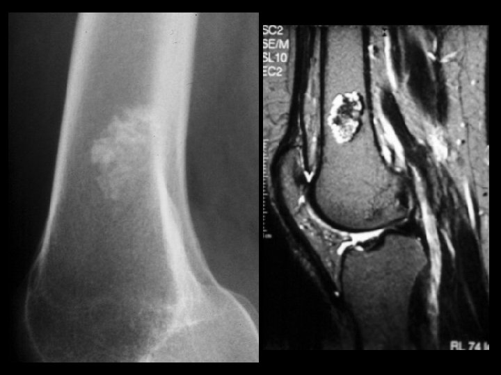
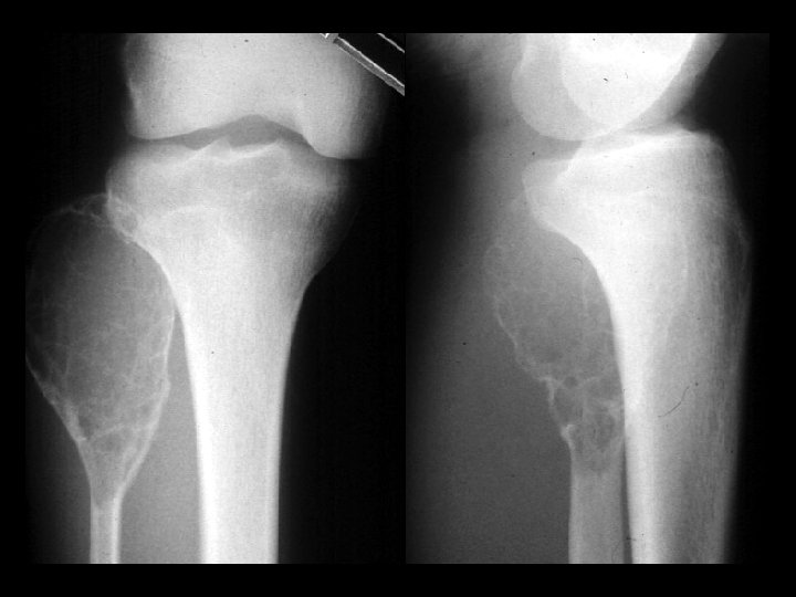
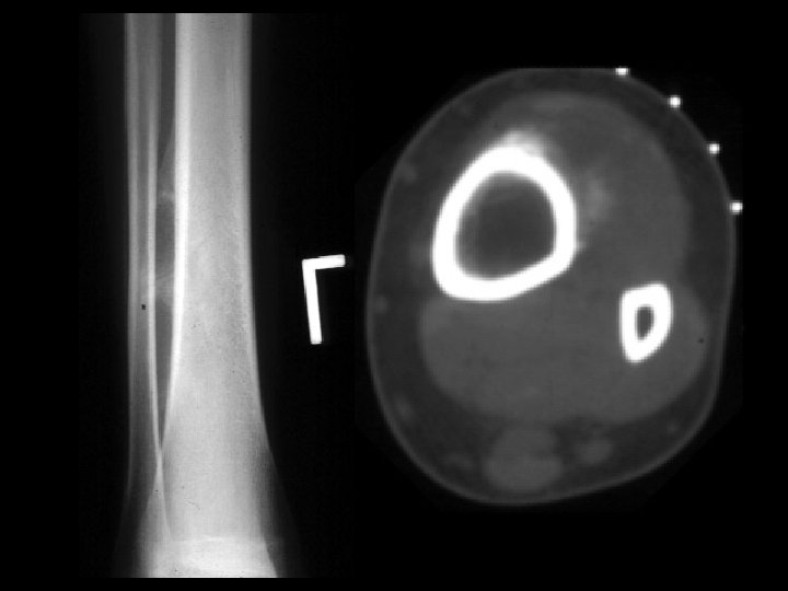
Aneurysmal bone cyst Findings Eccentric metadiaphyseal lucent lesion

Aneurysmal bone cyst • Findings: – Eccentric metadiaphyseal lucent lesion with a thin sclerotic margin and fine internal septa • ddx: – Fibrous dysplasia – Chondromyxoid fibroma – GCT, chondroblastoma (if physis closed)

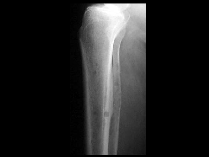
Adamantinoma • Findings: – Expansile mixed lytic and sclerotic lesion of the tibial midshaft – Cortical disruption and periosteal reaction • ddx: – Fibrous dysplasia

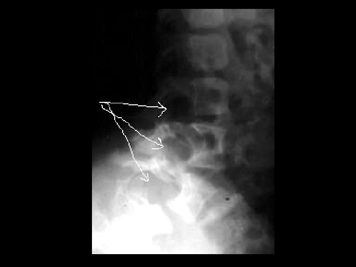
Avulsion injury • Findings: – Soft tissue calcification along the superior anterior acetabulum – No cortical involvement – Normal joint space • ddx: – Myositis ossificans – Parosteal osteosarcoma

Brodie’s abscess • Findings: – Well-defined lytic lesion in the metaphysis – Large zone of surrounding sclerosis – CT is diagnostic of a fluid filled lesion • ddx: – Osteoblastoma – Langerhan’s cell histiocytosis

Calcaneal Ewing’s sarcoma • Findings: – Permeative lesion in the calcaneous – Cortical disruption and slight periosteal reaction • ddx: – Lymphoma – Metastasis/myeloma – Infection – Langerhan’s cell histiocytosis

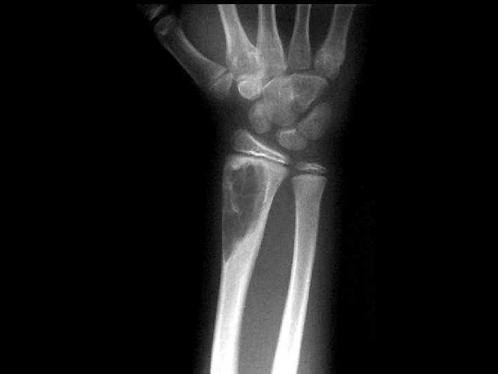
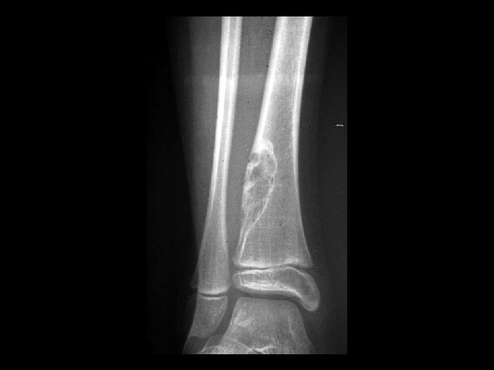
Chondromyxoid fibroma • Findings: – Well-defined eccentric lucent lesion – Cortical thinning – Narrow zone of transition • ddx: – ABC – Infection – Fibrous dysplasia – Giant cell tumor (if physis closed) – Non-ossifying fibroma (if sclerotic margin)
 distal femur")
Cortical desmoid • Findings: – Irregular cortical thickening of the posterior (medial) distal femur – Avulsion injury related to the medial gastrocnemius • ddx: – Parosteal osteosarcoma – Myositis ossificans

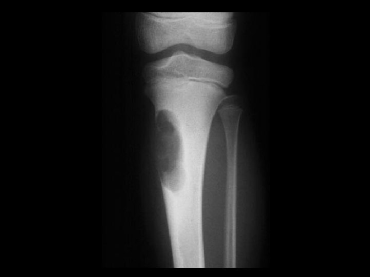
Langerhan’s cell histiocytosis • Findings: – Predominantly lytic lesion of the tibia with cortical thickening, periosteal reaction, and soft tissue swelling and edema • ddx: – Infection – Ewing’s sarcoma – Lymphoma

Enchondroma • Findings: – Intramedullary lesion containing calcified chondroid matrix – Endosteal scalloping • ddx: – Chondrosarcoma – Bone infarct
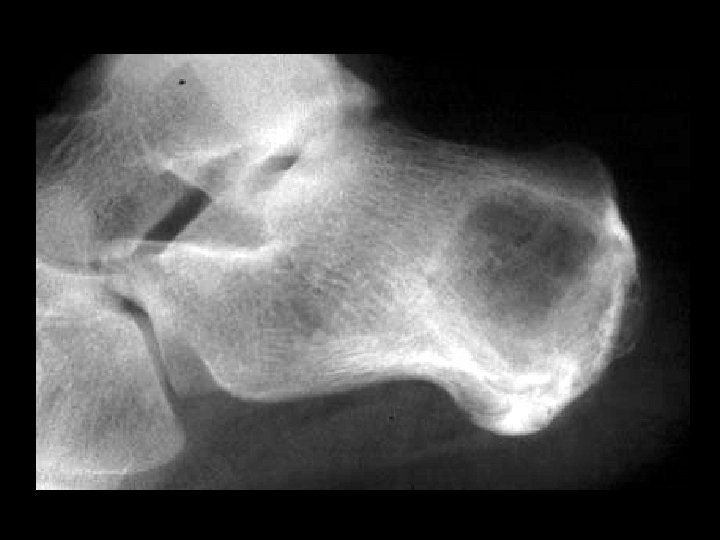

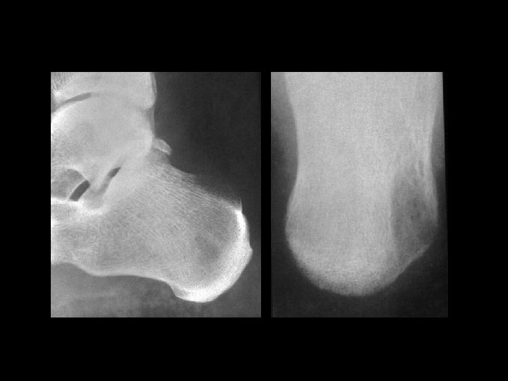
Calcaneal UBC • Findings: – Central lucent lesion within the calcaneous – Fine sclerotic margin • ddx: – Intraosseous lipoma

Ewing’s sarcoma • Findings: – Permeative diaphyseal lesion with intense periosteal reaction and soft tissue swelling • ddx: – Osteomyelitis – Langerhan’s cell histiocytosis – Lymphoma
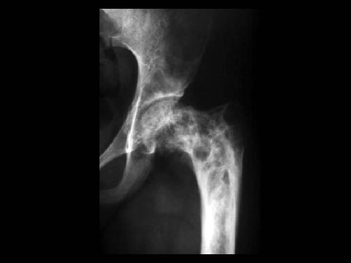

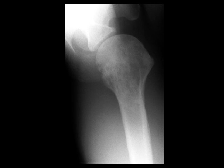
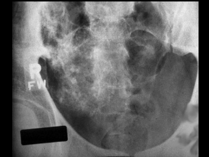
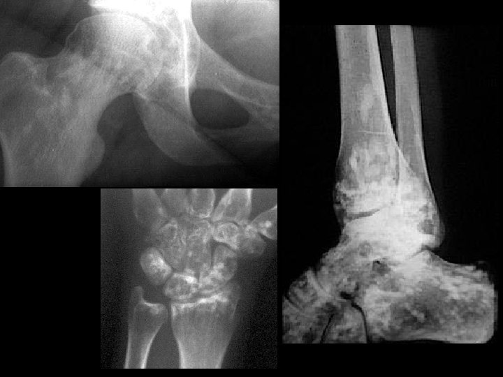
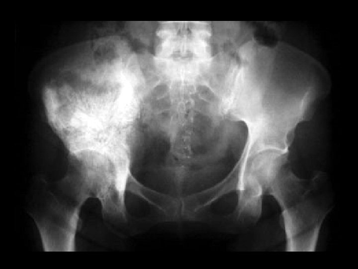
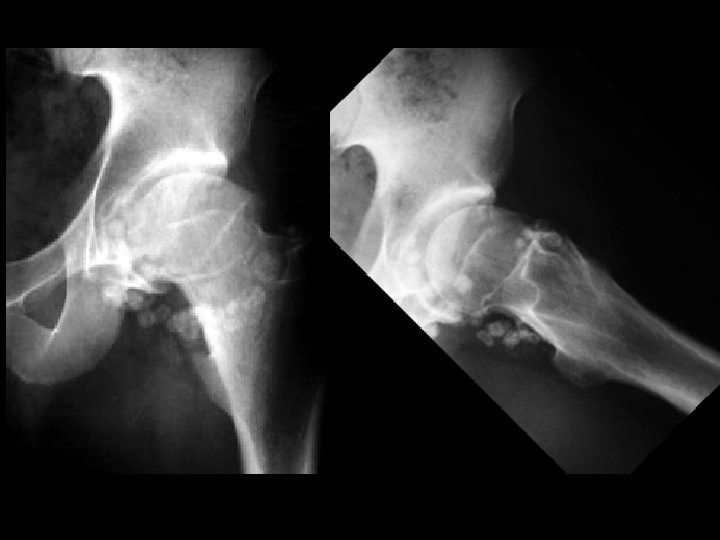
Fibrous dysplasia • Findings: – Mixed sclerotic and lytic lesion of the left hemipelvis and proximal femur – “shepherd's crook sign” • ddx: – NONE! – This is an Aunt Minnie!

Gaucher’s disease • Findings: – Flask-shaped long bones = undertubulation – Varied appearance includes multiple lucent lesions and bone infarcts • ddx: – other glycogen storage dz
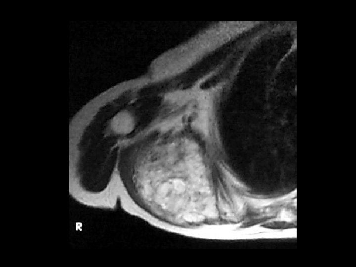

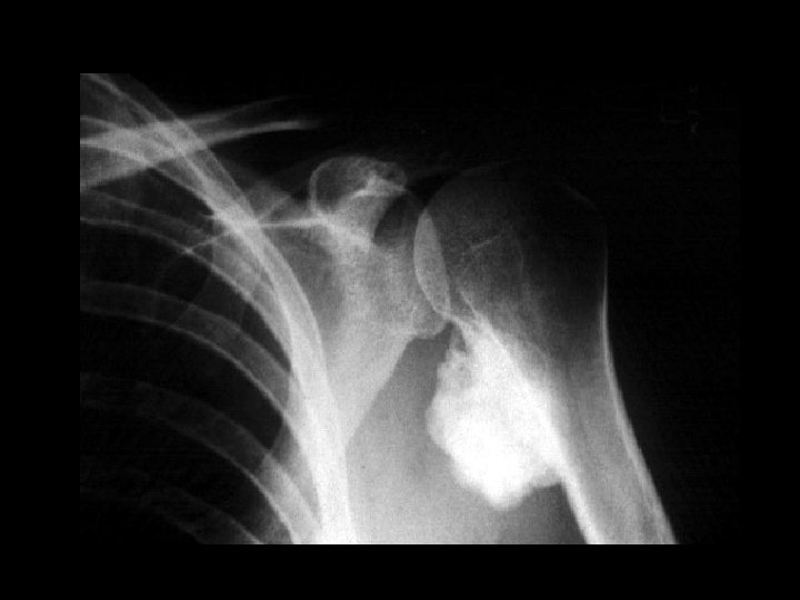
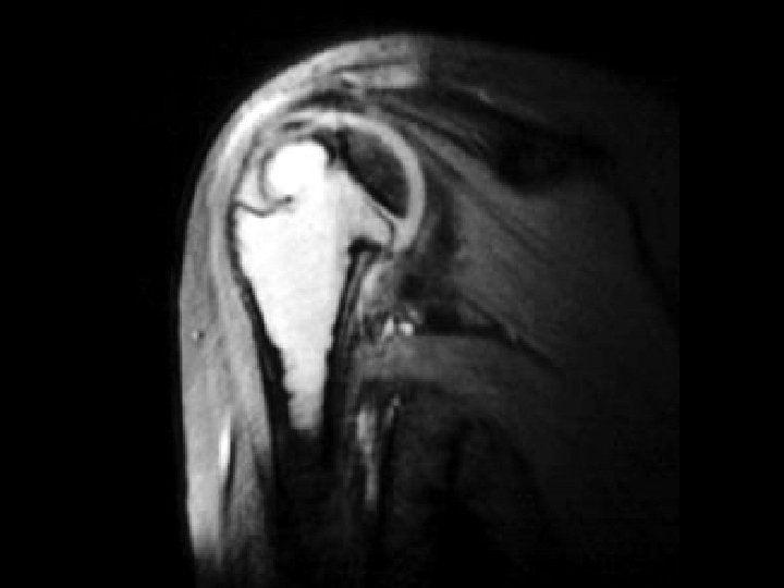
Scapular metastasis • Findings: – Soft tissue mass of the right shoulder with destruction of the adjacent scapula • ddx: – Soft tissue sarcoma

Mafucci syndrome • Findings: – Multiple enchondromas and soft tissue hemangiomas • ddx: – NONE! – This is an Aunt Minnie!

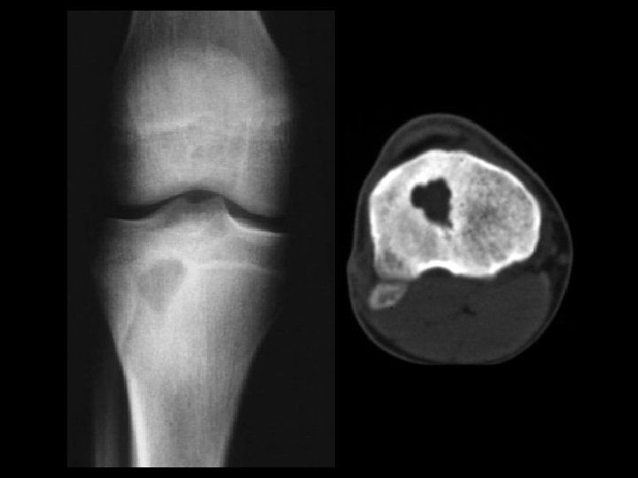
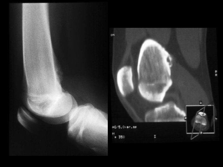
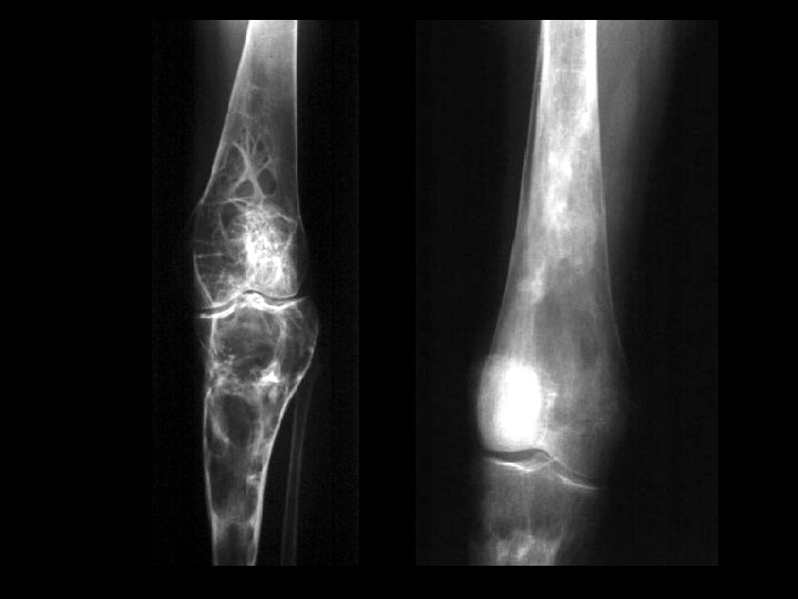
Giant cell tumor • Findings: – End of bone lucent and expansile lesion with a narrow zone of transition – No periosteal reaction or soft tissue mass • ddx: – ABC – metastasis – chondroblastoma

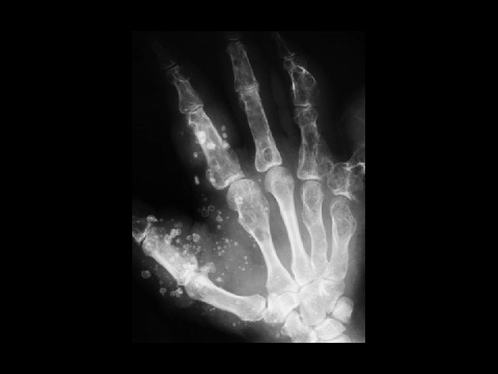
Multiple myeloma • Findings: – Numerous punched out lytic lesions involving multiple bones • ddx: – Metastases

Osteoma • Findings: – Well-defined lesion of compact bone involving the left ileum – No change over many years • ddx: – Sclerotic metastasis

Lymphoma • Findings: – Permeative lesion of the proximal humerus • ddx: – Multiple myeloma – Metastasis – MFH – Infection – Langerhan’s cell histiocytosis

Non-ossifying fibroma • Findings: – Well-defined lucent cortical lesion – Sclerotic margin – No periosteal reaction or soft tissue mass • ddx: – Fibrous dysplasia – Langerhan’s cell histiocytosis – ABC

Neurofibromatosis Type I • Findings: – Enlargement of multiple neural foramina and scalloping of the posterior vertebral bodies • ddx: – NONE! – This is an aunt Minnie!

Malignant fibrous histiocytoma • Findings: – Permeative lesion of the superior pubic ramus • ddx: – Lymphoma – Multiple myeloma – Metastasis – Osteomyelitis

Fibrous dysplasia • Findings: – Long lytic lesion in a long bone – Cortical thickening – “ground glass matrix” • ddx: – NONE! – This is an Aunt Minnie!

Chondrosarcoma • Findings: – Large bone-forming soft tissue mass centered in the right SI joint – “ring and arc Ca 2+” = calcified chondroid matrix • ddx: – NONE! – This is an Aunt Minnie!

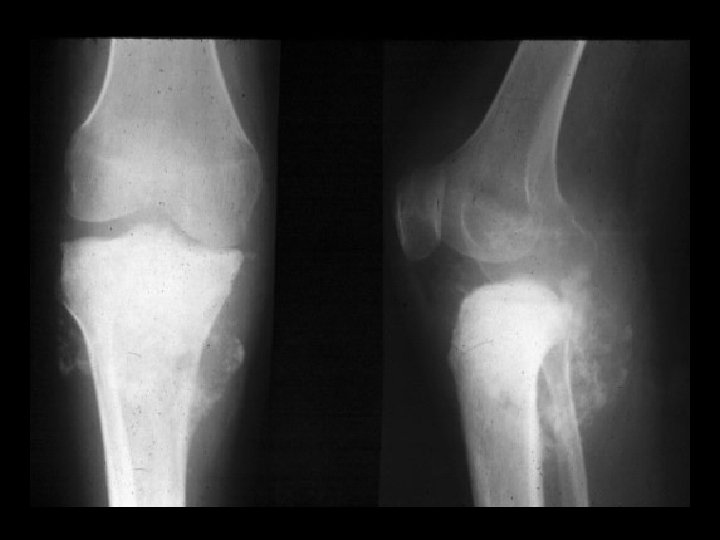
Osteosarcoma • Findings: – Aggressive lesion of the distal tibia in a skeletally immature person – “cloud-like” appearance = calcified osteoid matrix – Intense periosteal reaction – Soft tissue mass • ddx: – NONE! – This is an Aunt Minnie!

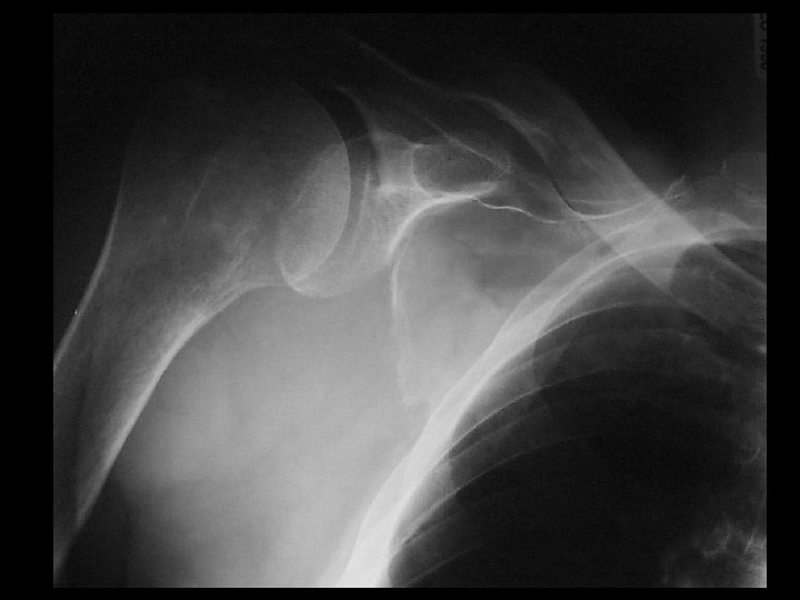
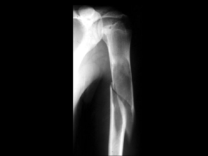
Parosteal osteosarcoma • Findings: – Dense lobular lesion along the medial proximal humerus – No apparent cortical destruction (check w/ CT) • ddx: – NONE! – This is an Aunt Minnie!

Osteopoikelosis • Findings: – Innumerable round and oval sclerotic lesions involving multiple bones – No cortical destruction, periosteal reaction, or soft tissue mass • ddx: – NONE! – This is an Aunt Minnie!
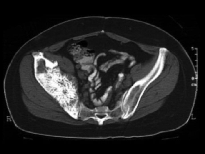

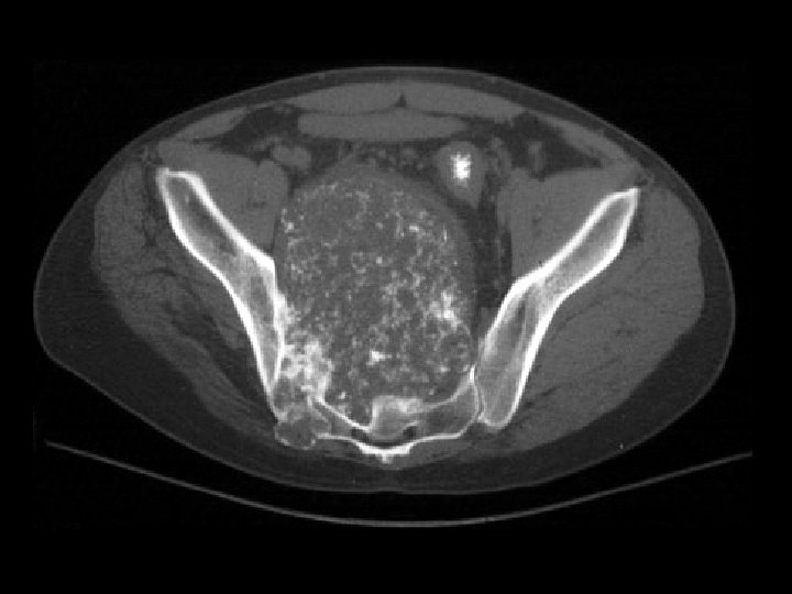
Hemangioma • Findings: – Sclerotic expansile lesion of the right hemipelvis – “Irish lace” appearance on CT • ddx: – NONE! – This is an Aunt Minnie!
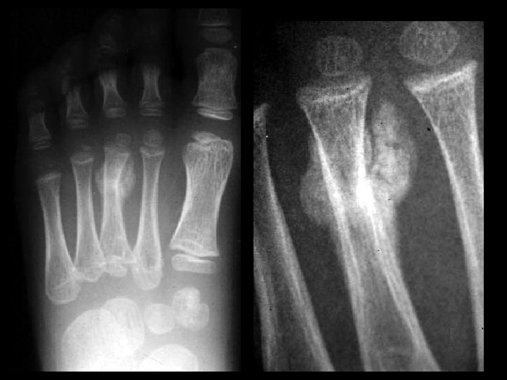

Stress fracture • Findings: – Slightly angulated fracture of the distal 3 rd metatarsal – Periosteal reaction – Surrounding calcified osteoid matrix = callus • ddx: – Osteosarcoma with pathologic fracture

Periosteal osteosarcoma • Findings: – Lucent juxtacortical fusiform mass along the diaphyseal surface – Spiculated perpendicular periosteal reaction = “sunburst” – Lack of medullary involvement • ddx: – Osteomyelitis – Langerhan’s cell histiocytosis – Ewing’s sarcoma

Synovial osteochondromatosis • Findings: – Multiple round calcified masses in the joint space – Erosions on both sides of the joint – Single joint involvement • ddx: – Primary (idiopathic) – Secondary to OA

Giant cell tumor • Findings: – End of bone, eccentric, lucent lesion with narrow zone of transition but no sclerotic margin • ddx: – Fibrous dysplasia – ABC – Chondroblastoma

Osteosarcoma • Findings: – lytic lesion of the anterior proximal tibia – cortical breakthrough and a soft tissue mass • ddx: – metastasis – infection – Langerhan’s cell histiocytosis
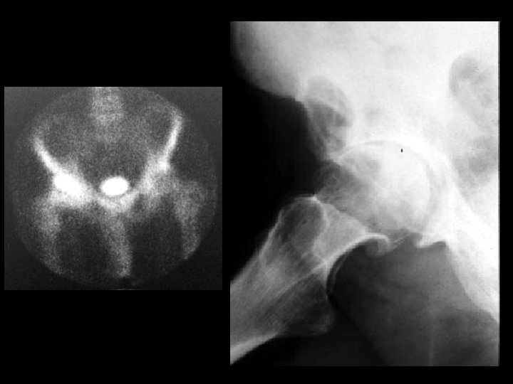

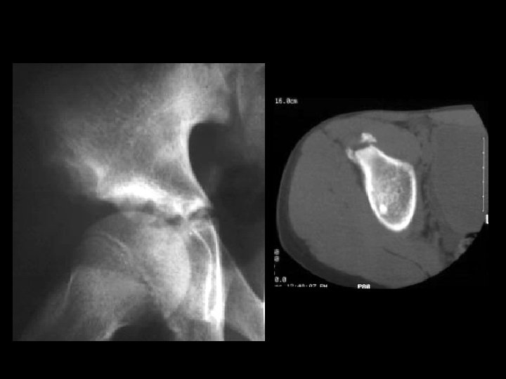
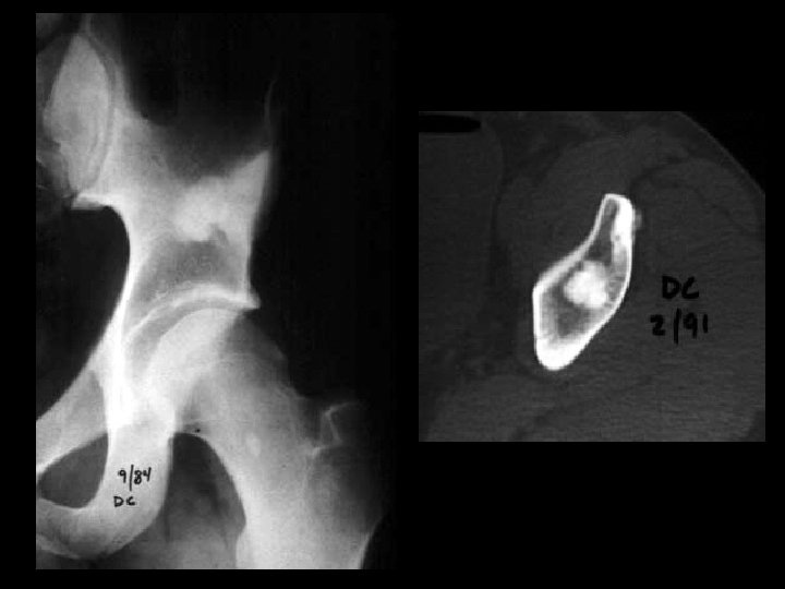
Severe OA & Geode • Findings: – severe osteoarthritis of the right hip – lytic lesion in the adjacent superior acetabulum – hot on bone scan • ddx: – fibrous dysplasia – ABC – infection – erosion
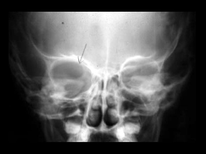

Neurofibromatosis type I • Findings: – partially empty orbit = sphenoid wing dysplasia • ddx: – lytic metastasis – osteomyelitis
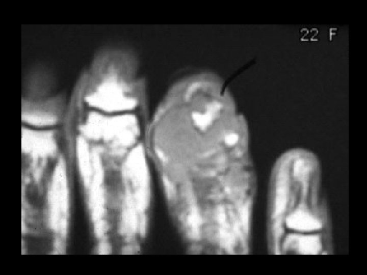

Giant cell tumor • Findings: – soft tissue mass destroying the tuft • ddx: – osteomyelitis – metastasis – chondrosarcoma
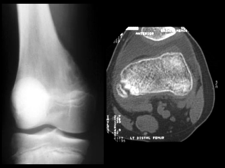
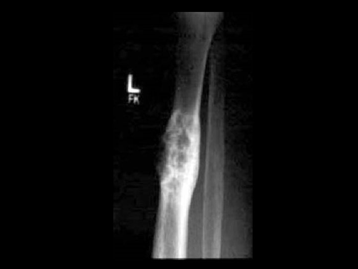
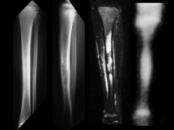
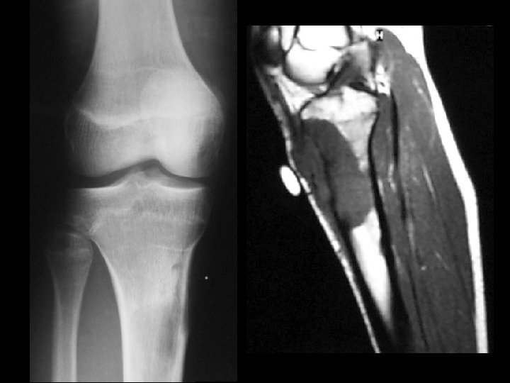
 • Findings: – Cortical thickening and irregularity along the posterior")
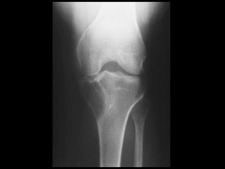
Tug lesion (avulsion injury) • Findings: – Cortical thickening and irregularity along the posterior medial distal femur – CT is diagnostic • ddx: – parosteal osteosarcoma – myositis ossificans

Unicameral bone cyst • Findings: – expansile central lesion of the proximal humerus – Fluid signal, no cortical breakthrough, or soft tissue mass • ddx: – NONE! – This is an Aunt Minnie!
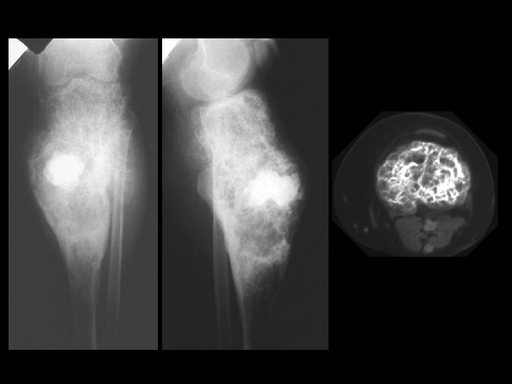

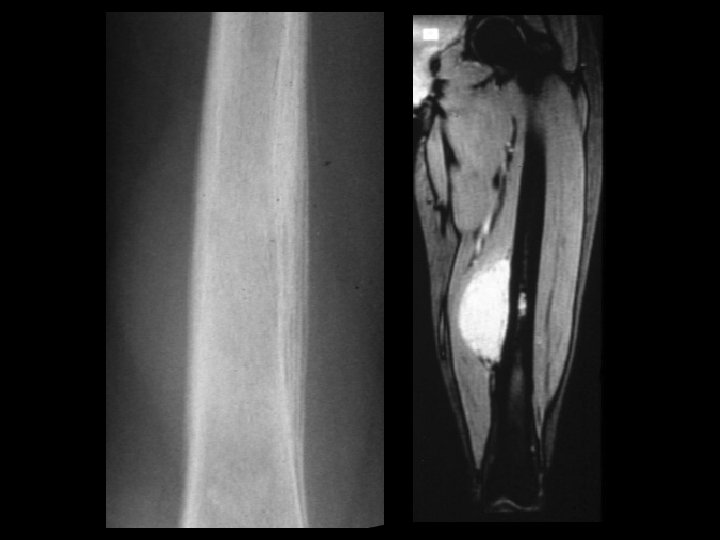
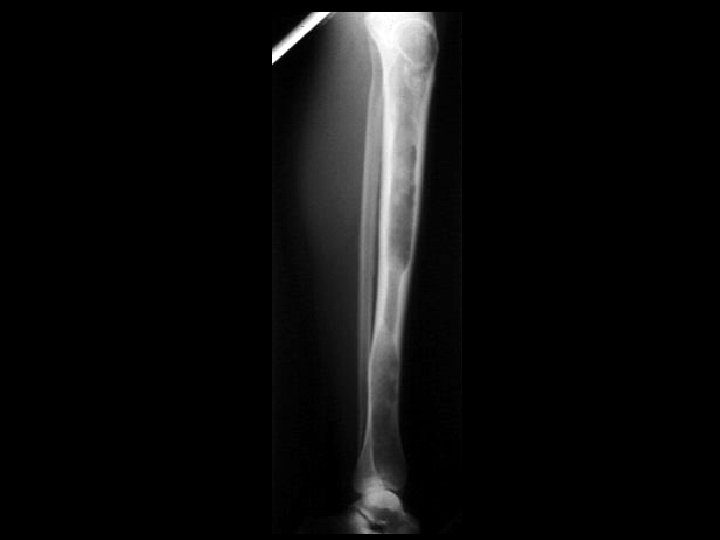
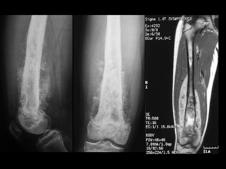
Hemangioma • Findings: – expansile lesion of the proximal tibia containing fine bony septations and a large course calcification – CT scan shows a lacey appearance • ddx: – Paget’s dz – ABC
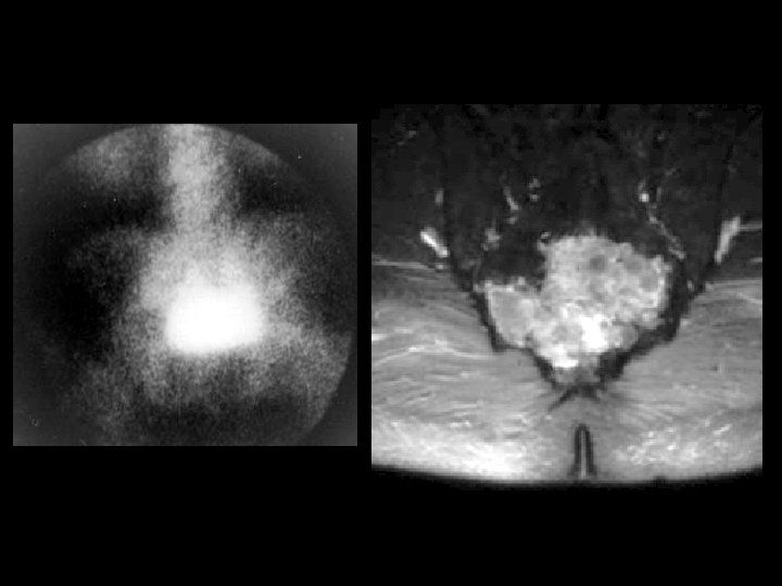

Chordoma • Findings: – Large destruction lesion in the deep pelvis – high T 2 signal – hot on bone scan • ddx: – chondrosarcoma – teratoma – metastasis
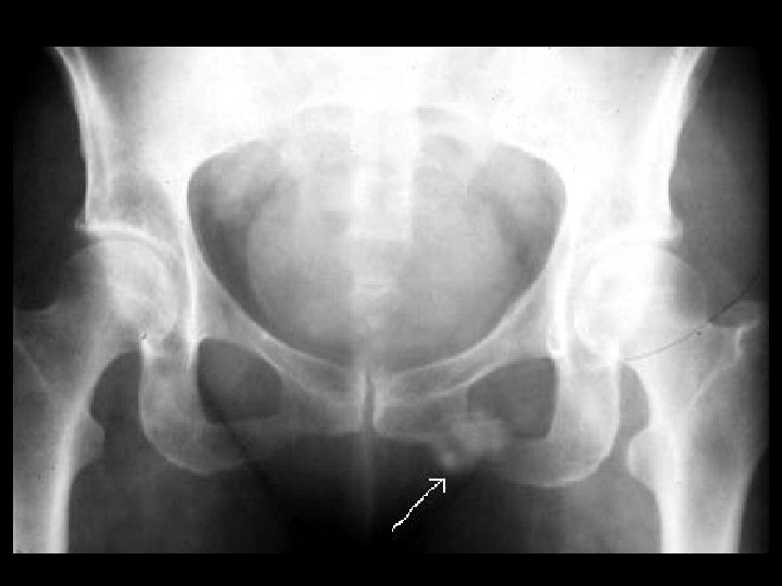

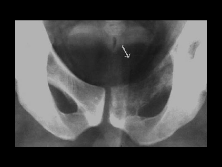
Healing stress fracture • Findings: – Fluffy density overlying the left inferior pubic ramus • ddx: – chondrosarcoma – parosteal osteosarcoma – myositis ossificans

Osteosarcoma • Findings: – Bone forming lesion of the proximal tibia in a skeletally mature patient – fluffy and cloud-like calcified matrix • ddx: – NONE! – This is an Aunt Minnie!

UBC & pathologic fracture • Findings: – central lucent lesion with a narrow zone of transition complicated by a pathologic fracture • ddx: – NONE! – This is an Aunt Minnie!
- Slides: 89