Anesthetic Considerations for Diastolic Dysfunction Suneel P R


: 707– 21 (2007")


 • Myocardial")


- Slides: 66
Anesthetic Considerations for Diastolic Dysfunction Suneel. P. R Associate Professor SCTIMST Trivandrum
Dysfunction: systolic vs. diastolic • Systolic function is intuitively meaningful • Diastology is a relative newcomer
Diastolic damages • Nearly 50% of all cardiac failures • Prognosis and mortality same as systolic • Mortality is four times when compared with normal population
Diastolic heart failure • The Ejection Fraction will be normal • Called Heart failure with normal EF (HFnl. EF) • Diastolic dysfunction can occur along with systolic dysfunction
Diastology When does diastole begin ? • Anatomical -when aortic valve closes • Molecular level- dissociation of the actinmyosin cross-bridges • The heart begins the relaxation process in systole !!
Relaxation-requires energy BJA 98 (6): 707– 21 (2007
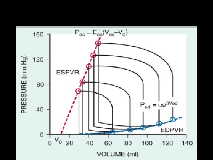
Diastolic dysfunction definition Inability of the ventricles to fill at low pressure The end-diastolic pressure is 16 -26 mm Hg (normal EDP is < 12 mm Hg) The atrial pressures that are needed to complete filling are even higher
Pathophysiology- two key terms Increased filling pressures are due to 1. Abnormality of relaxation 2. Decreased compliance
Physiology: The stages 1. 2. 3. 4. Isovolumic relaxation Rapid filling Diastasis Atrial contraction
Physiology
Isovolumetric relaxation Ao. VC MVO
Isovolumetric contraction • • Occurs between two closed valves Active relaxation occurs during this time The ventricular pressures continue to fall Mitral valve opening creates “suction effect”
Physiology
Rapid filling phase
Diastasis
Atrial “kick”
Active diastolic dysfunction Abnormality of relaxation Failure of energy dependent part of diastole • • Myocardial ischemia Hypertension Aortic stenosis Hypertrophic cardiomyopathy
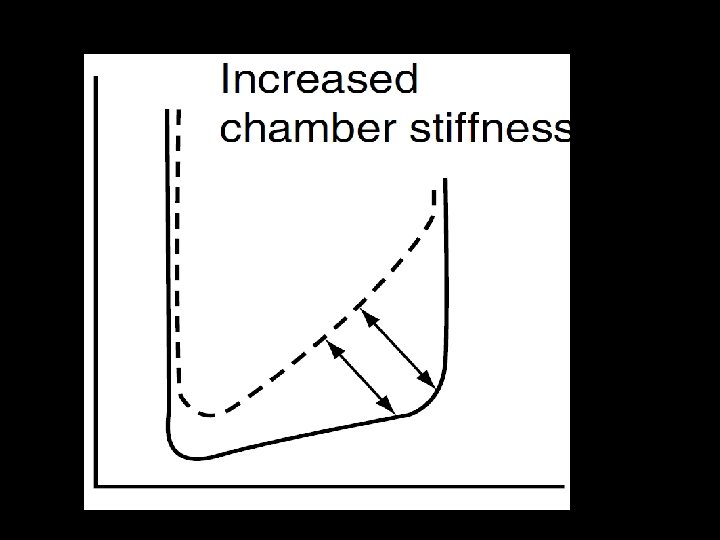
Passive diastolic dysfunction Increase in chamber stiffness • Infiltrative disorders ( amyloidosis) • Myocardial fibrosis • Progression from impaired relaxation
Physiology End systole End Diastole
Physiology
Impaired relaxation
Diagnosis of diastolic dysfunction • Echocardiography
Transmitral Pulse Wave Doppler
Transmitral Pulse Wave Doppler E A
Stage I of diastolic dysfunction 1. Impaired relaxation
Stage II diastolic dysfunction • Pseudonormalization
Stage III of diastolic dysfunction • Restrictive filling
Improvement to a worse grade • Tachycardia • Loss of atrial contraction • Volume excess
Improvement to a milder grade Reduction in preload • Reverse Trendelenburg • Diuresis • Amyl nitrate inhalation • Valsalva maneuver Relief of tachycardia Return from AF to Sinus
Stage IV diastolic dysfunction • Irreversible restrictive filling pattern
Pulmonary venous Doppler
Pulmonary venous Doppler
Pulomnary venous Doppler Impaired relaxation • D wave decreases in size • S/D ratio >1 Pseudonormal and Restrictive filling • Increase in D • S/D < 1 • Increase in A wave duration
Other echocardiographic tools • Tissue Doppler imaging to assess mitral annular movement • Color M mode of the Mitral valve to assess the propagation velocity
Diastolic dysfunction vs. failure • Dysfunction is a physiologic or preclinical state • Abnormal relaxation and increased chamber stiffness compensated by increased LAP • The LV preload is maintained • When these mechanisms are stressed, diastolic heart failure ensues
Braunwald 8 th edition
Diastolic heart failure Definite • C/F of heart failure Within 72 hours • Echo evidence of normal LVEF • Echo evidence of diastolic dysfunction
Most likely diastolic heart failure • • SBP >160 mm Hg DBP> 100 mm Hg Concentric LVH Worsened by – Tachycardia – Volume bolus • Improved by – Reducing HR – Restoring sinus rhythm
When to suspect diastolic dysfunction • • • History of previous diastolic heart failure Age > 70 years Female sex Uncontrolled hypertension Myocardial ischemia Diabetes mellitus • Comorbidities: Obesity, renal failure
Echo • Specifically documented If not then, look for – LVH –absence does not rule out! – LA enlargement – RV enlargement – Pulmonary hypertension
Perioperative worsening Deterioration in diastolic dysfunction • Myocardial ischemia – Directly affects relaxation – Induces rhythm disturbances • Hypovolemia • Tachycardia • Rhythms other than sinus
Perioperative worsening • • Shivering Anemia Hypoxia Electrolyte imbalances
Perioperative worsening • Post-op sympathetic stimulation • Post-op hypertensive crisis
Periop-risks • Delayed weaning from mechanical ventilation • Difficulty weaning from CPB • More use of vasoactive agents • Prolonged ICU stay & mortality
Conducting the anesthetic Pre-operative evaluation Functional status & exercise tolerance Optimizing the perioperative drugs
Perioperative drugs • • • Diuretics Beta blockers, calcium channel blockers ACEI & ARBs Statins Antiplatlets
Monitoring - Major surgeries • Standard monitoring tools • Invasive arterial pressures • Monitoring volume status is important • Central venous pressures or Pulmonary artery catheter or TEE ?
GA or Regional • No definite recommendation either way • Epidural vs. spinal ? Epidural wins
General anesthesia • IV induction & maintained with volatile agents and opioids • Greater hemodynamic instability
General anesthesia Good induction practices • Consideration for age • Titrate to effect • Smooth take over from spontaneous-bag mask • Hpoxia, hypercarbia worsens PHT
GA-control of BP • Systolic BP within 20 % of baseline • Maintain diastolic BP • Keep pulse pressure < DBP
Control of BP Rule of the 70 s • Age >70 years • Pulse rate around 70 s • DBP >70 • Pulse pressure < 70
Drug combination for hemodynamics • Low dose nitroglycerin and titrated phenylephrine • Either agent alone can worsen the hemodynamics
Nitroglycerine + Titrated phenylephrine 1. 2. 3. 4. Preserves vascular distensibility Avoids reduction in preload Maintains coronary perfusion pressure Maintains stroke volume with minimal cardiac work
Management of hypertensive crisis Sound anesthetic practices Plan for post-op analgesia Prevention of shivering Intravenous calcium channel blocker IV nitroglycerin
Post-op diastolic heart failure • • • Reduce preload Diuretics Use of nitrates CPAP Use of adrenaline, dobutamine, dopamine
Specific drugs for diastole Milrinone • Phosphodiesterase III inhibitor • Inotropic, vasodilatory with minimal chronotropy • Increases calcium ion uptake to SR
Milrinone • Lusitropic effect more evident in heart failure • Bolus dose of 50µgm/Kg over 60 minutes • Infusion of 0. 5 to 0. 75µgm/Kg/min
Specific drugs for diastole Levosimendan • Sensitizes the contractile elements to calcium • Has a vasodilator effect • Improves both systolic and diastolic function
Thank you