Anemia with pregnancy Dr Ahlam AlKharabsheh Anemia A

Anemia with pregnancy Dr. Ahlam Al-Kharabsheh

● Anemia: A pathological condition in which the oxygen –carrying capacity of red blood cells is insufficient to meet the body’s needs. ▪ World health organization( WHO ) : Hb < 11 g/dl. ▪ Centers for disease control and prevention of North America (CDC) : Hb < 11 g/dl in the first and third trimesters and Hb < 10. 5 g/dl in the second trimester. Postpartum anemia is a Hb < 10 g /dl. ● Anemia is the most common medical disorder of pregnancy. Around 30 -50% of women become anemic during pregnancy, with iron deficiency being responsible in more than 90% of cases. The incidence of folate deficiency is around 5%. Rare types include : vitamin B 12 deficiency, sickle cell disease, thalassemia and others.

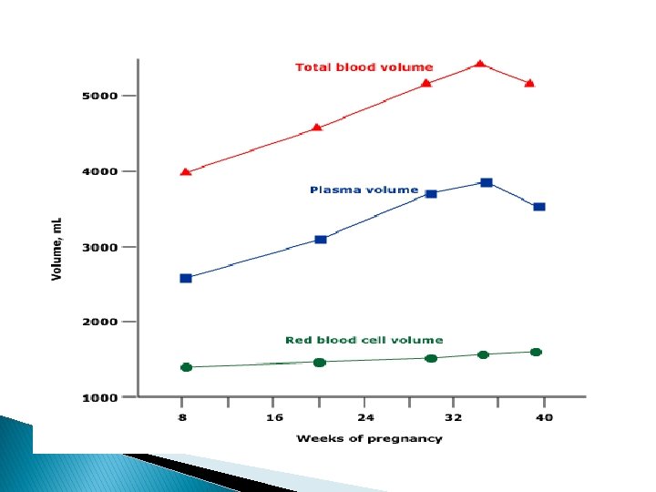
Physiological changes ▪ Plasma volume increases by 50%. ▪ Red cell mass increases by up to 25%. ▪ There is a consequent fall in Hb concentration, hematocrit and red cell count because of hemodilution. ▪ Mean cell volume (MCV) increases secondary to erythropoiesis. ▪ Mean cell Hb concentration (MCHC) remains stable. ▪ Serum iron and ferritin concentartion decreases secondary to increased utilization. ▪ Total iron binding capacity increases. ▪ Iron requirements increase(due to expanding red cell mass and fetal requirements ) from 2. 5 mg/day in the first trimester to 6. 6 mg/day in the third trimester (700 -1400 mg total pregnancy). ▪ There is a moderate increase in iron absorption. ▪ Folate requirements increase in pregnancy ( due to the fetus, placenta, uterus and expanded maternal red cell mass). ▪ There is no major effect on B 12 stores, although levels decrease (preferential active transport to the fetus).

Severe chronic anemia is associated with : Fetal 1 -Increased risk of preterm delivery. 2 -Fetal growth restriction(FGR) and low birth weight. 3 -Intrauterine fetal death (IUFD). 4 -Increased risk of spontaneous abortion. 5 -Increased risk of perinatal mortality. Maternal 1 -Increased risk of infection. 2 -Poor tolerance to blood loss at time of delivery. 3 -Cardiovascular stress. 4 -Increased risk of postnatal depression. 5 -Increased risk of maternal mortality.

Screening All pregnant women should be offered screening for anaemia, by estimating the Hb concentration by means of a full blood count (CBC) : ● In first trimester (or at booking) ● With the next screening bloods (usually performed between 28 -32 weeks) ● And at 36 weeks gestation.
 ● Diagnosis : ▪ IDA is classically described as microcytic")
Iron deficiency anemia (IDA) ● Diagnosis : ▪ IDA is classically described as microcytic , hypochromic anemia because of reduced MCV and MCHC. But the diagnosis should still be confirmed. ▪ The diagnostic test for iron deficiency is a serum ferritin concentration. This is not affected by recent ingestion of iron, and a concentration of < 12 mcg/L is diagnostic. (the greatest sensitivity and specificity → Iron def. can be present in the absence of anemia ( low Hb is a late event of iron deficiency ) and other parameters of the full blood count that usually give a clue to this ( reduced MCV, MCH and MCHC ) are not as accurate during pregnancy. ● Prevention : ▪ CDC guidelines : Pregnant women should begin taking low dose (30 mg/day) elemental oral iron at the first prenatal visit as primary prevention of iron deficiency. ▪ Education about the diet. ▪ Treatment of hook worm infestations in non-industrialized countries.

Lab test � Parameters �↓ � ferritin, hemoglobin, mean corpuscular volume, mean corpuscular hemoglobin �↑ � total iron-binding capacity, transferrin, red blood cell distribution width
 Blood transfusion")
● Treatment Oral iron Parenteral iron (IM & IV ) Blood transfusion

Oral iron ▪ If there is enough time, oral iron is the first line treatment (ferrous salts = elemental iron). ▪ The recommended dose is 120 -240 mg of elemental iron per day ( maximum increase in Hb is 0. 8 g/dl per week ). ▪ Side effects of oral iron are related to the amount of elemental iron contained. There is a 40 % risk of side effects with oral iron preparations, mainly gastrointestinal , and this can have a direct effect on tolerance and compliance. ▪ Vitamin C taken simultaneously aids absoption, hence common advice to take iron with fresh orange juice. ▪ Avoid consuming tea and coffee during or shortly after medication, preferably taken on an empty stomach one hour before meals. Iron should be given two hours before, or four hours after, ingestion of antacids.

Therapeutic Trial of Iron Oral iron therapy • Increase in reticulocytes in 5 – 10 days • Rise in Hb at a rate of 0. 8 gm/dl every 3 • weeks till normal If no response or incomplete response, • do additional tests


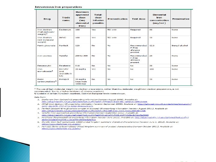
▪ Choice of preparation : The most appropriate oral iron therapy is use of a tablet containing ferrous salts, such as : ●Ferrous fumarate – 106 mg elemental iron/tablet ●Ferrous sulfate – 65 mg elemental iron/tablet ●Ferrous gluconate – 28 to 36 mg iron/tablet ▪ Follow up : Re-check complete blood picture every 4 weeks. Once the [Hb] is in the normal range, continue oral iron treatment until completion of breastfeeding (with lower dose). Parenteral iron ( IM & IV ) ▪ Indication : For those can not be managed with oral therapy because of lack of compliance, severe gastrointestinal side effects, malabsorption. ▪ Risk of anaphylactic-type reactions. So the recommendation that resuscitation equipment and personnel trained in the detection and treatment of anaphylactic-type reactions be readily available during administration of all parenteral iron preparations.

Blood transfusion ● Indications: 1 - Towards the end of pregnancy, no time to achieve reasonable Hb concentration before delivery using iron therapy (oral & parenteral). 2 - For the patient who is hemodynamically unstable. 3 - Severe iron deficiency anemia (Hb < 7 g/dl). ● Transfusion is the most rapid way to increase Hb concentration, but is a relatively slow way to increase iron stores. ● Each unit of packed RBCs with a volume of 300 m. L contains approximately 200 m. L of red cells and 200 mg of iron in the form of hemoglobin heme. Transfusion of one unit of packed RBCs to an adult will raise the hematocrit by roughly 3 percentage points (and the hemoglobin by approximately 1 g/d. L).

Folate deficiency ● Aetiology : ▪ There is a significant increase in folate requirements during pregnancy because of the increased cell replication that is taking place in the fetus , uterus and bone marrow ( increase in red cell mass ). Plasma folate concentrations decrease throughout pregnancy , reaching half the non-pregnant levels by term. ▪ The incidence is higher in multiple pregnancies and closely spaced successive pregnancies ** Daily requirement is 800 ug. ● Consequences : 1) Neural tube defects : there a clear links between periconceptional folate deficiency and neural tube defects, hence the advice that all women planning pregnancy should take 400 ug/day of folic acid and continue this for the first 12 weeks of pregnancy until the neural tube is closed. 2) Cleft lip and palate. 3) Megaloblastic anemia. ● Treatment : 5 mg folic acid per day and continue throughout the pregnancy.

Conditions that require folate supplements: 1 - All pregnant epileptic women who take anti-convulsants. 2 - Women with hemolytic anemia, such as hemoglobinopathies, red cell membrane and enzyme disorders. 3 - Lactating mothers?

Vitamin B 12 deficiency � Red cell and serum Vitamin B 12 concentrations decrease during pregnancy, due to increase tissue uptake under the influence of estrogen and preferential transport to the fetus. � � � Adult stores are 300 ug or more and these are affected by pregnancy as minimal amount is required for fetal development, so fetal risks are rare. It is rare Occurs in patients with gastrectomy , ileitis, distal illeal resection, pernicious anaemia, intestinal parasites (diphilobothrium latum), Crohn’s disease. Dietary deficiency is seen in strict vegetarian. Clinical manifestations: numbness & paresthesia of the fingers & toes, followed by weakness, ataxia and poor concentration. Changes in mental status may occur. Diagnosis: Peripheral smear (hypersegmented neutrophils, oval macrocytes, Howell-Jolly bodies �Vitamin B 12 level < 80 pico g/ml.

Diagnosis in pregnancy: if suspected, look for the etiology. Treatment of vit. B 12 Deficiency: Cyanocobalamin or hydroxycobalmin 1 mg, 3 times a week for 2 weeks and then every 3 months.

Normal Hb � Normal Hb is composed � 96 -97% Hb A � 2 -3% Hb A 2 hemoglobin alpha 2 � Less than 1% fetal hemoglobin
 ● SCD is a group of")
Pregnancy in women with sickle cell disease (SCD) ● SCD is a group of inherited single-gene autosomal recessive disorders caused by the ‘sickle’ gene, which affects haemoglobin structure. SCD has its origins in sub. Saharan Africa and the Middle East, hence it is most prevalent in individuals of African descent as well as in the Caribbean, Middle East, parts of India and the Mediterranean, and South and Central America. ● SCD is the most common inherited condition worldwide. About 300 000 children with SCD are born each year; two-thirds of these births are in Africa. ● Although most pregnancies complicated by maternal SCD are likely to result in live birth, these pregnancies are at increased risk of obstetrical and fetal complications, as well as medical complications of SCD. These risks are due, at least in part, to the metabolic demands, hypercoagulable state, and vascular stasis associated with pregnancy.

Diagnosis � Normocytic normochromic anemia � The reticulocyte count increased 3 -15 % � Lactate dehydrogenase elevated � Hepatoglobin is decreased � Peripheral blood : sickle cell, target cell , Howell-Jolly bodies ***Screening and diagnosis by hb electrophoresis ( Hb S 85 -100%, absent Hb A , normal Hb A 2 , Hb F elevated more than 15 %)
 2) 3) 4) Detailed evaluation of patient Genetic counseling Management of")
Preconception care 1) 2) 3) 4) Detailed evaluation of patient Genetic counseling Management of medications and immunization Counseling about medical, obstetrical, and infant outcomes

Detailed evaluation of patient 1 - Detailed history and full clinical examination. ( history of pain events and hospitalizations, Measurement of baseline blood pressure ( to rule out hypertension ). 2 - Retinal evaluation to detect early proliferative sickle retinopathy, which may worsen during pregnancy. 3 - Chemistry panel, urinalysis, and 24 -hour protein excretion to determine baseline organ function, particularly sickle nephropathy. 4 - Hemoglobin/hematocrit and ferritin level. Women with SCD often have excessive iron stores, but a small proportion is iron deficient. Women with excessive iron stores should not receive prenatal vitamins with iron and should consider delaying pregnancy until they have been treated with iron chelators, which are contraindicated in pregnancy. 5 - Baseline urine culture, because of the increased frequency of asymptomatic bacteriuria. Urinary tract infections are more common in sickle cell disease and more difficult to treat because of underlying renal papillary necrosis.

6 - Baseline pulmonary function tests, including pulse oximetry, are recommended because of the increased risk of pulmonary embolism, acute chest syndrome, and broncho-reactive lung disease in pregnancy. 7 - Hepatitis B and C screening to assess risk of perinatal transmission. 8 - Echocardiogram as a screening test for pulmonary hypertension and early cardiac dysfunction, which are associated with increased mortality in SCD and pregnancy. 9 - Serologic red cell phenotyping and screening for red cell alloimmunization to identify patients with multiple red cell alloantibodies who may be difficult to match for transfusion and may be at risk for hemolytic disease of the fetus and newborn. -Ask about transfusion history. 10 - Testing partner for hemoglobinopathy. The risk of SCD in offspring is 50 % if the biologic father is heterozygous; the risk is 100 % if he is homozygous. 11 - LFT: Elevated bilirubin may indicate hemolysis, or chololithasis as a result of chronic hemolysis or chronic hepatitis.

Genetic counseling ●Discuss the type and risk of inherited disease in offspring, and the variability of phenotype. ●Discuss the range of pregnancy options, as appropriate, including: ** Pre-implantation genetic diagnosis (PGD) for selection of embryos without SCD. ** Prenatal diagnosis. ** A gestational surrogate pregnancy allows the patient to avoid both the maternal and fetal risks associated with pregnancy complicated by SCD. **Use of donor sperm from a male without hemoglobinopathy. **Adoption.

Management of medications and immunization 1 - Immunization : Polyvalent pneumococcal, Hemophilus influenza type B, and meningococcal vaccines are recommended for pregnant patients with SCD. Pregnancy should be avoided for at least four weeks after administration of a live vaccine. 2 - Folic acid supplementation ( 5 mg / day ). 3 - Hydroxyurea : An increase in major congenital defects has been reported in animal 4 - Iron studies. it is prudent to discontinue hydroxyurea three months before conception. chelators : (Deferoxamine), it should be discontinued. It is associated with congenital anomalies in some animal studies. Data from exposure in humans are limited. Delay pregnancy if on treatment for excessive iron stores. 5 - ACE inhibitors and ARBs : teratogenic 6 - Prophylactic penicillin: It may be continued during pregnancy. 7 - Analgesia: NSAIDs are generally avoided after 30 weeks of gestation because of the risk of premature narrowing or closure of the ductus arteriosus.

Counseling about medical, obstetrical, and infant outcomes SCD course during pregnancy -There is consistent evidence that anemia and vasoocclusive or acute painful episodes occur more often in pregnancy and are the most common maternal SCD complications associated with pregnancy, occurring in over 50 % of pregnant women with SCD. Painful episodes are more common with advancing pregnancy and postpartum. -Increased maternal mortality : 72 deaths per 100, 000 deliveries versus 12. 7 deaths per 100, 000 deliveries in women without SCD -Increased risk of transfusion, Systemic inflammatory response syndrome, Pneumonia , sepsis, Genitourinary tract infection , Cerebral vein thrombosis and Deep vein thrombosis. Pregnancy outcome Increased risk of Miscarriages, IUGR, Eclampsia , Gestational hypertension and preeclampsia , Preterm labor, Postpartum infection , Abruption and Antepartum bleeding. Perinatal Mortality: 1 -8%.

Management during pregnancy Prenatal care 1 -Ideally, prenatal, intrapartum, and postpartum care provided by a multidisciplinary team experienced in caring for women and pregnancies complicated by SCD. 2 -If some or all of the baseline evaluation was omitted pre-pregnancy, the missing assessments should be performed in early pregnancy. 3 -Monthly determination of hemoglobin level and chemistry panel. 4 - Iron supplementation is avoided unless iron deficiency is documented by a low serum ferritin level. 5 -Folic acid supplementation should be continued during pregnancy ( 5 mg/day). 6 -Nausea and vomiting of pregnancy is common in all pregnant women. Control of symptoms, especially prevention of dehydration from anorexia or vomiting, may help to decrease the incidence of acute painful episodes. 7 -Baseline and serial screening with urinalysis and culture.

8 -Close monitoring for development of preeclampsia. Some suggest daily use of low dose (75 mg) aspirin to reduce this risk. Start from the beginning of 2 nd trimester till -10 days before expected day of delivery. 9 -Monitoring fetal growth with ultrasound and fetal well-being with NST or BPP scoring is reasonable during the third trimester. 10 -Dehydration, hypoxia, acidosis, infection, and cold may precipitate painful crisis; therefore, these conditions should be avoided, if possible. 11 -Prenatal diagnosis : Evaluation of the fetus for sickle hemoglobin, as well as other hemoglobinopathies, can be performed in at-risk pregnancies using invasive techniques, such as CVS at 11 to 14 weeks of gestation or amniocentesis as early as 15 to 16 weeks. 12 -All patients with SCD should receive venous thromboembolism (VTE) prophylaxis with a low molecular weight heparin or unfractionated heparin.

13 - RBC antibody screen, in 1 st prenatal visit, if negative, repeat test at 24 -28 weeks. 14 - Anti-D : same recommendation. 15 - Influenza vaccine: should be recommended if it has not been administered. RCOG Green-top Guideline No. 61, page 9, Table 2: Specific antenatal care for women with SCD

Labor and vaginal delivery -There are no medical contraindications to vaginal delivery. -Induction of labor and cesarean delivery are performed only for the usual obstetrical indications. -During labor and delivery the parturient should be kept well oxygenated (O 2 saturation ≥ 95 percent), warm, and hydrated to prevent sickling. Continuous fetal heart rate monitoring since these pregnancies are at higher risk of complications. -Neuraxial anesthesia is useful to reduce maternal cardiac demands secondary to labor pain and anxiety.

Postpartum management -Antibiotic prophylaxis per local standards. -Adequate fluid intake so the mother is well hydrated. This may necessitate IV fluid administration and anti-emetic therapy until oral intake is adequate. -Adequate oxygenation (O 2 saturation ≥ 95 percent). Supplemental oxygen should be given, as needed. Continuous positive airway pressure (CPAP) should be considered if chest signs and/or symptoms develop, or oxygen saturation falls below 92 percent. -Early ambulation and thromboembolism prophylaxis. -Breastfeeding : Hemoglobinopathy is not a contraindication to breastfeeding, which should be encouraged for its maternal and infant health benefits, except in mothers taking medications that are transferred into breast milk and considered potentially harmful to the infant (hydroxyurea). -Contraception : All methods of combined (estrogen-progestin) and progestin-only hormonal contraception and the copper-releasing IUD safe and effective for women with SCD.

Thalassemia: ** Quantitative disorders of globin chain production that affect either alpha or Beta globin chains. Alpha thalassemia: There are normally two pairs (four) of functional α globin genes on chromosome 16. If one or two are missing α-thalassemia trait. These traits are not detected on hemoglobin electrophoresis because no abnormal hemoglobin is made. In addition, there is neither excess nor lack of any normal hemoglobins. Deletion of three genes hemoglobin H (Hb. H) disease. Chronic hemolytic anemia, with moderate anemia, hypochromia, and marked microcytosis and normal life expectancy. On hemoglobin electrophoresis, Hb. H peak appears on the high-performance liquid chromatography trace and the presence of Hb. H inclusion bodies in the red cell that appear like “golf ball” cells on supravital staining.
, hemoglobin")
α-thalassemia major no α chain production, tetramers of fetal gamma chains (γ 4), hemoglobin Barts. Characterized by: Severe anemia, failure of oxygen delivery to tissues, cardiac failure, and abnormal organogenesis. The condition is incompatible with life and causes intrauterine hydrops. -Serious obstetric complications often occur, including preeclampsia and delivery difficulties because of the large fetus and placenta. --Antenatal screening for α-thalassemia is directed at preventing hemoglobin Barts hydrops.

Diagnosis and Management Options: Pre-pregnancy * Women who have α-thalassemia trait should be identified before pregnancy so that they can be alerted to the one in four chance of having a hydropic fetus if their partner carries the same trait. � Women with Hb. H disease should be encouraged to take regular folate supplementation outside of pregnancy to meet the demands of increased bone marrow turnover. Prenatal At-risk couples should be counseled about the risks and offered antenatal diagnosis with CVS or amniocentesis. � Oral iron supplements should not be prescribed on the basis of red cell indices alone ( hypochromia and microcytosis). They are indicated when ferritin levels are reduced. � In Hb. H disease, folate supplementation (5 mg daily) is recommended, and transfusion may be needed for women with severe symptomatic anemia or early signs of fetal compromise. Labor and Delivery and Postpartum no specific management recommendations. �

β-Thalassemia � � β-Thalassemia trait, or heterozygote state: is important to detect for antenatal screening purposes. Racial groups at greatest risk include those of Mediterranean origin and some Asian populations, but it can occur in any racial group. � Partner screening should be performed to determine the risk of having a child affected with a major hemoglobinopathy. � The physiologic stress of pregnancy may exacerbate symptoms of thalassemia. The transfusion regimen needs careful monitoring because blood requirements tend to increase in pregnancy. Iron chelation therapy also needs to be reviewed.

Diagnosis and Management Options: β-Thalassemia trait is indicated by: ** Hypochromic, microcytic red cell indices. ** Finding of increased hemoglobin A 2 on hemoglobin electrophoresis. β-thalassemia major: Patients are often small in stature, and affected women have small pelvic bones. This finding might be the reason for the increased rate of cesarean delivery reported in these women. The fetus risks: Fetal hypoxia may occur and has been associated with: IUGR, pregnancy loss, and preterm labor. These complications do not occur when maternal anemia is managed well. Women with iron overload are at increased risk for maternal diabetes, which can lead to an increased risk of birth defects and prenatal and maternal complications.

**Fertility is often reduced in women with transfusion-dependent thalassemia major owing to iron overload and central hypogonadism, but pregnancy is possible for some. **Many require regular transfusion programs and iron chelation therapy. Unnecessary iron loading should be avoided. Oral and intravenous iron supplements are contraindicated. If possible, iron chelation therapy should be optimized prior to pregnancy and then discontinued during pregnancy. Iron chelation can be restarted after delivery. Desferrioxamine is safe to use if breast-feeding. **Assessment of the function of organs affected by iron overload (heart, liver, and endocrine system) should be carried out regularly throughout medical follow-up. Assessment includes evaluation of cardiac status, liver function tests, thyroid and parathyroid function tests, and glucose testing.

**Bone problems with osteopenia and osteoporosis often occur in transfusion-dependent thalassemics, and these can worsen during pregnancy. Vitamin D and calcium supplements are advisable if bone density is reduced, but bisphonates should be discontinued. **Transfusion requirements tend to increase in pregnancy. Labor and Delivery and Postpartum There are no specific management recommendations.
- Slides: 40