
ANEMIA IN CHILDREN Sumadiono Dept of Pediatric Fac
- Slides: 39
ANEMIA IN CHILDREN Sumadiono Dept. of Pediatric Fac. Of Medicine GMU Yogyakarta
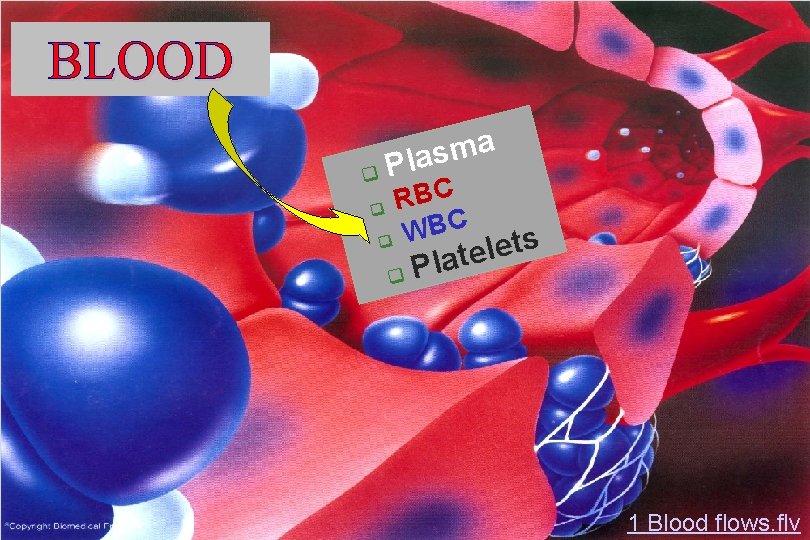
BLOOD a m s la P C B R C B W s t e l late P 1 Blood flows. flv
Plasma consists of: 90% water 10 % solutes: albumin, globulin, clotting factors, antibodies, electrolytes
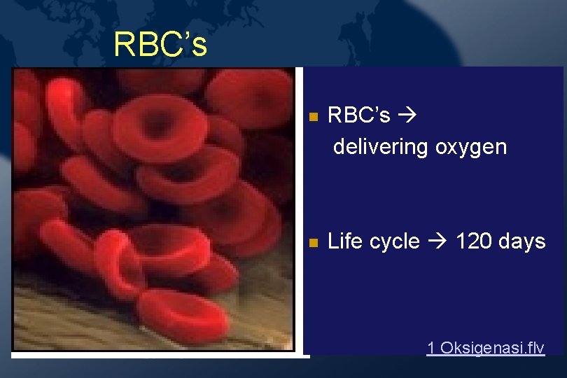
RBC’s delivering oxygen Life cycle 120 days 1 Oksigenasi. flv
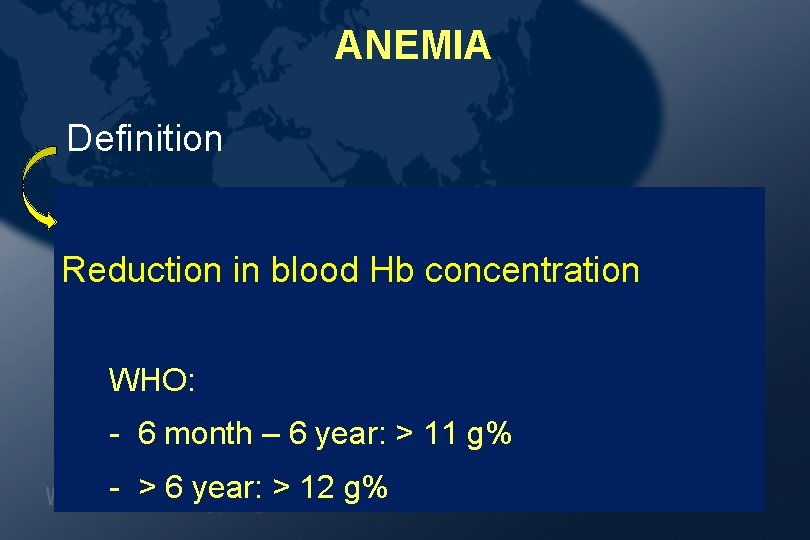
ANEMIA Definition Reduction in blood Hb concentration WHO: - 6 month – 6 year: > 11 g% - > 6 year: > 12 g%
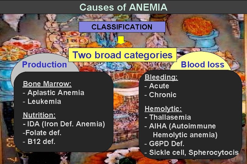
Causes of ANEMIA CLASSIFICATION Production Two broad categories Bone Marrow: - Aplastic Anemia - Leukemia Nutrition: - IDA (Iron Def. Anemia) -Folate def. - B 12 def. Blood loss Bleeding: - Acute - Chronic Hemolytic: - Thallasemia - AIHA (Autoimmune Hemolytic anemia) - G 6 PD Def. - Sickle cell, Spherocytocis
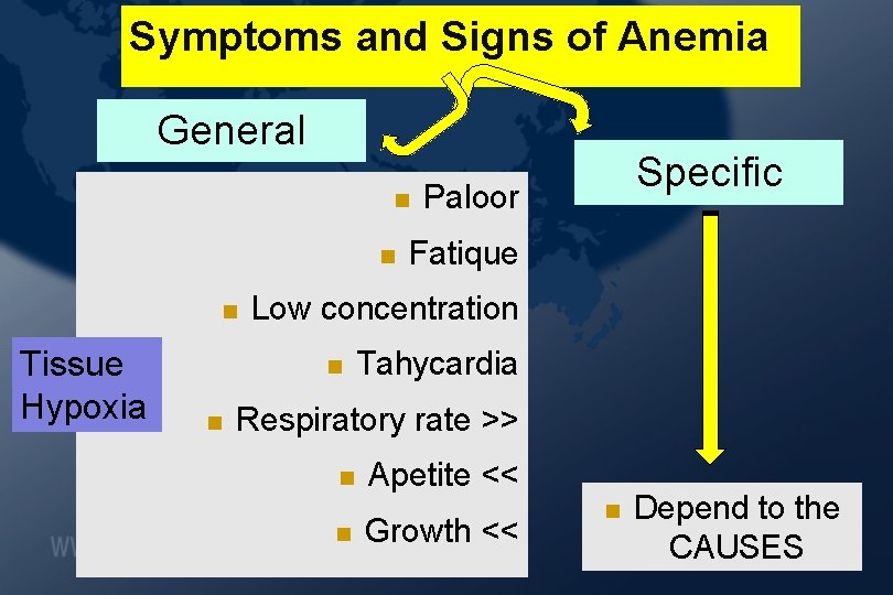
Symptoms and Signs of Anemia General Tissue Hypoxia Paloor Fatique Low concentration Specific Tahycardia Respiratory rate >> Apetite << Growth << Depend to the CAUSES
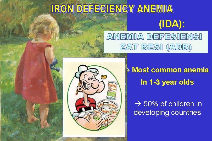
(IDA): ANEMIA DEFESIENSI ZAT BESI (ADB) Most common anemia In 1 -3 year olds 50% of children in developing countries
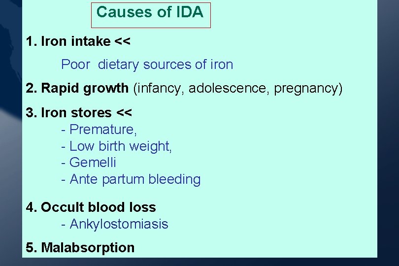
Causes of IDA 1. Iron intake << Poor dietary sources of iron 2. Rapid growth (infancy, adolescence, pregnancy) 3. Iron stores << - Premature, - Low birth weight, - Gemelli - Ante partum bleeding 4. Occult blood loss - Ankylostomiasis 5. Malabsorption
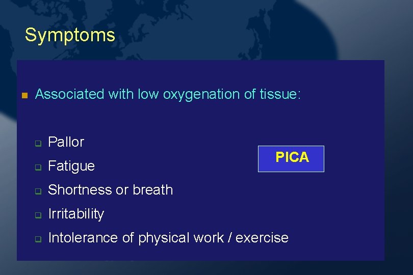
Symptoms Associated with low oxygenation of tissue: Pallor PICA Fatigue Shortness or breath Irritability Intolerance of physical work / exercise
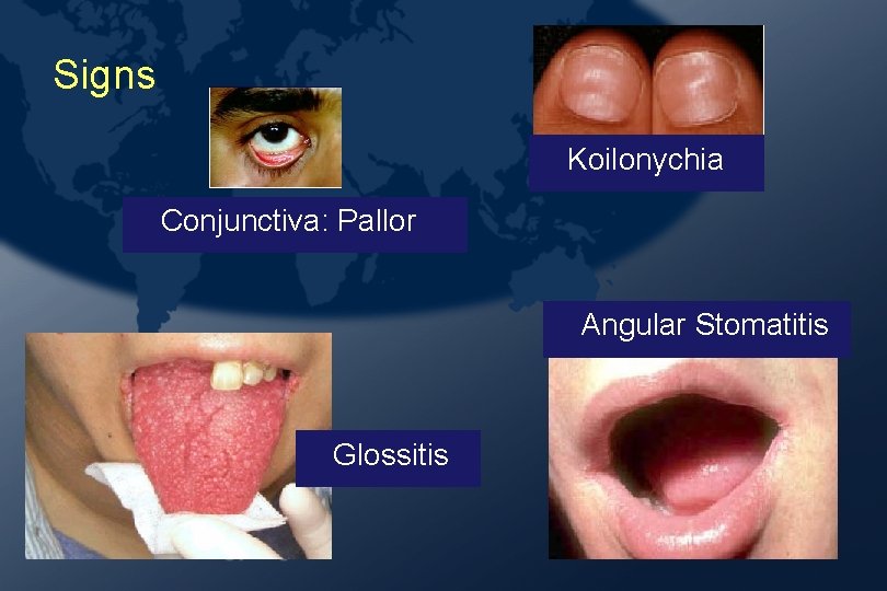
Signs Koilonychia Conjunctiva: Pallor Angular Stomatitis Glossitis
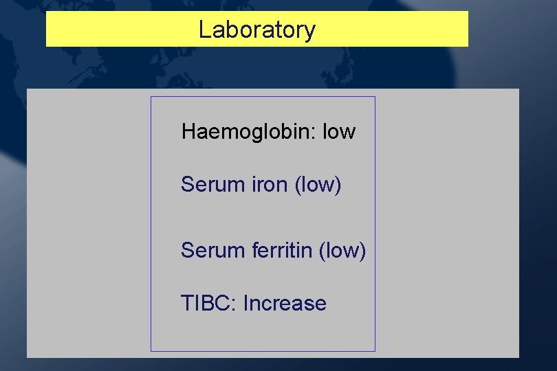
Laboratory Haemoglobin: low Serum iron (low) Serum ferritin (low) TIBC: Increase
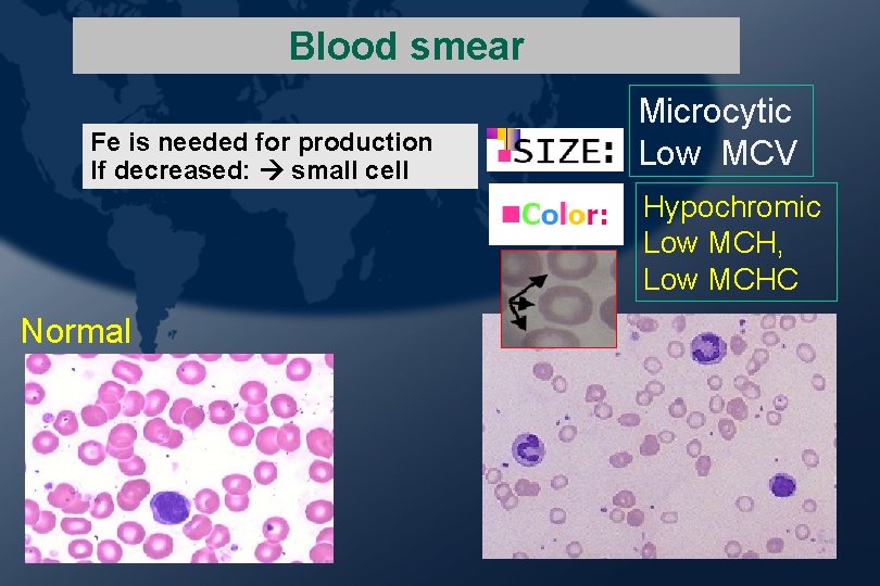
Blood smear Fe is needed for production If decreased: small cell Microcytic Low MCV Hypochromic Low MCH, Low MCHC Normal
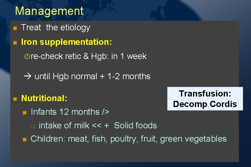
Management Treat the etiology Iron supplementation: · re-check retic & Hgb: in 1 week until Hgb normal + 1 -2 months Nutritional: Infants 12 months /> Transfusion: Decomp. Cordis intake of milk << + Solid foods Children: meat, fish, poultry, fruit, green vegetables
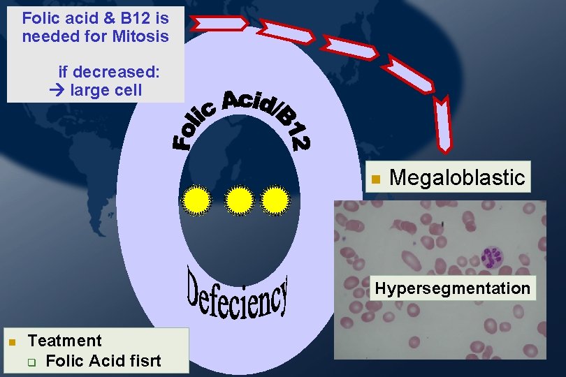
Folic acid & B 12 is needed for Mitosis if decreased: large cell Megaloblastic Hypersegmentation Teatment Folic Acid fisrt
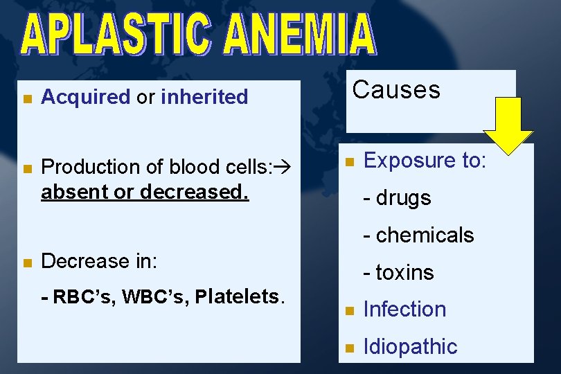
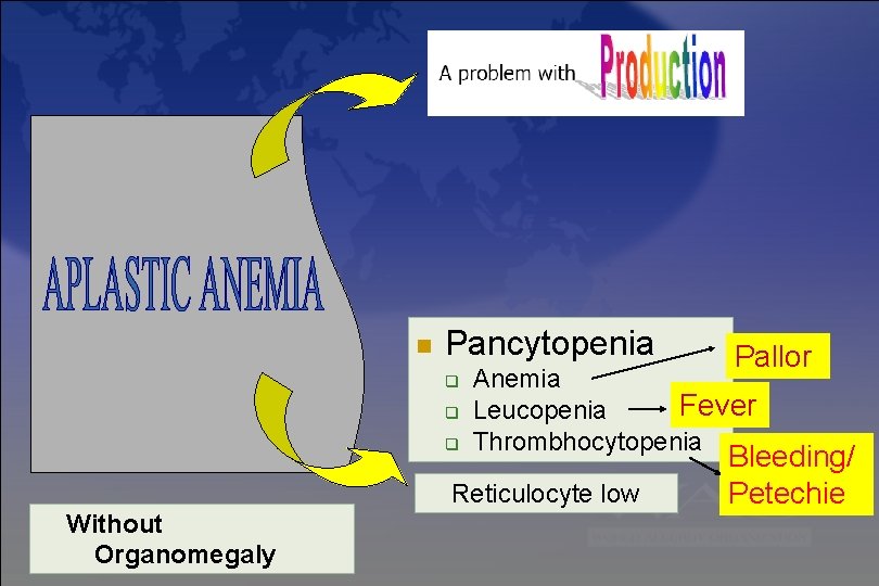
Acquired or inherited Production of blood cells: absent or decreased. Causes Exposure to: - drugs - chemicals Decrease in: - RBC’s, WBC’s, Platelets. - toxins Infection Idiopathic
Pancytopenia Anemia Fever Leucopenia Thrombhocytopenia Reticulocyte low Without Organomegaly Pallor Bleeding/ Petechie
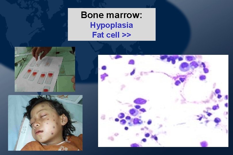
Bone marrow: Hypoplasia Fat cell >>
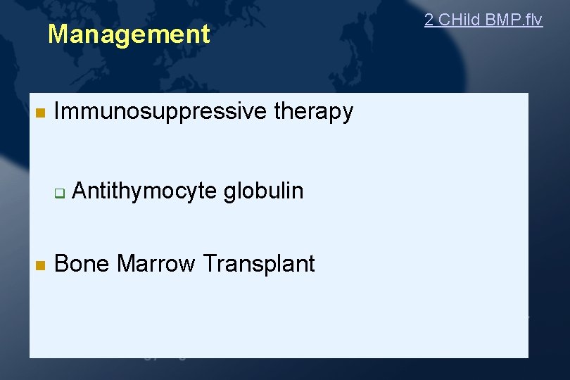
Management Immunosuppressive therapy Antithymocyte globulin Bone Marrow Transplant 2 CHild BMP. flv
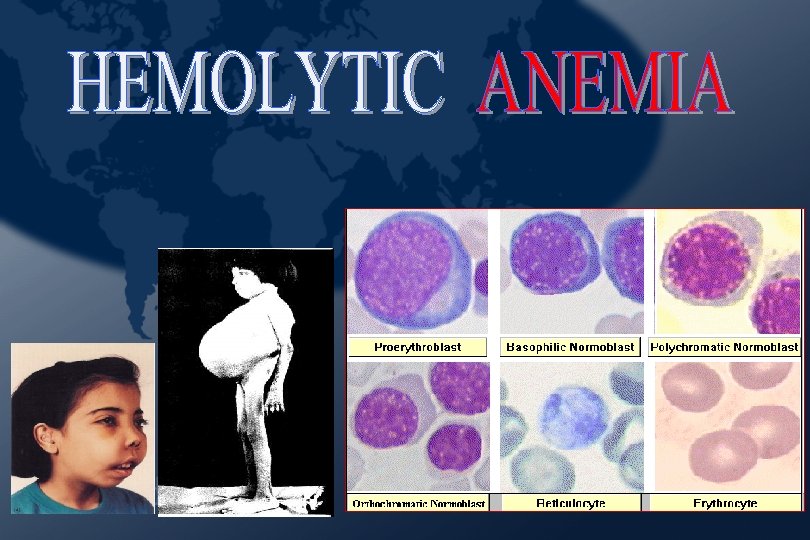
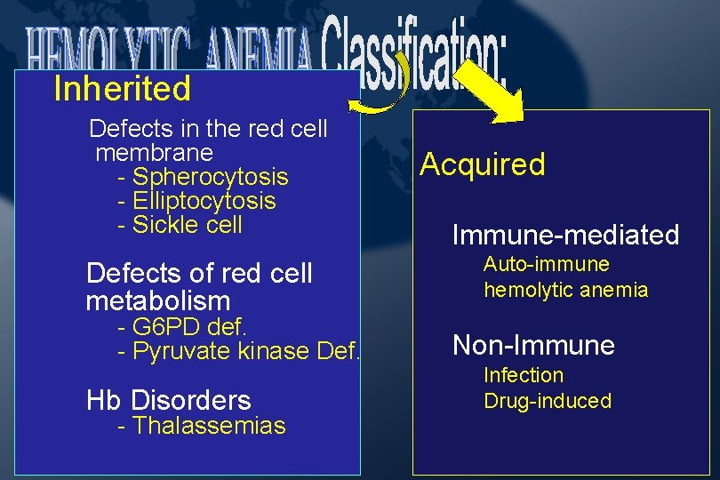
Inherited Defects in the red cell Providing membrane - Spherocytosis - Elliptocytosis - Sickle cell Defects of red cell metabolism - G 6 PD def. - Pyruvate kinase Def. Hb Disorders - Thalassemias Acquired Immune-mediated Auto-immune hemolytic anemia Non-Immune Infection Drug-induced
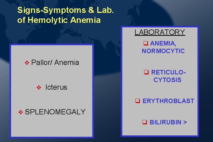
Signs-Symptoms & Lab. of Hemolytic Anemia LABORATORY ANEMIA, NORMOCYTIC v Pallor/ v Anemia Icterus RETICULOCYTOSIS ERYTHROBLAST v SPLENOMEGALY BILIRUBIN > 2000 -2001 22
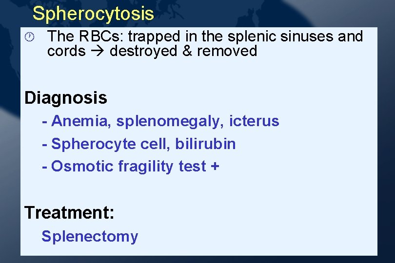
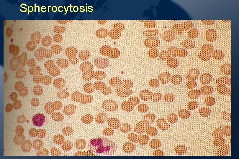
Spherocytosis · The RBCs: trapped in the splenic sinuses and cords destroyed & removed Diagnosis - Anemia, splenomegaly, icterus - Spherocyte cell, bilirubin - Osmotic fragility test + Treatment: Splenectomy
Spherocytosis
Glucose-6 -Phosphate Dehydrogenase Deficiency · >> in males · Hemolysis: episodic following to drugs, infection or ingestion of fava beans · The important drugs: - anti-malarials - sulfonamides - some antipyretics/analgesics - vitamin K, nalidixic acid
G 6 PD Deficiency Contd. · Diagnosis ·Hemolysis · Qualitative with a triggering agent and quantitative measurement of G 6 PD · Management · Supportive care · Blood transfusion ·Education to prevent further episodes.
Auto-immune Hemolytic Anemia (AIHA) Antibodies to the RBC antigens (autoantibodies). - Warm autoimmune hemolytic anemia: Ig. G: optimal at 370 C - Cold autoimmune hemolytic anemia: Ig. M: optimally below 370 C
. b La The direct & Indirect Coombs test t n e tm Glucocorticoids a e r T Splenectomy Immunosuppressive drugs.
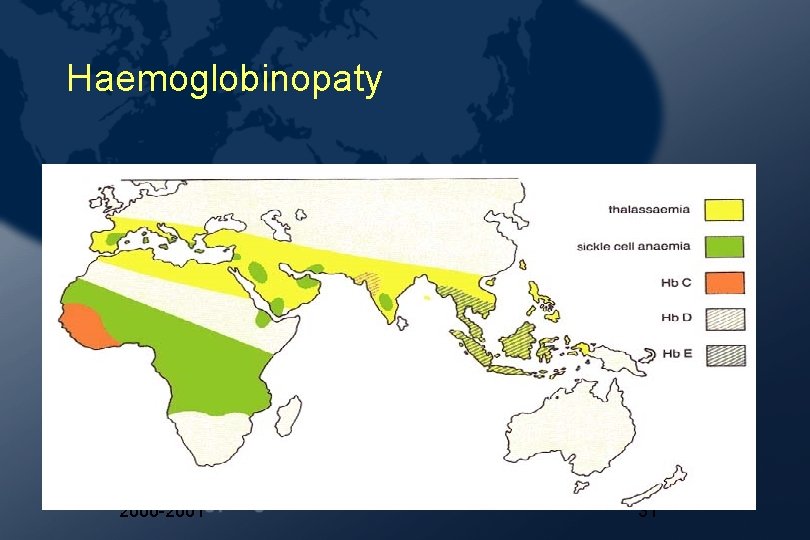
Thalassemias Decreased or absent synthesis of globin chains · Autosomal recessive · Mediterranean, Middle East, India, South East Asia and Africa
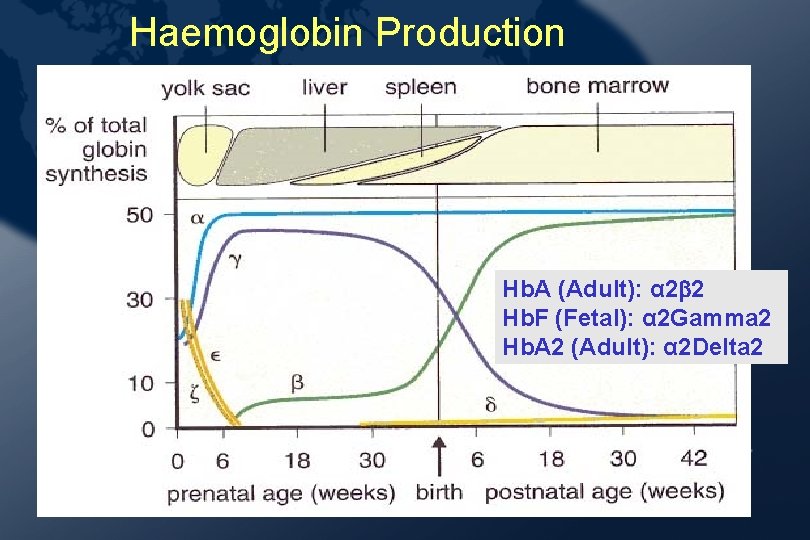
Haemoglobin Production Hb. A (Adult): α 2 2 Hb. F (Fetal): α 2 Gamma 2 Hb. A 2 (Adult): α 2 Delta 2 2000 -2001 30
Haemoglobinopaty 2000 -2001 31
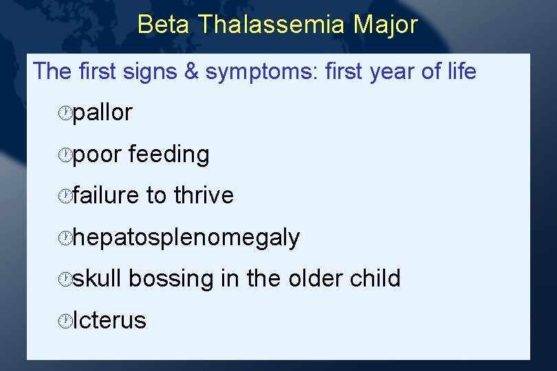
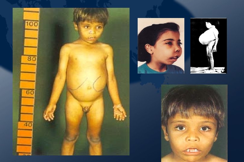
Beta Thalassemia Major The first signs & symptoms: first year of life ·pallor ·poor feeding ·failure to thrive ·hepatosplenomegaly ·skull bossing in the older child ·Icterus
2000 -2001 33
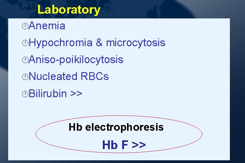
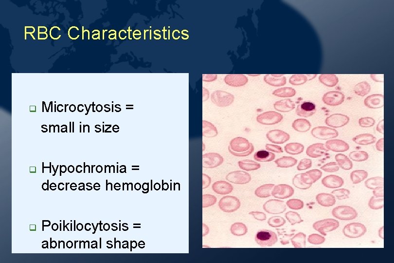
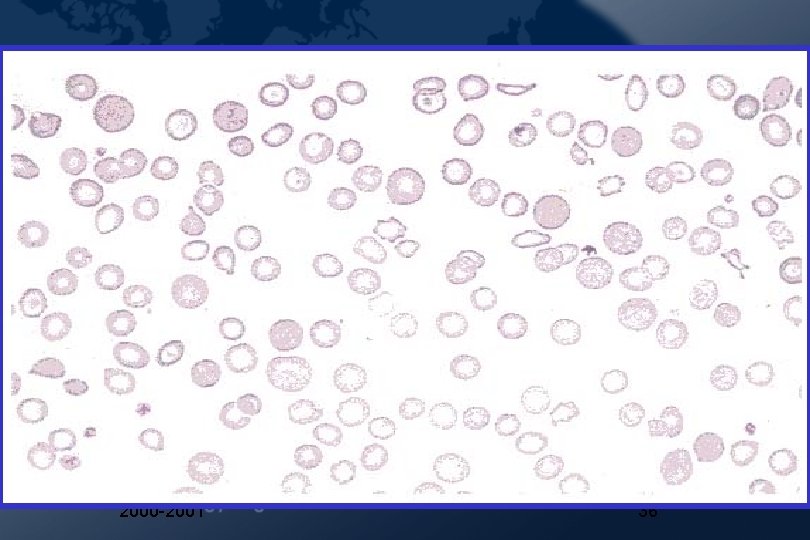
Laboratory ·Anemia ·Hypochromia & microcytosis ·Aniso-poikilocytosis ·Nucleated ·Bilirubin RBCs >> Hb electrophoresis Hb F >>
RBC Characteristics Microcytosis = small in size Hypochromia = decrease hemoglobin Poikilocytosis = abnormal shape
2000 -2001 36
Management · Transfusion: PRC · Splenectomy · for · Iron hypersplenism , increased blood transfusion chelation therapy · with · Stem desferrioxamine: SC, IV cell transplantation
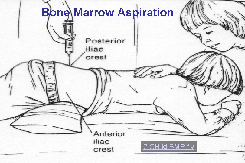
Bone Marrow Aspiration 2 CHild BMP. flv
T H A N K Y O U