Anatomy SkeletalMuscular System The Skeletal System divided into


: *frontal")
: *mandible (1) *maxilla (2)")
: *sternum (1) *true ribs")
: *cervical vertebrae (7) *thoracic")

 *humerus (2) *radius")
 3 fused pairs *ilium")
 *femur (2) *patella (2)")


 containing synovial")


- Slides: 57
Anatomy Skeletal/Muscular System
The Skeletal System: divided into two groups, the axial and appendicular skeleton for a total of 206 bones in the body. I. The Axial Skeleton: consists of 80 bones that revolve around the vertical axis of the skeleton. *skull, ribs, sternum and vertebral column
A. Bones of the Axial Skeleton 1. The Skull A. Cranial bones (8): *frontal (1) *parietal (2) *temporal (2) *sphenoid (1) *ethmoid (1) *occipital (1) http: //www. learnbones. com
A. Bones of the Axial Skeleton B. Facial Bones (14): *mandible (1) *maxilla (2) *zygomatic bone (2) *nasal bone (2) *lacrimal (2) *palatine (2 *inferior nasal concha (2) *volmer (1)
A. Bones of the Axial Skeleton 2. Thoracic cage (25): *sternum (1) *true ribs (14) articulate with the sternum directly. *false ribs (6) *floating ribs (4)
A. Bones of the Axial Skeleton 3. Vertebral Column (26): *cervical vertebrae (7) *thoracic vertebrae (12) *lumbar vertebrae (5) *sacrum (5 fused) *coccyx (4 fused)
II. The Appendicular Skeleton: consists of 126 bones that append to the axial skeleton. A. Bones of the Appendicular Skeleton 1. Pectoral girdle (4) *clavicle (2) *scapula (2)
A. Bones of the Appendicular Skeleton 2. The Upper Limb (60) *humerus (2) *radius (2) *ulna (2) *carpals (16) *metacarpals (10) *phalanges (28)
A. Bones of the Appendicular Skeleton 3. Pelvic Girdle (2) 3 fused pairs *ilium *ischium *pubis
A. Bones of the Appendicular Skeleton 4. Lower limb (60) *femur (2) *patella (2) *tibia (2) *fibula (2) *tarsal (14) *metatarsals (10) *phalanges (28)
III. Functions of the Skeletal System A. Axial Skeleton Functions: *protects the brain/heart/lungs/spi nal cord and digestive organs. *provides structure and support for upright position *transfers weight to lower extremities
III. Functions of the Skeletal System B. Appendicular Skeleton Functions: *protection of digestive/excretive and repro. organs *attachment of ligament and muscles allowing movement *blood cell formation
IV. Types of Bones 1. Long Bones: those that are longer than they are wide. *femur, tibia, fibula *humerus, radius, ulna *clavicle *metacarpals *metatarsals *phalanges
2. Short Bones: are as wide as they are long. *tarsals of the foot *carpals of the hand
3. Flat Bones: broad flat plates used for protection and muscular attachment. Most RBC’s are produce in flat bones. *skull *rib cage *sternum *scapula *pelvis *os coxae (hip bone)
4. Irregular Bones: have peculiar shapes and cannot be grouped in the other bone categories. *verterbrae *sacrum *coccyx *mandible *maxilla *hyoid
long bone structure
V. Anatomical Terminology
V. Anatomical Terminology 1. Anterior: towards the front. 2. Posterior: towards the back. 3. Superior: towards the head. 4. Inferior: towards the feet. 5. Proximal: nearest; closer to any point of reference. 6. Distal: remote; farther from any point of reference. 7. Lateral: point that is more distant from the median plane. 8. Medial: point that is closer to the median plane
VI. Connective Tissue 1. Cartilage: a flexible connective tissue found between the bones of joints. It acts as a cushion between joints and reduces friction in movement.
VI. Connective Tissue 2. Ligament: dense, flexible tissue that connects bone to bone at a joint.
VI. Connective Tissue 3. Tendon: consists of dense collagen fibers and connect muscle to bone.
VII. Joint: point where two or more bones articulate allowing movement and mechanical support. A. Types of Joints 1. Fibrous: held together by fibrous connective tissue. Usually immovable or slightly moveable. Ex. Suture joints between cranial bones and distal joint of tibia and fibula.
2. Cartilaginous: joints held together by cartilage. May be immovable or slightly movable.
3. Synovial: freely movable joints characterized by a synovial cavity (joint cavity) containing synovial fluid. Features of Synovial Joints: a. Articular cartilage: (hyaline cartilage) covers the ends of bones. b. Synovial membrane: surrounds the synovial cavity and secretes synovial fluid.
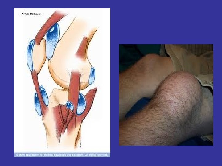
c. Articular Capsule: is composed of the synovial membrane and the fibrous capsule. d. Bursae: fluid filled sac that cushions and reduces friction between moving structures. e. Meniscus: disperses the weight of the body, reduces friction and stabilizes the joint.
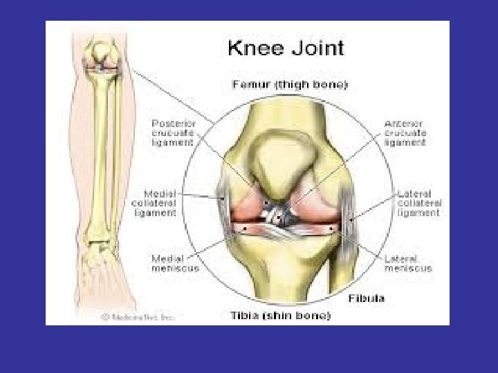
Types of Synovial joints 1. Hinge: allow flexion and extension along one plane. *elbow *knee *ankle *between phalangeal bones.
Types of Synovial joints 2. Ball and Socket: allow all movements except gliding. *hip *shoulder
Types of Synovial joints 3. Condyloid: allow flexion, extension, abduction, adduction and circumduction. *wrist (radius and carpals) *between the metacarpals and phalanges.
Types of Synovial joints 4. Pivot: one bone rotates about another. *the neck *radius and ulna
Types of Synovial joints 5. Gliding: permits slight sliding and gliding movements. *spine *wrist *ankles *clavicle
Types of Synovial joints 6. Saddle: same movement as condyloid joints. *thumb
The Muscular System I. Characteristics of muscle tissue *contractibility-ability of the muscle to shorten. *extensibility: ability of the muscle to lengthen. *elasticity: ability of the muscle to return to its normal size.
I. Characteristics of muscle tissue * atrophy-a decrease in the size of muscle tissue. * hypertrophy-an increase in the size of muscle tissue. * Controlled by nerve tissue and fed by capillaries.
II. Types of Muscle Tissue A. Smooth: Involuntary muscle that lines the walls of hollow organs, blood vessels and the digestive tract.
II. Types of Muscle Tissue B. Cardiac: involuntary striated muscle responsible for rhythmic contractions of the heart.
II. Types of Muscle Tissue C. Skeletal: Voluntary striated muscle that is responsible for movement.
III. Structure of Skeletal Muscle
III. Structure of Skeletal Muscle
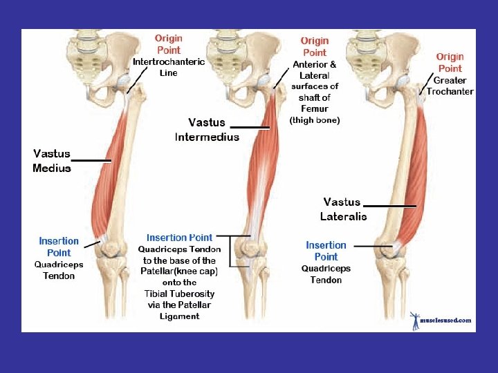
IV. Muscle Attachment 1. Origin: the point of attachment of a muscle tendon to a stationary bone. 2. Insertion: point of attachment of a muscle tendon to a movable bone.
Muscles of the anterior portion of the body Deltoid http: //www. getbodysmart. com/
Pectoralis
iliopsoas
Sartorius
Quadriceps femoris
Tibialis anterior
Abdominus rectus and External obliques
Biceps brachii
Muscles of the posterior portion of the body
Triceps brachii
Gluteus maximus
Hamstrings
Gastrocnemius and Soleus
Erector spinae