Anatomy Physiology The ONLY fluid tissue in the


 � Erythrocytes sink to the bottom (45%")

")


 � Main function is to")


")


 and interleukins prompt bone marrow to")


- Slides: 28
Anatomy & Physiology
� The ONLY fluid tissue in the human body � Classified as a “connective tissue” � Components of blood �Living cells Formed elements �Non-living Plasma matrix
� If blood is centrifuged (Spun rapidly) � Erythrocytes sink to the bottom (45% of blood, a percentage known as the hematocrit) � Buffy coat contains leukocytes and platelets (less than 1% of blood) Buffy coat is a thin, whitish layer between the erythrocytes and plasma � Plasma rises to the top (55% of blood)
� Color: �Bright red means blood is rich in oxygen �Dull red means its poor in oxygen � Temperature: � 100. 4 degrees Farenheiht � Contribution � 8% of our BW to body weight?
� Comprised �Liquid of 90% water portion of blood � Components: �Nutrients �Salts (electrolytes) �Respiratory gases �Hormones �Plasma proteins �Waste products Composition varies depending on what the body needs and diet � Used as a medium to transport materials throughout the body
� Plasma proteins �Most abundant solutes in plasma �Most plasma proteins are made by liver �Various plasma proteins include Albumin—regulates osmotic pressure Clotting proteins—help to stem blood loss when a blood vessel is injured Antibodies—help protect the body from pathogens
� Acidosis �Blood becomes too acidic � Alkalosis �Blood � In becomes too basic each scenario, the respiratory system and kidneys help restore blood p. H to normal
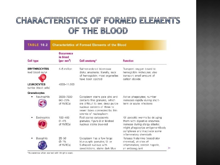
� A. K. A. (red blood cells or RBCs) � Main function is to carry oxygen � Anatomy of circulating erythrocytes Biconcave disks Essentially bags of hemoglobin (carry oxygen) Anucleate (no nucleus) Contain very few organelles � RBCs outnumber WBC 1000 -1!!!
�Iron-containing protein in RBCs �Binds strongly, but reversibly, to oxygen �Each hemoglobin molecule has four oxygen binding sites �ATP through anaerobic glycolysis Sickle Cell Anemia
�Anemia is a decrease in the oxygen-carrying ability of the blood �Sickle cell anemia (SCA) results from abnormally shaped hemoglobin �Polycythemia is an excessive or abnormal increase in the number of erythrocytes Caused by: Bone marrow cancer And living in high altitudes � Anemia
�A. K. A. “White Blood Cells” WBCs �Crucial in the body’s defense against disease �The only complete cells in blood, with a nucleus and organelles �Able to move into and out of blood vessels (diapedesis) �Can move by ameboid motion Cytoplasm extensions move them across �Can respond to chemicals released by damaged tissues
� Leukocytosis: Abnormal numbers of leukocytes WBC count above 11, 000 leukocytes/mm 3 Generally indicates an infection � Leukopenia: Abnormally � low leukocyte level Commonly caused by certain drugs such as corticosteroids and anticancer agents �Leukemia Bone marrow becomes cancerous, turns out excess WBC
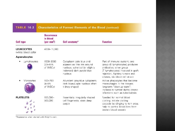
� Types of leukocytes �Granulocytes Include neutrophils, eosinophils, and basophils �Agranulocytes (-a; meaning without) Lack visible cytoplasmic granules Include lymphocytes and monocytes
� List of the WBCs from most to least abundant �Neutrophils �Lymphocytes �Monocytes �Eosinophils �Basophils � Easy way to remember this list �Never �Let �Monkeys �Eat �Bananas
Hemocytoblast stem cells Lymphoid stem cells Myeloid stem cells Secondary stem cells Basophils Erythrocytes Platelets Eosinophils Lymphocytes Monocytes Neutrophils
� Types of granulocytes �Neutrophils Multilobed nucleus with fine granules Act as phagocytes at active sites of ACUTE infections, mainly bacteria and fungi �Eosinophils Large brick-red cytoplasmic granules Found in response to allergies and infections from parasitic worms �Basophils Have histamine-containing granules Initiate inflammation by thinning blood vessels so other WBCs can penetrate to needed site easily
�Lymphocytes Nucleus fills most of the cell Play an important role in the immune response 2 nd most common leukocyte �Monocytes Largest of the white blood cells Function as macrophages Fight chronic infection
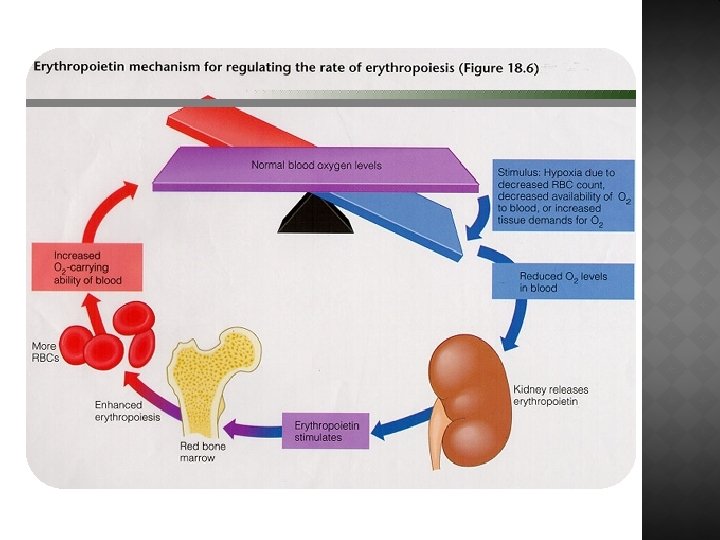
� Means, RBC formation � Occurs in red bone marrow � Wear out in 100 to 120 days � When worn out, RBCs are eliminated by phagocytes in the spleen or liver � Lost cells are replaced by division of hemocytoblasts in the red bone marrow � Rate is controlled by a hormone (erythropoietin) � Kidneys produce most erythropoietin as a response to reduced oxygen levels in the blood � Homeostasis is maintained by negative feedback from blood oxygen levels
� Controlled �Colony by hormones stimulating factors (CSFs) and interleukins prompt bone marrow to generate leukocytes �Thrombopoietin stimulates production of platelets �WBC growth occurs in red bone marrow �If there is a decrease in WBC they will take a sample of red bone marrow for further evaluation
� Large losses of blood have serious consequences �Loss of 15– 30% causes weakness �Loss of over 30% causes shock, which can be fatal � Transfusions are the only way to replace blood quickly � Transfused blood must be of the same blood group
� Blood contains genetically determined proteins � Antigens (a substance the body recognizes as foreign) may be attacked by the immune system � Antibodies are the “recognizers” � Blood is “typed” by using antibodies that will cause blood with certain proteins to clump (agglutination)
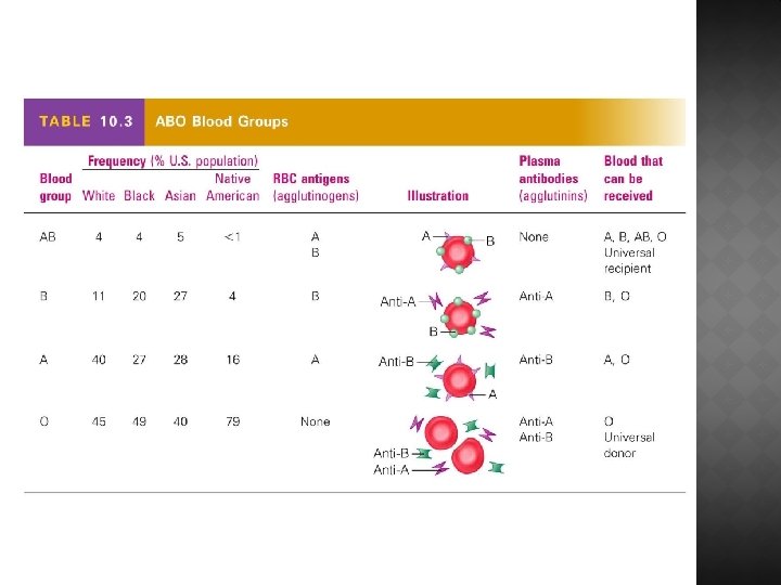
� Based on the presence or absence of two antigens �Type A �Type B � The lack of these antigens is called type O � The presence of both antigens A and B is called type AB � The presence of antigen A is called type A � The presence of antigen B is called type B � The lack of both antigens A and B is called type O
� Blood type AB can receive A, B, AB, and O blood �Universal recipient � Blood type B can receive B and O blood � Blood type A can receive A and O blood � Blood type O can receive O blood �Universal donor
� Named because of the presence or absence of one of eight Rh antigens (agglutinogen D) � Most Americans are Rh+ (Rh positive) � Problems can occur in mixing Rh+ blood into a body with Rh– (Rh negative) blood