
Anatomy Physiology of erection and ejaculation The penile



- Slides: 43
Anatomy &Physiology of erection and ejaculation
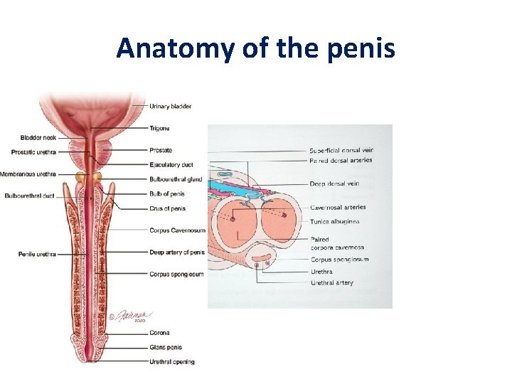
The penile shaft is composed of 3 erectile columns, the 2 corpora cavernosa and the corpus spongiosum, The single corpus spongiosum lies in the ventral groove between the 2 corpora cavernosa. The urethra passes through the corpus spongiosum
The erectile tissue within the corpora contains arteries, nerves, muscle fibers, and venous sinuses lined with flat endothelial cells, and it fills the space of the corpora cavernosa.
Anatomy of the penis
Arterial supply The blood supply to deep structures of the penis is derived from a continuation of the internal pudendal artery, after it gives off the perineal branch. Three branches of the internal pudendal artery flow to the penis, as follows:
The artery of the bulb (bulbourethral artery) passes through the deep penile (Buck) fascia to enter and supply the bulb of the penis and penile (spongy) urethra The dorsal artery travels along the dorsum of the penis between the dorsal nerve and deep dorsal vein and gives off circumflex branches that accompany the circumflex veins; the terminal branches are in the glans penis The deep penile (cavernosal) artery is usually a single artery that arises on each side and enters the corpus cavernosum at the crus and runs the length of the penile shaft, giving off the helicine arteries, which are an integral component of the erectile process
Blood supply to the skin of the penis is from the left and right superficial external pudendal arteries, which arise from the femoral artery
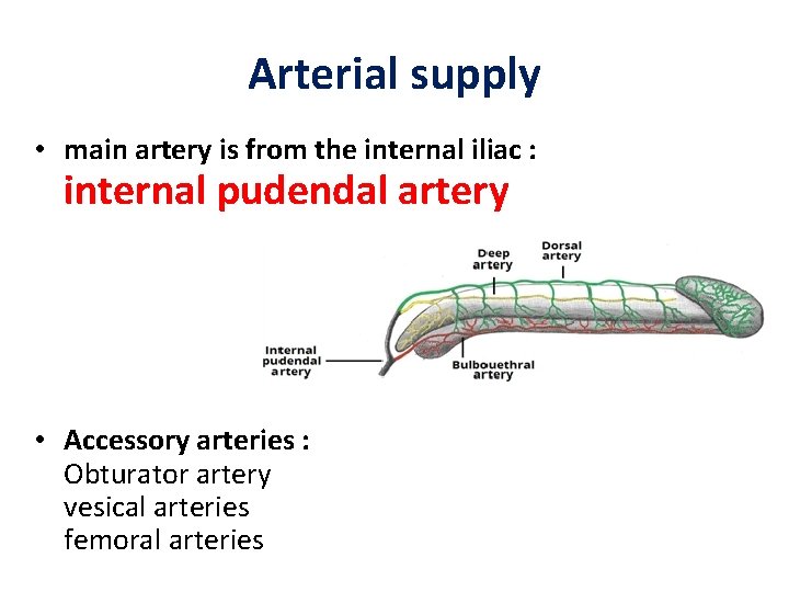
Arterial supply • main artery is from the internal iliac : internal pudendal artery • Accessory arteries : Obturator artery vesical arteries femoral arteries
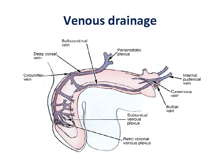
venous drainages The penis is drained by 3 venous systems, the superficial, intermediate, and deep Superficial veins are contained in the dartos fascia on the dorsolateral surface of the penis and coalesce at the base to form a single superficial dorsal vein, which usually drains into the great saphenous veins via the superficial external pudendal veins.
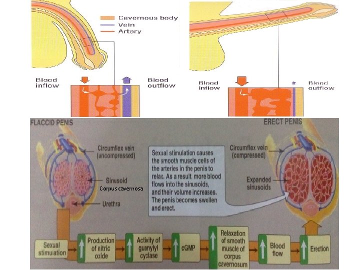
Dorsal penile veins ? ? These are the veins are going to be compressed to prohibit blood from draining
Venous drainage
Lymphatic drainage from the glans penis drains into large trunks in the area of the frenulum. These lymphatic vessels then circle to the dorsum of the corona and unite, coursing proximally beneath the deep penile (Buck) fascia, terminating mostly in the deep inguinal nodes of the femoral triangle. Some lymphatic drainage is to the presymphyseal lymph nodes and to the lateral lymph nodes of the external iliac lymphatics.
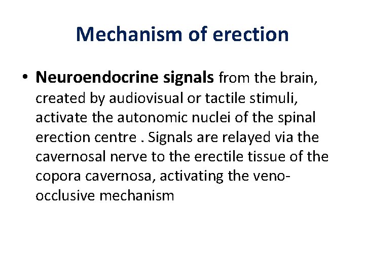
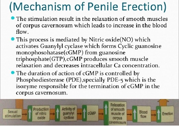
Mechanism of erection • Neuroendocrine signals from the brain, created by audiovisual or tactile stimuli, activate the autonomic nuclei of the spinal erection centre. Signals are relayed via the cavernosal nerve to the erectile tissue of the copora cavernosa, activating the venoocclusive mechanism
Corpus cavernosa
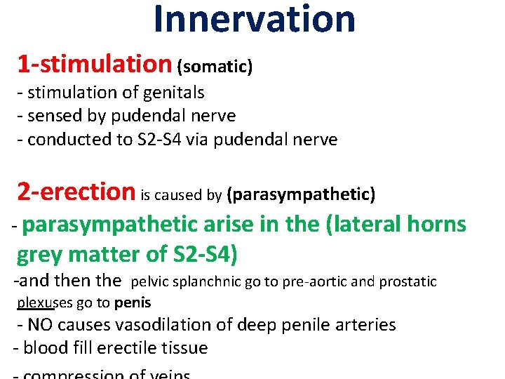
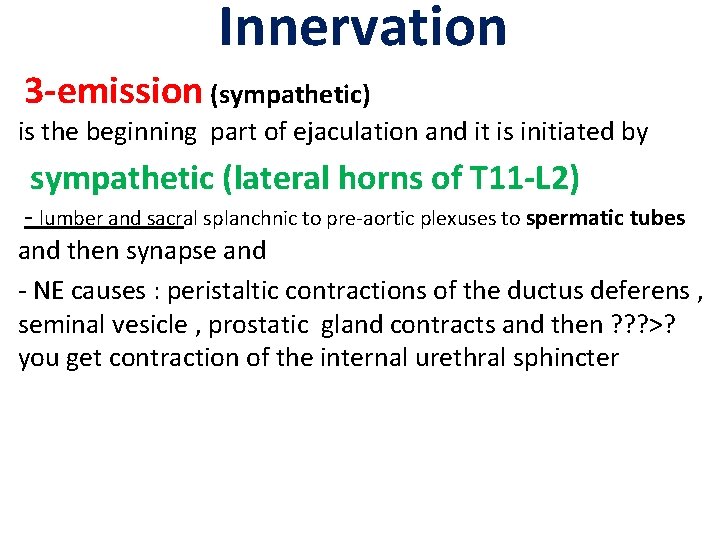
Innervation 1 -stimulation (somatic) - stimulation of genitals - sensed by pudendal nerve - conducted to S 2 -S 4 via pudendal nerve 2 -erection is caused by (parasympathetic) - parasympathetic arise in the (lateral horns grey matter of S 2 -S 4) -and then the pelvic splanchnic go to pre-aortic and prostatic plexuses go to penis - NO causes vasodilation of deep penile arteries - blood fill erectile tissue
Innervation 3 -emission (sympathetic) is the beginning part of ejaculation and it is initiated by sympathetic (lateral horns of T 11 -L 2) - lumber and sacral splanchnic to pre-aortic plexuses to spermatic tubes and then synapse and - NE causes : peristaltic contractions of the ductus deferens , seminal vesicle , prostatic gland contracts and then ? ? ? >? you get contraction of the internal urethral sphincter
- semen don’t mix with urine - semen don’t go retrogradely into the urinary bladder
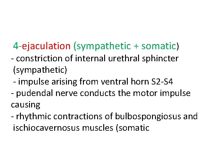
4 -ejaculation (sympathetic + somatic) - constriction of internal urethral sphincter (sympathetic) - impulse arising from ventral horn S 2 -S 4 - pudendal nerve conducts the motor impulse causing - rhythmic contractions of bulbospongiosus and ischiocavernosus muscles (somatic
Innervation • Central: medial preoptic area (MPOA) and paraventricular nucleus (PVN) in the hypothalamus are important centres for sexual function and penile erection.
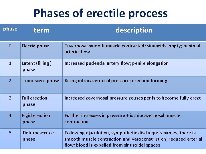
Phases of erectile process phase term description 0 Flaccid phase Cavernosal smooth muscle contracted; sinusoids empty; minimal arterial flow 1 Latent (filling ) phase Increased pudendal artery flow; penile elongation 2 Tumescent phase Rising intracavernosal pressure; erection forming 3 Full erection phase Increased cavernosal pressure causes penis to become fully erect 4 Rigid erection phase Further increases in pressure + ischiocavernosal muscle contraction 5 Detumescence phase Following ejaculation, sympathetic discharge resumes; there is smooth muscle contraction and vasoconstriction; reduced arterial flow; blood is expelled from sinusoidal spaces
Thank you
Erectile dysfunction evaluation & treatment Fadi Abu-Tair
Definition Erectile dysfunction (ED) (also called impotence) describes the ‘consistent or recurrent inability to attain and/or maintain a penile erection sufficient for sexual intercourse
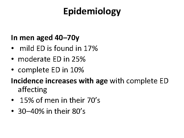
Epidemiology In men aged 40– 70 y • mild ED is found in 17% • moderate ED in 25% • complete ED in 10% Incidence increases with age with complete ED affecting • 15% of men in their 70’s • 30– 40% in their 80’s
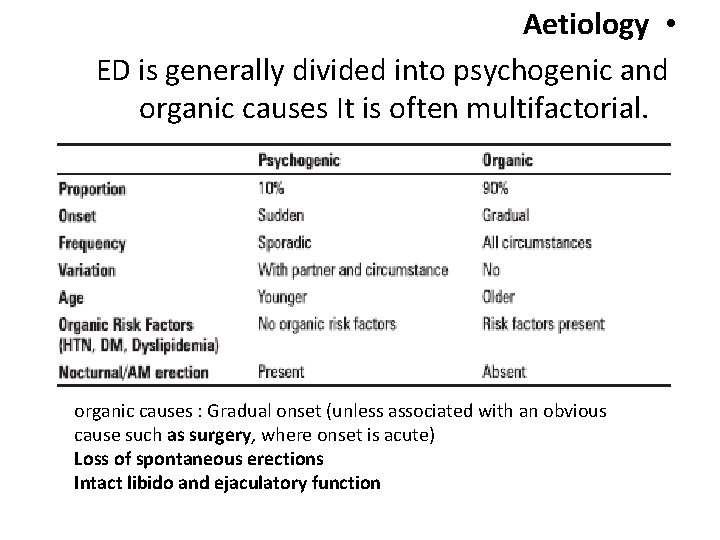
Aetiology • ED is generally divided into psychogenic and organic causes It is often multifactorial. organic causes : Gradual onset (unless associated with an obvious cause such as surgery, where onset is acute) Loss of spontaneous erections Intact libido and ejaculatory function
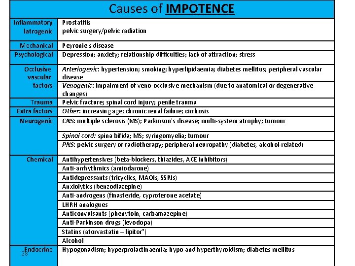
Causes of IMPOTENCE Inflammatory Iatrogenic Prostatitis pelvic surgery/pelvic radiation Mechanical Psychological Peyronie's disease Depression; anxiety; relationship difficulties; lack of attraction; stress Occlusive vascular factors Trauma Extra factors Neurogenic Arteriogenic: hypertension; smoking; hyperlipidaemia; diabetes mellitus; peripheral vascular disease Venogenic: impairment of veno-occlusive mechanism (due to anatomical or degenerative changes) Pelvic fracture; spinal cord injury; penile trauma Other: increasing age; chronic renal failure; cirrhosis CNS: multiple sclerosis (MS); Parkinson's disease; multi-system atrophy; tumour Spinal cord: spina bifida; MS; syringomyelia; tumour PNS: pelvic surgery or radiotherapy; peripheral neuropathy (diabetes, alcohol-related) Chemical Endocrine 28 Antihypertensives (beta-blockers, thiazides, ACE inhibitors) Anti-arrhythmics (amiodarone) Antidepressants (tricyclics, MAOIs, SSRIs) Anxiolytics (benzodiazepine) Anti-androgens (finasteride, cyproterone acetate) LHRH analogues Anticonvulsants (phenytoin, carbamazepine) Anti-Parkinson drugs (levodopa) Statins (atorvastatin – lipitor®) Alcohol Hypogonadism; hyperprolactinaemia; hypo and hyperthyroidism; diabetes mellitus
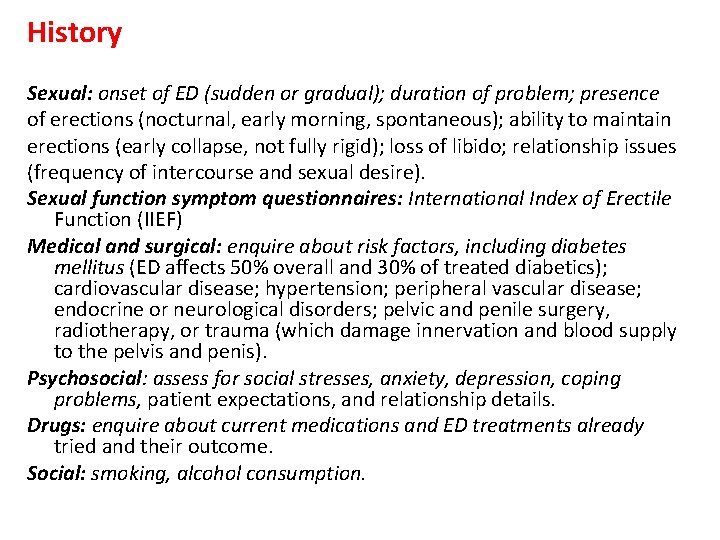
History Sexual: onset of ED (sudden or gradual); duration of problem; presence of erections (nocturnal, early morning, spontaneous); ability to maintain erections (early collapse, not fully rigid); loss of libido; relationship issues (frequency of intercourse and sexual desire). Sexual function symptom questionnaires: International Index of Erectile Function (IIEF) Medical and surgical: enquire about risk factors, including diabetes mellitus (ED affects 50% overall and 30% of treated diabetics); cardiovascular disease; hypertension; peripheral vascular disease; endocrine or neurological disorders; pelvic and penile surgery, radiotherapy, or trauma (which damage innervation and blood supply to the pelvis and penis). Psychosocial: assess for social stresses, anxiety, depression, coping problems, patient expectations, and relationship details. Drugs: enquire about current medications and ED treatments already tried and their outcome. Social: smoking, alcohol consumption.
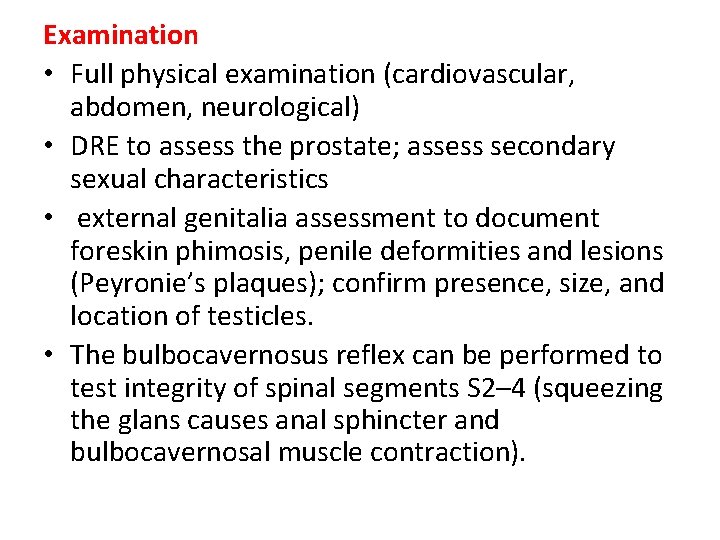
Examination • Full physical examination (cardiovascular, abdomen, neurological) • DRE to assess the prostate; assess secondary sexual characteristics • external genitalia assessment to document foreskin phimosis, penile deformities and lesions (Peyronie’s plaques); confirm presence, size, and location of testicles. • The bulbocavernosus reflex can be performed to test integrity of spinal segments S 2– 4 (squeezing the glans causes anal sphincter and bulbocavernosal muscle contraction).
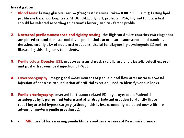
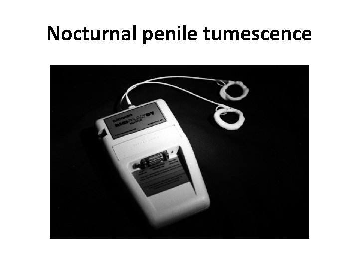
Investigation 1. Blood tests: fasting glucose; serum (free) testosterone (taken 8. 00– 11. 00 a. m. ); fasting lipid profile are basic work-up tests. SHBG; U&E; LH/FSH; prolactin; PSA; thyroid function test should be selected according to patient’s history and risk factor profile. 2. Nocturnal penile tumescence and rigidity testing: the Rigiscan device contains two rings that are placed around the base and distal penile shaft to measure tumescence and number, duration, and rigidity of nocturnal erections. Useful for diagnosing psychogenic ED and for illustrating this diagnosis to patients. 3. Penile colour Doppler USS: measures arterial peak systolic and end diastolic velocities, preand post-intracavernosal injection of PGE 1. 4. Cavernosography: imaging and measurement of penile blood flow after intracavernosal injection of contrast and induction of artificial erection, used to identify venous leaks. 5. Penile arteriography: reserved for trauma-related ED in younger men. Pudendal arteriography is performed before and after drug-induced erection to identify those requiring arterial bypass surgery (although this is less commonly indicated now with the advent of modern penile prostheses). 6. - MRI: useful for assessing penile fibrosis and severe cases of Peyronie’s disease.
Nocturnal penile tumescence
treatment • Correct any reversible causes (i. e. alter lifestyle, stop smoking, change medication, etc. ) • Psychosexual therapy q Aims to understand address underlying psychological issues and provides information and treatment in the form of sex education, psychosexual counselling q instruction on improving partner communication skills q cognitive therapy and behavioural therapy (programmed relearning of couple’s sexual relationship). q Pharmacotherapy may be a useful adjuvant.
1 -Phosphodiesterase type-5 (PDE 5) inhibitors: first-line therapies sildenafil (Viagra. R), tadalafil (Cialis. R), vardenafil (Levitra. R) • enhance cavernosal smooth muscle relaxation • by blocking the breakdown of c. GMP by phosphodiesterase. • Sexual stimulus is still required to initiate events. • Success is reported in up to 80%. • Early use of PDE 5 inhibitors following radical prostatectomy can help optimize the return of spontaneous erections (penile rehabilitation). Contraindications: Ø patients taking nitrates Ø recent myocardial infarction Ø recent stroke Ø hypotension Ø unstable angina, non-arteritic anterior ischaemic Ø optic nerve neuropathy (NAION). Ø Cautions: intermediate and high risk cardiovascular disease requires cardiac review prior to treatment use with A-blockers, groups with predisposition to priapism.
2 -Dopamine receptor agonist: Ø apomorphine (Uprima. R) Ø taken sublingually Ø acts centrally on dopaminergic receptors in the paraventricular nucleus of the hypothalamus Ø enhance and coordinate the effect of sexual stimuli. Ø Side effects: nausea, headache, dizziness Ø Not commonly used
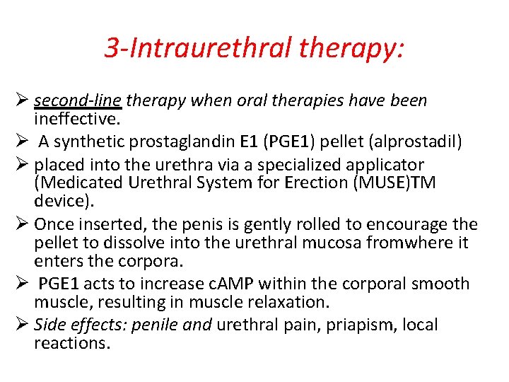
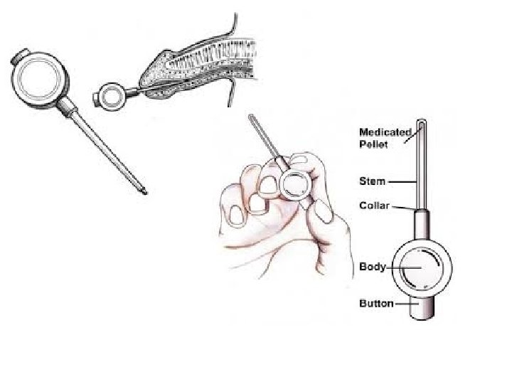
3 -Intraurethral therapy: Ø second-line therapy when oral therapies have been ineffective. Ø A synthetic prostaglandin E 1 (PGE 1) pellet (alprostadil) Ø placed into the urethra via a specialized applicator (Medicated Urethral System for Erection (MUSE)TM device). Ø Once inserted, the penis is gently rolled to encourage the pellet to dissolve into the urethral mucosa fromwhere it enters the corpora. Ø PGE 1 acts to increase c. AMP within the corporal smooth muscle, resulting in muscle relaxation. Ø Side effects: penile and urethral pain, priapism, local reactions.
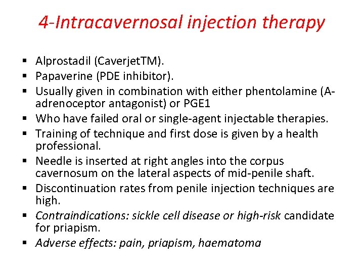
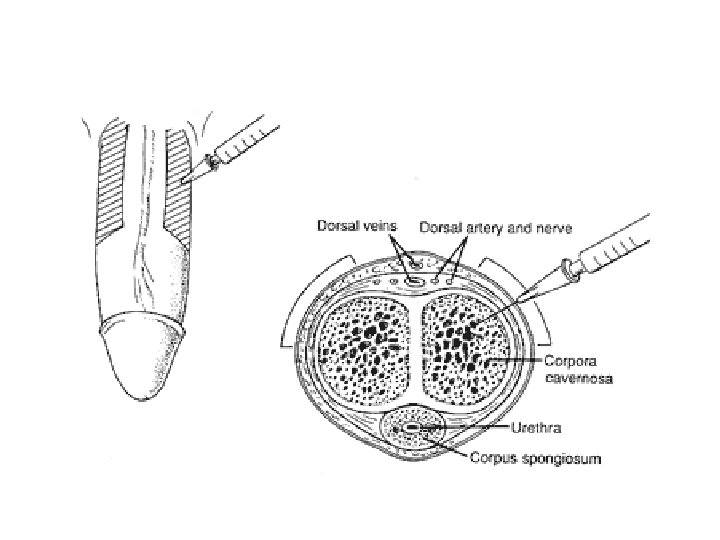
4 -Intracavernosal injection therapy § Alprostadil (Caverjet. TM). § Papaverine (PDE inhibitor). § Usually given in combination with either phentolamine (Aadrenoceptor antagonist) or PGE 1 § Who have failed oral or single-agent injectable therapies. § Training of technique and first dose is given by a health professional. § Needle is inserted at right angles into the corpus cavernosum on the lateral aspects of mid-penile shaft. § Discontinuation rates from penile injection techniques are high. § Contraindications: sickle cell disease or high-risk candidate for priapism. § Adverse effects: pain, priapism, haematoma
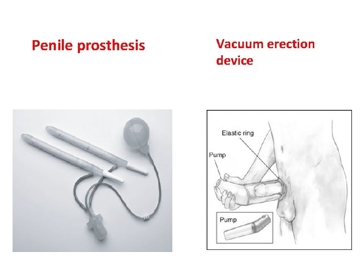
Vacuum erection device Ø Used when pharmacotherapies have failed. Ø It contains three components: a vacuum chamber, pump, and constriction band Ø The penis is placed in the chamber and the vacuum created by the pump increases blood flow to the corpora cavernosa to induce an erection. Ø The constriction band is placed onto the base of the penis to retain blood in the corpora and maintain rigidity. Ø Relative contraindication: anticoagulation therapy. Ø Side effects: penile coldness, bruising. Microvascular arterial bypass and venous ligation surgery Used in: specialist centres where there is a clear-cut diagnosis of a vascular disorder. Acts to increase arterial inflow and decrease venous outflow. Rarely used now as it is uncommon for success rates to exceed 50%.
Penile prosthesis Ø Semi-rigid, malleable, and inflatable penile prostheses Ø otherapies have failed or are unsuitable Ø Also indicated for Peyronie’s disease, trauma, and penile fibrosis (i. e. secondary to priapism). Ø The device is surgically implanted into the corpora to provide penile rigidity and generally has high satisfaction rates, up to 90% Ø Side effects: infection, erosion, mechanical failure, penile shortening, glans may not fully engorge. Testosterone replacement therapy Ø Indicated for hypogonadism Ø available in oral, buccal, intramuscular, pellet, transdermal patch, and gel forms. Ø Most guidelines recommend PSA, Hb, and LFT checks before and after starting treatment Ø It can improve the results of PDE 5 inhibitors in hypogonadal men.
Penile prosthesis Vacuum erection device
Peyronie’s disease An acquired benign penile condition characterized by deformity of the penile shaft secondary to the formation of a fibrous inelastic scar on the tunica albuginea. Epidemiology Prevalence is 3– 9%, predominantly affecting men aged 40– 60 y. Pathophysiology excessive connective tissue (fibrosis) and increased cellularity with random orientation of collagen fibres. Dorsal penile plaques are most common (66%). The corpus cavernosus underlying the lesion cannot lengthen fully on erection, resulting in penile curvature. The disorder has two phases: - Active phase (1– 6 months) - Quiescent (stable) phase (9– 12 months Presented as • penile pain, a palpable lump (plaque), • penile curvature, ED (in 40%),