Anatomy Physiology I Skeletal System Joints Pro Manhal


 Weakest parts of the skeleton. Articulation is the site where two or")
 The structural classification of joints Fibrous joints (bones held together by")
 Lack a synovial cavity The articulating bones are held very closely")
 Sutures Occur only between bones of the skull, little growth during")
 Lacks a synovial cavity Allows little or no movement Joint is")
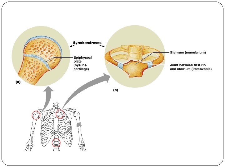
 Synchondroses Connecting tissue is hyaline cartilage Epiphyseal (growth) plate, all are")
 Synovial cavity allows a joint to be freely movable, diarthroses. Ligaments")

 Copyright 2009, John Wiley & Sons, Inc.")
 Accessory Ligaments and Articular Discs Collateral ligaments of the knee joint")
 Nerve and Blood Supply Nerve endings convey information about pain from")
: Friction-reducing structure Bursae Sac-like structures containing fluid similar to synovial fluid")


 Specific terminology is used to designate the")
 Gliding Simple movement back-and-forth and from side-to-side")
 Flexion Decrease in the angle between articulating")
 Circumduction Movement of a body part in")
 Copyright 2009, John Wiley & Sons, Inc.")
 Elevation Upward movement of a part of")
 Inversion Movement of the foot medially Its")
 Special Movements Elevation Depression Protraction Retraction Inversion")

 Copyright 2009, John Wiley & Sons, Inc.")
 Synovial joints are classified based on type of movement")
 Planar Joints Primarily permit back-and-forth and side-to-side movements Intercarpal")
 Pivot Joints Rounded end of one bone protrudes into")
 Saddle Joints Articular surface of one bone is saddle-shaped,")
 Copyright 2009, John Wiley & Sons, Inc.")
 Range of motion")
 Arrangement and tension")
 The selected joints described are: Temporomandibular joint Shoulder")
 Temporomandibular Joint Combined hinge and planar joint formed")
 Shoulder Joint Ball-and-socket joint formed by the head")


 Hip Joint Ball-and-socket joint formed by the femur")

 Elbow Joint Hinge joint formed by the humerus,")
 Knee Joint Largest and most complex joint of")


 Knee Joint Copyright 2009, John Wiley & Sons,")
 Copyright 2009, John Wiley & Sons, Inc.")
 Copyright 2009, John Wiley & Sons, Inc.")


 �Most common chronic arthritis; often called “wear- and-tear” arthritis �Affects women more")


 �Chronic, inflammatory, autoimmune disease of unknown cause, with an insidious onset")


 Arthroplasty Joints may be replaced surgically with artificial joints Most commonly replaced")
 Copyright 2009, John Wiley & Sons, Inc.")
 Copyright 2009, John Wiley & Sons, Inc.")
- Slides: 68
Anatomy & Physiology I Skeletal System: Joints Pro: Manhal Chbat
Joints Chapter 9 Joint Classifications Fibrous Joints Cartilaginous Joints Synovial Joints Types of Movements at Synovial Joints Types of Synovial Joints Factors Affecting Contact and Range of Motion at Synovial Joints Selected Joints of the Body Aging and Joints Arthroplasty
Joints (Articulations) Weakest parts of the skeleton. Articulation is the site where two or more bones meet.
Joints (Joint Classification) The structural classification of joints Fibrous joints (bones held together by dense collagen fibers) Cartilaginous joints (bones held together by cartilage) Synovial joints (bones held together by ligaments) The functional classification of joints Synarthrosis (an immovable joint) Amphiarthrosis (a slightly movable joint) Diarthrosis (a freely movable joint)
Joints (Fibrous Joints) Lack a synovial cavity The articulating bones are held very closely together by dense irregular connective tissue Fibrous joints permit little or no movement Three types of fibrous joints Sutures Syndesmoses Gomphoses
Joints (Fibrous Joints) Sutures Occur only between bones of the skull, little growth during youth, in middle age are called synostoses. Syndesmoses Permits slight movement Interosseous membrane Between tibia and fibula, ulna and radius. Gomphoses Immovable joint Joint in which a cone-shaped peg fits into a socket Articulations of the teeth with the alveolar sockets of the maxillae and mandible Copyright 2009, John Wiley & Sons, Inc.
Joints (Cartilaginous Joints) Lacks a synovial cavity Allows little or no movement Joint is tightly connected by either cartilage Two types of cartilaginous joints Synchondroses Symphyses Copyright 2009, John Wiley & Sons, Inc.
Joints (Cartilaginous Joints) Synchondroses Connecting tissue is hyaline cartilage Epiphyseal (growth) plate, all are synarthrotic. Symphyses Slightly movable joint, amphiarthrotic. Ends of the articulating bones are covered with hyaline cartilage, but a disc of fibrocartilage connects the bones Pubic symphysis Between the anterior surfaces of the hip bones Intervertebral joints between the vertebrae
Joints (Synovial Joints) Synovial cavity allows a joint to be freely movable, diarthroses. Ligaments hold bones together in a synovial joint All limb joints, and most joints of the body. Synovial joints all have the following Articular cartilage Joint (synovial) cavity Articular capsule Synovial fluid Reinforcing ligaments
Articular Capsule A sleeve-like capsule encloses the synovial cavity The articular capsule is composed of two layers an outer fibrous capsule an inner synovial membrane Synovial Fluid The synovial membrane secretes synovial fluid Functions to reduce friction by: lubricating the joint absorbing shocks supplying oxygen and nutrients to the cartilage removing carbon dioxide and metabolic wastes from the cartilage
Joints (Synovial Joints) Copyright 2009, John Wiley & Sons, Inc.
Joints (Synovial Joints) Accessory Ligaments and Articular Discs Collateral ligaments of the knee joint Anterior and posterior cruciate ligaments of the knee joint Menisci Pads of cartilage lie between the articular surfaces of the bones Allow bones of different shapes to fit together more tightly
Joints (Synovial Joints) Nerve and Blood Supply Nerve endings convey information about pain from the joint to the spinal cord and brain Nerve endings respond to the degree of movement and stretch at a joint Arterial branches from several different arteries merge around a joint before penetrating the articular capsule
Joints (Synovial Joints): Friction-reducing structure Bursae Sac-like structures containing fluid similar to synovial fluid and lined with synovial membrane. Located between tendons, ligaments, muscle, skin, and bones rub together. Cushion the movement of these body parts Tendon sheaths Wrap around tendons Reduce friction at joints
Synovial Joints: Stability �Stability is determined by: �Articular surfaces – shape determines what movements are possible �Ligaments – unite bones and prevent excessive or undesirable motion
Synovial Joints: Movement �The two muscle attachments across a joint are: �Origin – attachment to the immovable bone �Insertion – attachment to the movable bone �Described as movement along transverse, frontal, or sagittal planes
Synovial Joints: Range of Motion �Nonaxial – slipping movements only �Uniaxial – movement in one plane �Biaxial – movement in two planes �Multiaxial – movement in or around all three planes
Joints (Types of Movements at Synovial Joints) Specific terminology is used to designate the movements that occur at joints Movements are grouped into four main categories: 1) 2) 3) 4) Gliding Angular movements Rotation Special movements Copyright 2009, John Wiley & Sons, Inc.
Joints (Types of Movements at Synovial Joints) Gliding Simple movement back-and-forth and from side-to-side There is no significant alteration of the angle between the bones Limited in range Intercarpal joints, intertarsal joints, and the articular processes of the vertebrae. Angular Movements Increase or a decrease in the angle between articulating bones Angular movements include Flexion Extension Lateral flexion Hyperextension Abduction Adduction Circumduction
Joints (Types of Movements at Synovial Joints) Flexion Decrease in the angle between articulating bones Bending the trunk forward Extension Increase in the angle between articulating bones Flexion and extension are opposite movements Lateral flexion Movement of the trunk sideways to the right or left at the waist Hyperextension Continuation of extension beyond the normal extension Bending the trunk backward Abduction Movement of a bone away from the midline Moving the humerus laterally at the shoulder joint Adduction Movement of a bone toward the midline Movement that returns body parts to normal position from abduction
Joints (Types of Movements at Synovial Joints) Circumduction Movement of a body part in a circle Moving the humerus in a circle at the shoulder joint Rotation A bone revolves around its own longitudinal axis Turning the head from side to side as when you shake your head “no” Copyright 2009, John Wiley & Sons, Inc.
Joints (Types of Movements at Synovial Joints) Copyright 2009, John Wiley & Sons, Inc.
Joints (Types of Movements at Synovial Joints) Elevation Upward movement of a part of the body Closing the mouth Its opposing movement is depression Downward movement of a part of the body Opening the mouth Protraction Movement of a part of the body anteriorly Thrusting the mandible outward Its opposing movement is retraction Retraction Movement of a protracted part of the body back to normal
Joints (Types of Movements at Synovial Joints) Inversion Movement of the foot medially Its opposing movement is eversion Eversion Movement of the sole laterally Dorsiflexion Bending of the foot at the ankle in an upward direction Its opposing movement is plantar flexion Plantar flexion Bending of the foot at the ankle in a downward direction Supination Movement of the forearm so that the palm is turned upward Its opposing movement is pronation Pronation Movement of the forearm so that the palm is turned downward Opposition Movement of the thumb in which the thumb moves across the palm to touch the tips of the fingers on the same hand
Joints (Types of Movements at Synovial Joints) Special Movements Elevation Depression Protraction Retraction Inversion Eversion Dorsiflexion Plantar flexion Supination Pronation Opposition Copyright 2009, John Wiley & Sons, Inc.
Special Movements Figure 8. 6 a
Joints (Types of Movements at Synovial Joints) Copyright 2009, John Wiley & Sons, Inc.
Joints (Types of Synovial Joints) Synovial joints are classified based on type of movement Planar Hinge Pivot Condyloid Saddle Ball-and-socket
Joints (Types of Synovial Joints) Planar Joints Primarily permit back-and-forth and side-to-side movements Intercarpal joints Nonaxial joints. Hinge Joints Produce an opening and closing motion like that of a hinged door Permit only flexion and extension Knee, elbow, and interphalangeal joints. Copyright 2009, John Wiley & Sons, Inc.
Joints (Types of Synovial Joints) Pivot Joints Rounded end of one bone protrudes into a “sleeve, ” or ring, composed of bone (and possibly ligaments) of another. Joints that enable the palms to turn anteriorly and posteriorly (radioulnar, atlantoaxial) Condyloid Joints (ellipsoidal Joints) The projection of one bone fits into the oval-shaped depression of another bone Wrist Biaxial (radiocarpal, metacarpophalangeal) Copyright 2009, John Wiley & Sons, Inc.
Joints (Types of Synovial Joints) Saddle Joints Articular surface of one bone is saddle-shaped, and the articular surface of the other bone fits into the “saddle” Thumb (carpometacarpal joint of the thumb) Ball-and-Socket Joints Ball-like surface of one bone fitting into a cuplike depression of another bone Shoulder and hip Multiaxial. Copyright 2009, John Wiley & Sons, Inc.
Joints (Types of Synovial Joints) Copyright 2009, John Wiley & Sons, Inc.
Joints (Factors Affecting Contact and Range for Motion at Synovial Joints) Range of motion (ROM) Refers to the range, measured in degrees of a circle, through which the bones of a joint can be moved Factors contribute to keeping the articular surfaces in contact and affect range of motion: Structure or shape of the articulating bones Shape of bones determines how closely they fit together Strength and tension of the joint ligaments Ligaments are tense when the joint is in certain positions Tense ligaments restrict the range of motion
Joints (Factors Affecting Contact and Range for Motion at Synovial Joints) Arrangement and tension of the muscles Muscle tension reinforces the restraint placed on a joint by its ligaments , and thus restricts movement Contact of soft parts The point at which one body surface contacts another may limit mobility Movement be restricted by the presence of adipose tissue Hormones Flexibility may also be affected by hormones Relaxin increases the flexibility of the pubic symphysis and loosens the ligaments between the sacrum and hip bone toward the end of pregnancy Disuse Movement may be restricted if a joint has not been used for an extended period
Joints (Selected Joints of the Body) The selected joints described are: Temporomandibular joint Shoulder joint Elbow joint Hip joint Knee joint
Joints (Selected Joints of the Body) Temporomandibular Joint Combined hinge and planar joint formed by the mandible and the temporal bone Only movable joint between skull bones Only the mandible moves Copyright 2009, John Wiley & Sons, Inc.
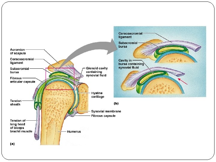
Joints (Selected Joints of the Body) Shoulder Joint Ball-and-socket joint formed by the head of the humerus and the scapula More freedom of movement than any other joint of the body Copyright 2009, John Wiley & Sons, Inc.
Figure 8. 11 a
Synovial Joints: Shoulder Stability Figure 8. 11 b
Joints (Selected Joints of the Body) Hip Joint Ball-and-socket joint formed by the femur and the hip bone Copyright 2009, John Wiley & Sons, Inc.
Synovial Joints: Hip Stability �Acetabular labrum �Iliofemoral ligament �Pubofemoral ligament �Ischiofemoral ligament �Ligamentum teres Figure 8. 12 a
Joints (Selected Joints of the Body) Elbow Joint Hinge joint formed by the humerus, the ulna, and the radius Copyright 2009, John Wiley & Sons, Inc.
Joints (Selected Joints of the Body) Knee Joint Largest and most complex joint of the body Modified hinge joint Three joints: femoropatellar, lateral and medial tibiofemoral Copyright 2009, John Wiley & Sons, Inc.
Synovial Joints: Knee Ligaments and Tendons – Anterior View �Tendon of the quadriceps femoris muscle �Lateral and medial patellar retinacula �Fibular and tibial collateral ligaments �Patellar ligament Figure 8. 8 c
Synovial Joints: Knee – Other Supporting Structures �Anterior cruciate ligament �Posterior cruciate ligament �Medial meniscus (semilunar cartilage) �Lateral meniscus
Synovial Joints: Knee – Other Supporting Structures Figure 8. 8 b
Joints (Selected Joints of the Body) Knee Joint Copyright 2009, John Wiley & Sons, Inc.
Joints (Selected Joints of the Body) Copyright 2009, John Wiley & Sons, Inc.
Joints (Selected Joints of the Body) Copyright 2009, John Wiley & Sons, Inc.
Sprains �The ligaments reinforcing a joint are stretched or torn �Partially torn ligaments slowly repair themselves �Completely torn ligaments require prompt surgical repair
Cartilage Injuries �The snap and pop of overstressed cartilage �Common aerobics injury �Repaired with arthroscopic surgery
Dislocations �Occur when bones are forced out of alignment �Usually accompanied by sprains, inflammation, and joint immobilization �Caused by serious falls and are common sports injuries �Subluxation – partial dislocation of a joint
Inflammatory and Degenerative Conditions �Bursitis �An inflammation of a bursa, usually caused by a blow or friction �Symptoms are pain and swelling �Treated with anti-inflammatory drugs; excessive fluid may be aspirated
Inflammatory and Degenerative Conditions �Tendonitis �Inflammation of tendon sheaths typically caused by overuse �Symptoms and treatment are similar to bursitis
Arthritis �More than 100 different types of inflammatory or degenerative diseases that damage the joints �Most widespread crippling disease in the U. S. �Symptoms – pain, stiffness, and swelling of a joint �Acute forms are caused by bacteria and are treated with antibiotics �Chronic forms include osteoarthritis, rheumatoid arthritis, and gouty arthritis
Osteoarthritis (OA) �Most common chronic arthritis; often called “wear- and-tear” arthritis �Affects women more than men � 85% of all Americans develop OA �More prevalent in the aged, and is probably related to the normal aging process
Osteoarthritis: Course �OA reflects the years of abrasion and compression causing increased production of metalloproteinase enzymes that break down cartilage �As one ages, cartilage is destroyed more quickly than it is replaced �The exposed bone ends thicken, enlarge, form bone spurs, and restrict movement �Joints most affected are the cervical and lumbar spine, fingers, knuckles, knees, and hips
Osteoarthritis: Treatments �OA is slow and irreversible �Treatments include: �Mild pain relievers, along with moderate activity �Magnetic therapy �Glucosamine sulfate decreases pain and inflammation
Rheumatoid Arthritis (RA) �Chronic, inflammatory, autoimmune disease of unknown cause, with an insidious onset �Usually arises between the ages of 40 to 50, but may occur at any age �Signs and symptoms include joint tenderness, anemia, osteoporosis, muscle atrophy, and cardiovascular problems �The course of RA is marked with exacerbations and remissions
Rheumatoid Arthritis: Course �RA begins with synovitis of the affected joint �Inflammatory chemicals are inappropriately released �Inflammatory blood cells migrate to the joint, causing swelling
Rheumatoid Arthritis: Course �Inflamed synovial membrane thickens into a pannus �Pannus erodes cartilage, scar tissue forms, articulating bone ends connect �The end result, ankylosis, produces bent, deformed fingers
Rheumatoid Arthritis: Treatment �Conservative therapy – aspirin, long-term use of antibiotics, and physical therapy �Progressive treatment – anti-inflammatory drugs or immunosuppressants �The drug Enbrel, a biological response modifier, neutralizes the harmful properties of inflammatory chemicals
Gouty Arthritis �Deposition of uric acid crystals in joints and soft tissues, followed by an inflammation response �Typically, gouty arthritis affects the joint at the base of the great toe �In untreated gouty arthritis, the bone ends fuse and immobilize the joint �Treatment – colchicine, nonsteroidal antiinflammatory drugs, and glucocorticoids
Joints (Arthroplasty) Arthroplasty Joints may be replaced surgically with artificial joints Most commonly replaced are the hips, knees, and shoulders Hip Replacements Partial hip replacements involve only the femur Total hip replacements involve both the acetabulum and head of the femur Knee Replacements Actually a resurfacing of cartilage and may be partial or total Potential complications of arthroplasty include infection, blood clots, loosening or dislocation of the replacement components, and nerve injury Copyright 2009, John Wiley & Sons, Inc.
Joints (Arthroplasty) Copyright 2009, John Wiley & Sons, Inc.
Joints (Arthroplasty) Copyright 2009, John Wiley & Sons, Inc.