Anatomy Physiology Chapter 14 The Brain And Cranial

Anatomy & Physiology Chapter 14 The Brain And Cranial Nerves SPR 06

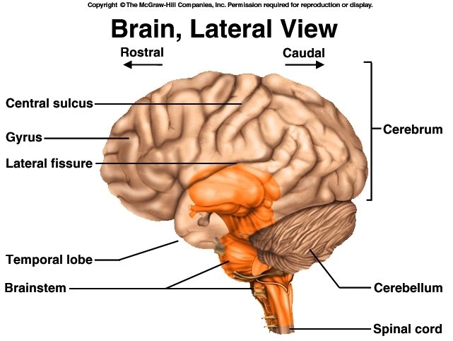
Overview of CNS Brain Size – proportional to human’s body size, not one’s intelligence Directional terms: 1. Rostral – toward forehead (nose) 2. Caudal – toward spinal cord (tail)

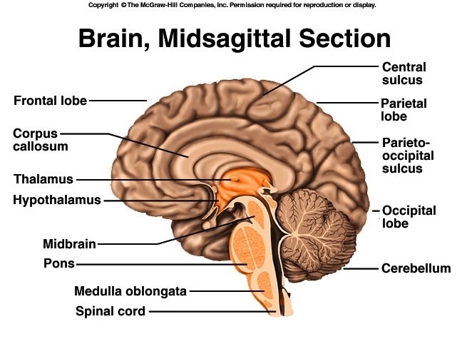
The Brain Divided into 3 major portions: 1. Cerebrum – 83% of brain volume 2. Cerebellum – 10% of volume, but contains 50% of neurons 3. Brainstem – ends at foramen magnum a. b. c. d. Diencephalon Midbrain Pons Medulla oblongata

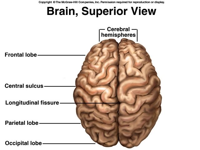
Cerebrum – – – Cerebral hemispheres Gyri – thick folds Sulci – shallow grooves Longitudinal fissure Corpus callosum – connects hemispherees


Gray & White Matter 1. Gray matter a. Composed of neuron cell bodies, dendrites, and synapses b. Forms surface cortex over cerebrum and cerebellum c. Deeper masses of gray matter = nuclei 2. White matter a. Lies deep to cortical gray matter b. Composed of tracts, as in spinal cord

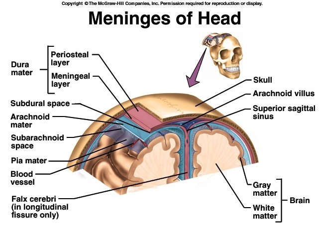
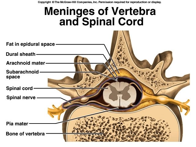
Meninges Similar to those of spinal cord 1. Dura mater 2. Arachnoid mater 3. Pia mater Dura has some differences: 1. 2. 3. 4. Two layers – fused, except at sinuses No epidural space Dural sinuses – collect blood Folds or septa - function to support and partition brain, and to limit movement of brain within the cranium

Dura Mater 1. Outer periosteal layer equivalent to periosteum 2. Inner meningeal layer a. Continues as dural sheath around spinal cord b. Folds form partitions = dural septa 3. Dural sinuses a. Spaces between two layers of dura b. Collect blood that circulates through brain 4. Subdural space a. Separates dura mater from arachnoid mater b. Considered a serous cavity

Dural Septa 1. Falx cerebri a. Dips into longitudinal fissure between cerebral hemispheres b. Attaches to crista galli of ethmoid bone 2. Falx cerebelli a. Runs along vermis of cerebellum b. Partially separates right and left halves 3. Tentorium cerebelli a. Separates cerebrum and cerebellum

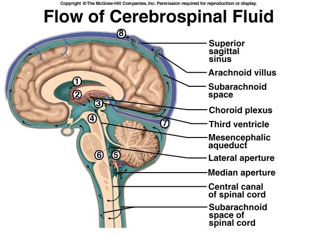
Arachnoid Mater 1. Middle layer 2. Subarachnoid space a. Separates arachnoid from pia b. Filled with CSF c. Contains largest blood vessels of brain, but are poorly protected 3. Arachnoid villi a. Cauliflower-like extensions of arachnoid mater that protrude through dura b. Absorb CSF into venous blood of sinus

Pia Mater 1. 2. 3. 4. “Gentle mother” Very vascular, translucent Follows contours of brain – “shrink wrap” Rich with blood vessels

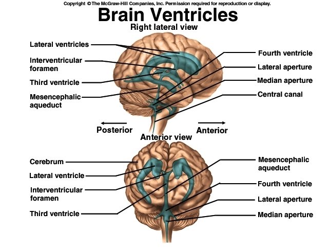
Ventricles Brain has four internal chambers = ventricles Two Lateral ventricles a. Large arc in each cerebral hemisphere b. Interventricular foramen – connects to 3 rd ventricle c. Septum pellucidum - “transparent wall” 3. Third Ventricle a. Inferior to c. callosum b. Mesencephalic (cerebral) aqueduct – connects 3 rd and 4 th ventricle

Ventricles 4. Fourth ventricle a. Between pons & cerebellum b. Apertures i. Openings in walls of 4 th ventricle ii. Connect ventricles to subarachnoid space iii. From this subarachnoid space, CSF is returned to blood in sinus by arachoid villi

Cerebrospinal Fluid 1. Ependymal cells a. Line the ventricles & canals b. Neuroglia similar to cuboidal epithelium c. Cilia help circulate CSF 2. Choroid plexus a. Capillary network in roof of each ventricle b. Filtration of blood plasma from capillaries 3. CSF formed by modification of plasma from choroid plexus by ependymal cells

Cerebrospinal Fluid Three functions: 1. Buoyancy a. Brain neither sinks nor floats, is suspended b. Prevents pressure necrosis due to weight 2. Protection – fluid cushion 3. Chemical stability a. Rinses wastes from CNS b. Regulates chemical environment

Cerebrospinal Fluid 1. Brain produces 500 ml CSF daily 2. Is constantly reabsorbed, so ventricles contain only about 150 ml (half cup) 3. CSF circulates, not static a. Driven by pressure b. Pulsations due to heartbeat c. Ciliated ependymal cells

Traumatic Brain Injury 1. Head injuries are leading cause of accidental death in U. S. 2. The coup injury - direct blow vs. recoup injury – ricochet effect 3. Concussion 4. Contusion 5. Intracranial hemorrhage 6. Cerebral edema

Brain Barrier System 1. Brain only 2% of body weight, but receives 15% of blood and 20% of O 2 and glucose 2. Blood supply critical to brain 10 second interuption - lose consciousness 2 minutes – impaired neural function 4 minutes – irreversible brain damage 3. Blood also source of potentially harmful substances Antibodies Macrophages Proteins/amino acids Fluctuating ion concentrations
")
Brain Barrier System • Two mechanisms that guard brain tissue 1. Blood-brain barrier (BBB) a. Guard blood capillaries throughout brain 2. Blood-CSF Barrier a. Guard capillaries of choroid plexus

Blood-Brain Barrier 1. Endothelial cells of capillaries joined by tight junctions a. Protoplasmic astrocytes stimulate formation of tight junctions b. Anything leaving blood must pass through endothelial cells, not between them c. Least permeable capillaries of the body 2. Exceptionally thick basal lamina (basement membrane)

Blood-CSF Barrier 1. Protection at choroid plexus 2. Tight junctions between ependymal cells 3. No tight junctions between ependymal cells elsewhere a. Important to allow exchange between CSF and brain Examples: i. Vomiting center of brainstem monitors blood for toxic substances ii. Hypothalamus regulates body temperature

Brain Barrier System 1. Selective barrier, not an absolute a. Highly permeable to glucose, water, and some electrolytes via facilitated diffusion through channel proteins in endothelial cell membranes b. Nonessential amino acids and potassium are actively pumped from brain into blood c. Barrier ineffective against lipid-soluble substances such as alcohol, nicotine, anesthetics, O 2 and CO 2 2. Is obstacle to delivery of some drugs Nasal spray – delivery up olfactory nerve

Brain Barrier System 3. Trauma/inflammation can damage BBS 4. Barrier incomplete in newborns a. Toxic substances can cause problems not seen in adults 5. Circumventricular organs (CVO’s) a. Found in 3 rd & 4 th ventricles b. Barrier system absent c. Allows brain to monitor blood glucose, p. H, osmolarity, etc. d. Route for HIV

Cerebrovascular Accidents 1. Single most common nervous system disorder & 3 rd leading cause of death in U. S. 2. Blood circulation is blocked and brain tissue dies 3. Most common cause is blockage of cerebral artery by blood clot 4. Other causes – compression by hemorrhage or edema, narrowingof vessels by atherosclerosis

Cerebrovascular Accidents 5. Typically, survivors are paralyzed on one side of body 6. < 35% of CVA survivors are alive 3 years later 7. Not all strokes are “completed” a. Transient ischemic attacks (TIA’s) b. Last 5 – 50 minutes c. Temporary paralysis, but red flags of potential impending more serious CVA’s

Cerebrovascular Accidents 7. Initial vascular blockage or ischemia doesn’t cause most damage a. Damaged neurons release “buckets” of glutamate (excitatory neurotransmitter) b. Acts as excitotoxin – changes ion transport, allows unregulated Ca+2 influx into neuron c. Generates free radicals that damage or kill thousands of surrounding healthy neurons d. Activates microglia and triggers inflammation which compounds damage

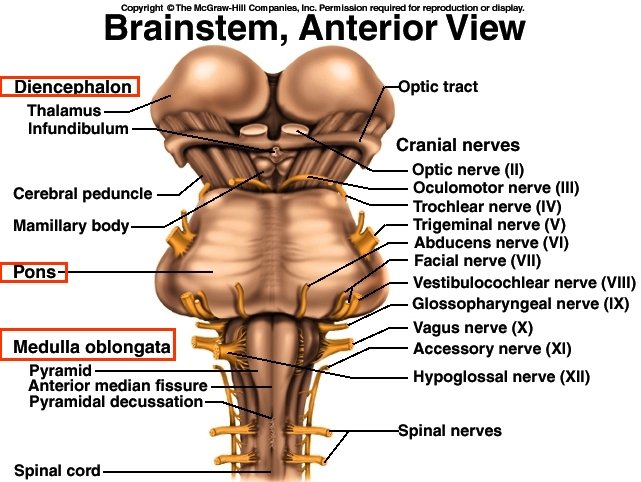
The Brainstem 1. Medulla oblongata 2. Pons 3. Midbrain
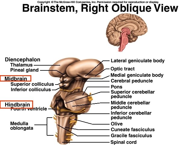

Medulla oblongata 1. Physically connects spinal cord to brain 2. Contains ascending & descending tracts passing through brain stem 3. Pyramids a. Ridges of corticospinal tracts (pyramidal) b. Taper at ends – “baseball bats” c. Decussation of pyramids – contralateral control 4. Sensory & motor nuclei associated with cranial nerves IX – XII.

Medulla oblongata 5. Motor nuclei: a. Cardiac center – rate and force of heartbeat b. Vasomotor center i. Vasoconstriction/dilation ii. Regulates blood pressure iii. Reroutes blood c. Respiratory centers – rate and depth

Pons 1. “Bridge” chiefly composed of conduction tracts a. Transverse fibers links cerebellum to rest of brain b. Longitudinal fibers connect spinal cord to higher centers of brain 2. Pons nuclei a. Many functions, includes pneumotaxic center 3. Forms ventral floor of 4 th ventricle 4. CN V (Trigeminal) arises from pons 5. Cranial nerve VI, VII, & VIII – arise from junction of pons and medulla

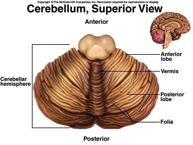
Cerebellum 1. Largest part of hindbrain 2. Cerebellar hemispheres a. Bilaterally symmetrical b. Bridged by vermis 3. Folia – slender folds 4. Thin outer cortex of gray matter, deep layer of white matter 5. Arbor vitae – white matter

Cerebellum 6. Transverse cerebral fissure – separates cerebellum and cerebrum 7. Functions in subconscious muscular coordination and agility 8. Cerebellar peduncles a. Connect cerebellum to brainstem b. Three pairs i. Inferior peduncles – to medulla ii. Middle peduncles – to pons iii. Superior peduncles – to midbrain

Cerebellar Peduncles 1. Superior cerebellar peduncles a. Cerebellum to midbrain b. Carry instructions from deep cerebellar nuclei to cerebral motor cortex via thalamus 2. Middle cerebellar peduncles a. One-way communication from pons advising cerebellum of voluntary motor activities 3. Inferior cerebellar peduncles a. Convey sensory info from proprioceptors and vestibular nuclei (equilibrium)
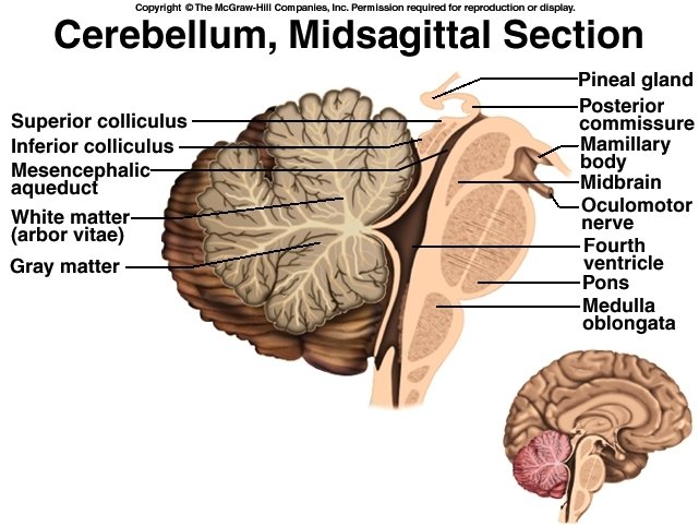

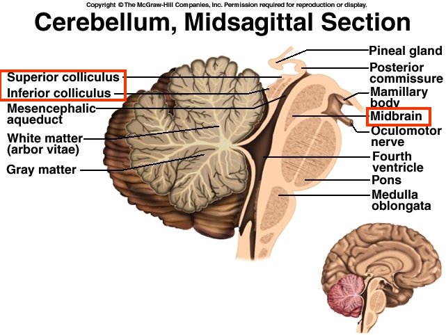
The Midbrain 1. Short segment that connects hindbrain & forebrain 2. Contains mesencephalic aqueduct 3. Gives rise to Cranial nerves III & IV 4. Cerebral peduncles – anchor cerebrum to brainstem, contain corticospinal tracts 5. Tegmentum – main mass of midbrain 6. Tectum – “roof” over aqueduct a. Corpora quadrigemina - nuclei
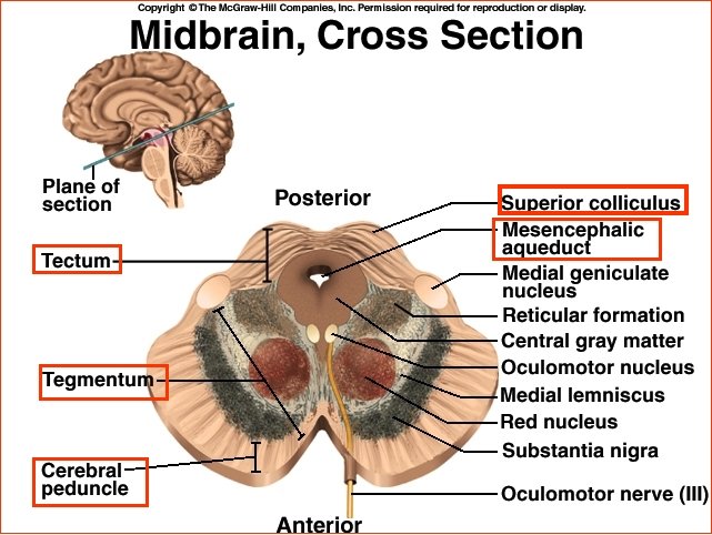

The Reticular Formation • Gray matter that runs through midbrain, pons, & medulla • Somatic motor control • Cardiovascular control • Pain modulation • Sleep & consciousness – Habituation

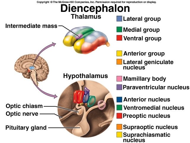
The Diencephalon 1. Thalamus - bulk of diencephalon a. “Gateway” to cerebrum – final relay point b. for ascending sensory information b. Interconnected with limbic system 2. Hypothalamus a. Part of walls & floor of 3 rd ventricle b. Pituitary gland attached by stalk c. *Major control center of autonomic nervous system & endocrine system 3. Epithalamus a. Pineal gland – melatonin b. Habenula – relay from limbic system
 a. Influences heart rate, blood")
Roles of Hypothalamus 1. Autonomic control center – (ANS) a. Influences heart rate, blood pressure, etc 2. 3. 4. 5. 6. 7. Center for emotional response Hormone secretion Body temperature regulation Regulation of food intake Regulation of water intake and thirst Regulation of wake-sleep cycles

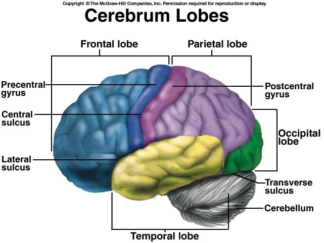
The Cerebrum 1. Gyri – extensive folding increases processing capabilities, one of greatest differences between humans and other mammals 2. 5 lobes: a. Frontal – voluntary motor control, thought processes b. Parietal – sensory reception, somesthetic & taste integration c. Occipital – principal visual center d. Temporal – hearing, smell, learning e. Insula – not well understood

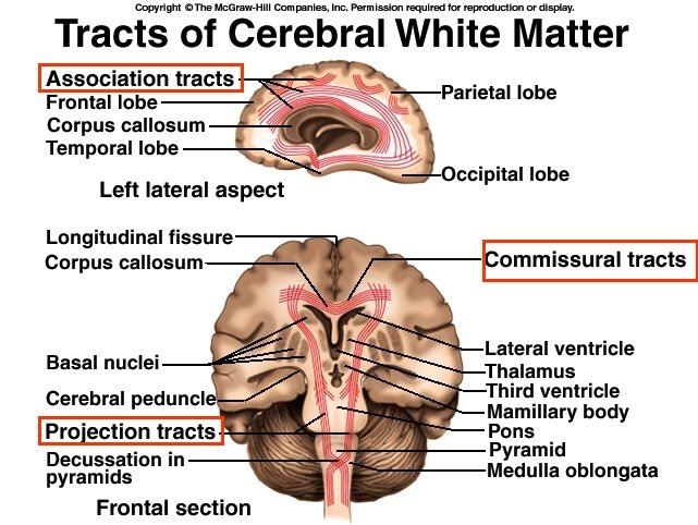
Cerebral White Matter 3 types of tracts: 1. Projection tracts a. Link cerebral cortex to brain stem, cerebellum, b. and spinal cord c. Carry information between cerebrum & rest of body 2. Comissural tracts a. Cross from one cerebral hemisphere to other b. Usually through corpus callosum 3. Association tracts a. Connect different regions of same hemisphere b. Long fibers connect different lobes c. Short fibers connect different gyri within lobe

Cerebral Gray Matter Neural integration carried out in gray matter: 1. Cerebral cortex 2. Basal nuclei 3. Limbic system

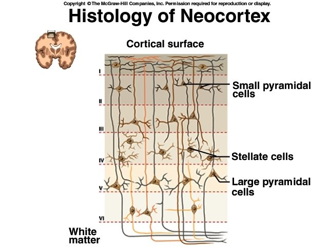
Cerebral Cortex 1. Cerebral cortex a. Thin layer of gray matter covering hemispheres 2. Two types of neurons a. Stellate cells – Receive sensory input & process information on local level b. Pyramidal cells – output neurons – transmit signals to other parts of CNS 3. Neocortex – 6 layered tissue

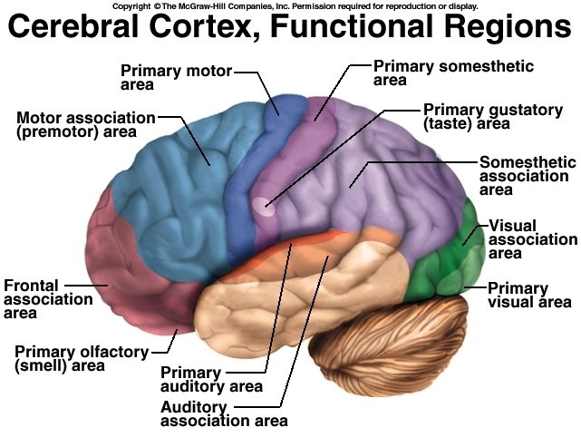
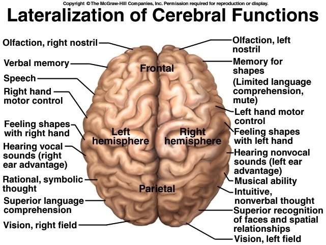
Cerebral Cortex 1. Cerebral cortex contains three kinds of functional areas a. Motor areas b. Sensory areas c. Association areas 2. Each hemisphere is concerned with sensory and motor functions of contralateral side of body 3. Two hemispheres are symmetrical in structure, but not in function

Amygdala – Emotion Hippocampus - Memory

Cognition 1. Refers to mental processes – awareness, thinking, perception, knowledge, & memory 2. 75% of brain = association areas – integration of information between sensory input & motor output 3. Parietal association cortex – perceiving & attending to stimuli 4. Temporal association cortex – identification 5. Frontal association cortex - planning

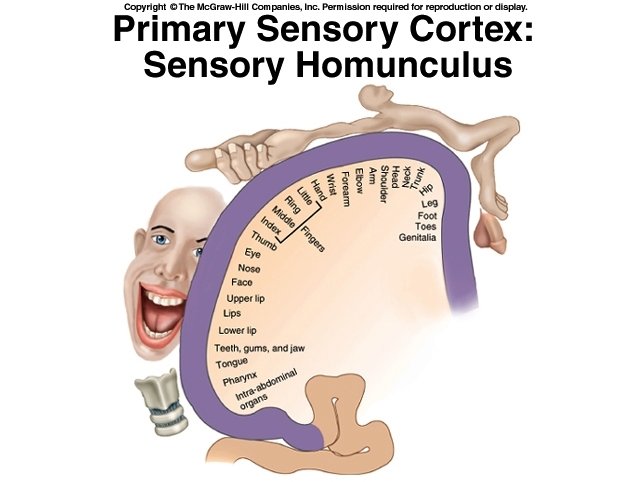
Sensation 1. Somesthetic – from widely distributed receptors, like touch, pressure, movement a. Routed to postcentral gyrus b. Primary somesthetic cortex c. Each gyrus is upside down sensory map of contralateral side of body – diagrammed as sensory homunculus 2. Special senses a. Primary sensory areas b. Sensory association areas
 area")
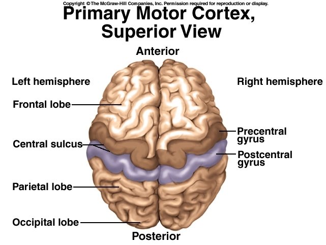
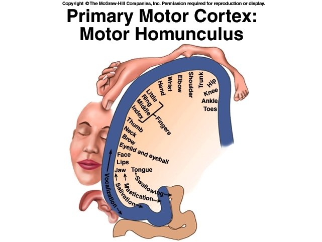
Motor Control 1. Intention of voluntary movement - begins in motor association (premotor) area of frontal lobes 2. Precentral gyrus ( primary motor area) – a. Send signals to brain stem and spinal cord b. Result in muscle contractions 3. Motor homunculus a. Distorted because amount of cortex devoted is proportional to number of muscles & motor units within that region, not to size of region

Upper & Lower Motor Neurons 1. Upper Motor Neurons a. b. c. c. d. Pyramidal cells (output neurons) of cerebral cortex b. May facilitate or inhibit lower motor neuron Controls muscles on contralateral side Synapse with lower motor neurons in brainstem or spinal cord (Corticospinal tract) 2. Lower Motor Neurons a. Innervate skeletal muscle b. Only axon extends outside of CNS c. Destruction or damage causes flaccid paralysis of affected motor unit
- Slides: 69