Anatomy of the Pharynx Dr Sheetal Rai Embryology

Anatomy of the Pharynx Dr Sheetal Rai
 Pharyngeal arches 2) Pharyngeal pouches 3) Pharyngeal clefts/grooves")
Embryology Components of branchial/pharyngeal apparatus: 1) Pharyngeal arches 2) Pharyngeal pouches 3) Pharyngeal clefts/grooves
 arches n n Derived from neural crest cells Resemble fish gills (branchia)")
Pharyngeal (branchial) arches n n Derived from neural crest cells Resemble fish gills (branchia) Begin to develop early in the 4 th week By end of 4 th week, four pairs of arches are visible on the surface (not 5 th and 6 th ) and a buccopharyngeal membrane ruptures forming communication between primitive oral cavity and foregut
 n n Contribute to the formation of the neck as")
Pharyngeal arches (cont. ) n n Contribute to the formation of the neck as well as the face. Visible structures at 42 weeks: 1 st arch: mandibular prominence, maxillary prominences, and the frontonasal prominence
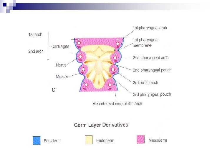
 n n n Core of mesenchymal tissue covered by surface")
Pharyngeal arches (cont. ) n n n Core of mesenchymal tissue covered by surface ectoderm (outside) and by endodermal epithelium (inside) Ectoderm -> skeletal Mesoderm -> muscles with accompanying nerve Arterial component (aortic arches) Therefore, each arch carries nerve, muscle, bone and blood supply
 ¨ Premaxilla, zygomatic bone, portion of temporal")
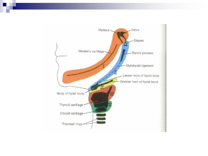
First pharyngeal arch n Maxillary process (dorsal) ¨ Premaxilla, zygomatic bone, portion of temporal bone n Mandibular process (ventral) ¨ Contains Meckel’s cartilage which disappears except for dorsal end (incus & malleus) and mandible
, mylohyoid, tensor tympani and tensor")
First pharyngeal arch Muscles of mastication, digastric (ant belly), mylohyoid, tensor tympani and tensor palatini n Therefore, the accompanying motor nerve is the mandibular branch of trigeminal (V 2) and sensory are V 1, V 2, and V 3 n 1 st aortic arch practically disappears but forms the maxillary artery n

Second pharyngeal arch n n Reichert’s cartilage – stapes, styloid process, stylohyoid ligament, lesser horn and upper part of the hyoid Muscles include: stapedius, stylohyoid, digastric (post belly), auricular, and those of facial expression Facial nerve (CN VII) 2 nd aortic arch – stapedial & hyoid arteries

Third pharyngeal arch n n Cartilaginous contributions include greater horn and lower part of hyoid Sole muscle: stylopharyngeus CN IX (Glossopharyngeal nerve) 3 rd aortic arch (quite large): common carotid, 1 st portion of internal carotid (remainder dorsal aorta), and external carotid

Fourth & sixth pharyngeal arch n n n Cartilaginous contributions to larynx derived from fusion: thyroid, cricoid, arytenoid, corniculate, and cuneiform Muscles of 4 th: cricothyroid, levator palatini, and pharyngeal constrictors are innervated by SLN (CN X) Muscles of 6 th: intrinsics of larynx are innervated by RLN (CN X) 4 th aortic arch: L->arch of aorta & R->subclavian 6 th aortic arch: L & R pulmonary with ductus arteriosus on left
 n n n 1 st: tubotympanic recess-> middle ear & eustacian")
Pharyngeal pouches (5) n n n 1 st: tubotympanic recess-> middle ear & eustacian tube -> TM 2 nd palatine tonsil/fossa 3 rd: inferior parathyroid (dorsal), thymus (ventral) 4 th: superior parathyroid 5 th: ultimobranchial body -> calcitonin producing C cells (parafollicular)
 n n 1 st: external auditory meatus 2 nd-4 th :")
Pharyngeal clefts/grooves (4) n n 1 st: external auditory meatus 2 nd-4 th : epicardial ridge and cervical sinus (disappears)

Anatomy of the pharynx
 Extends from base of skull to inferior border of cricoid cartilage")
Anatomy (cont. ) Extends from base of skull to inferior border of cricoid cartilage anteriorly and inferior border of C 6 posteriorly n Widest portion (5 cm) at hyoid n Narrowest portion (1. 5 cm) at caudal end n Divided into 3 parts: nasopharynx, oropharynx, and laryngo(hypo)pharynx n
 n Posterior: pharyngobasilar membrane and")
Nasopharynx Respiratory function n Anterior: choana (posterior nasal aperture) n Posterior: pharyngobasilar membrane and superior constrictor muscle n Superior: basilar portion of occipital bone n Inferior: soft palate n

Oropharynx Digestive function n Anterior: anterior tonsillar pillar n Posterior: superior constrictor n Superior: soft palate n Inferior: base of tongue, superior epiglottis n Laterally: palatoglossal and palatopharyngeal arches n
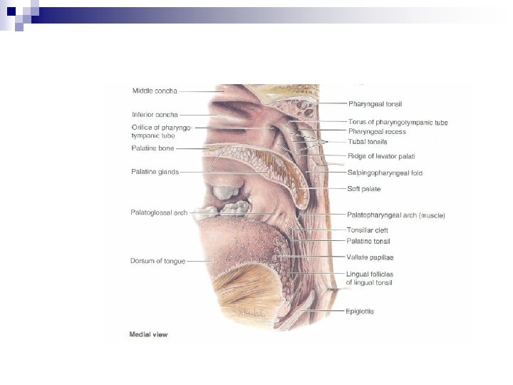

Hypopharynx Lies posterior to the larynx n Superior: superior border of epiglottis and pharyngoepiglottic folds n Inferior: inferior border of the cricoid n Posterior/lateral: middle & inferior constrictors, bodies of C 4 -C 6 n Anterior: laryngeal inlet n

Pharyngeal muscles

Pharyngeal muscles n n n External circular and internal longitudinal (opposite in remainder of GI tract) External: 3 constrictors (CN XI via X and ELN/RLN for middle and inferior) function to constrict wall of pharynx during swallow Internal: palatopharyngeus and salpingopharyngeus (CN XI via X) and stylopharyngeus (CN IX) act to elevate pharynx and larynx during speech/swallow
 tenses soft palate & opens ET during")
Pharyngeal muscles Tensor veli palatini (V 3) tenses soft palate & opens ET during yawn/swallow n Levator veli palatini (CN XI via X) elevates palate during swallow/yawn n Palatoglossus (CN XI via X) approximates tongue and soft palate n

Pharyngeal muscles

Pharyngeal lymphatic drainage n Oral cavity ¨ n I, III Oro/hypopharynx ¨ deep n II, IV Nasopharynx ¨ II, V, III

Pharyngeal vessels

Afferent innervation of pharynx

TUMOURS OF HYPOPHARYNX n CA Hypopharynx - very common in our country. MC tumours are squamous cell type. Subsites involved are: n pyriform sinus n postcricoid region n posterior pharyngeal wall

Carcinoma Pyriform Sinus n 60% of all hypopharyngeal cancers n Remain asymptomatic for a long time – due to large size of the pyriform sinus. n Metastatic neck nodes (levels II, IV) – earliest presentation.

Clinical Features n n n Sticking sensation in the throat Referred otalgia Metastatic neck nodes Hoarseness Stridor

Treatment n n Early growth without nodes – radiotherapy Growth limited to pyriform fossa –TL+PP + ND Growth extends to postcricoid region – TLPO + Gastric pull up Post op RT to all patients.

Carcinoma Postcricoid Region n One - third of patients of postcricoid carcinoma suffer from Paterson-Brown-Kelly (Plummer. Vinson) syndrome characterised by hypochromic microcytic anaemia.

Clinical Features n n Progressive dysphagia progressive malnutrition and weight loss Hoarseness and/or aphonia Laryngeal crepitus – absent

Treatment n Radiotherapy n Poor prognosis with both RT as well as surgery

Carcinoma Posterior Pharyngeal Wall C/F n Dysphagia n Neck nodes Treatment n Early lesions – RT n Small lesions may be excised.

THANK YOU
- Slides: 36