Anatomy of the pharynx and oesophagus Prof Abdulameer

Anatomy of the pharynx and oesophagus Prof. Abdulameer Al-Nuaimi E-mail: a. al-nuaimi@sheffield. ac. uk abdulameerh@yahoo. com
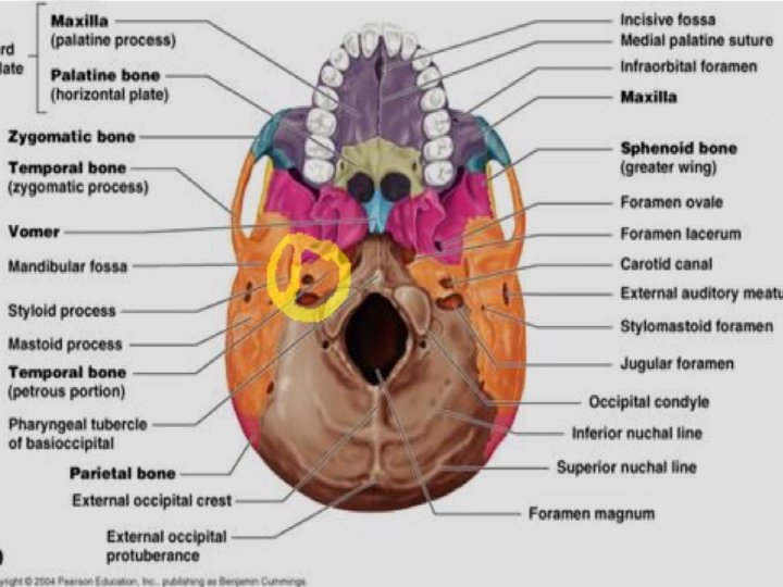

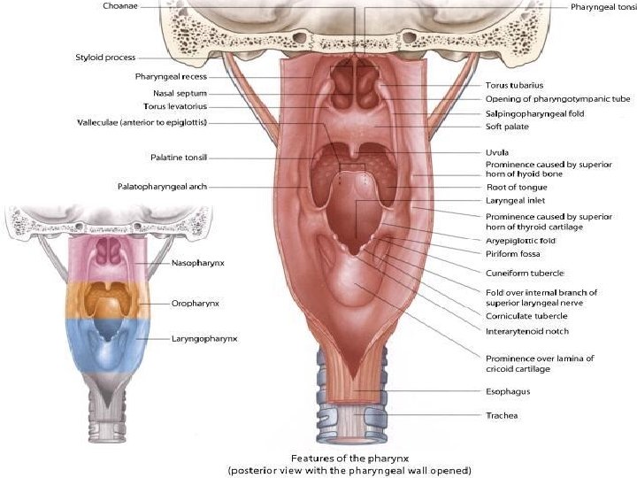
Pharynx

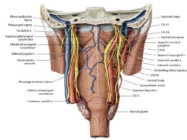
Pharyngobasilar fascia

Small muscles of pharynx

Small muscles of the pharynx

Pharyngeal recess Inside of nasopharynx

Cricopharyngeal sphincter Inside of oropharynx and laryngopharynx

Oesophagus

Histology of the oesophagus The oesophagus has a folded appearance. When swallowing food, it is able to distend, and accomodate the food being swallowed on its way to the stomach Epithelial lining is non keratinized stratified squamous epithelium

the mucosa consists of the epithelium, underlying lamina propria and muscularis mucosa. The lamina propria contains lymphatic capillaries, blood capilaries, and loose connective tissue. The darkly staining cells in the lamina propria are lymphoid aggregations The muscularis mucosa is a thin, double layer of smooth muscle, more substantial in the lower part of the oesophagus. Mucosa Muscularis mucosa

The submucosa is highly vascular, and contains loose connective tissue. It contains oesophageal glands, that secrete mucus to help ease the passage of swallowed food. The muscularis externa layer in the top third of the oesophagus contains skeletal muscle, in the middle, it is a mixture of smooth and skeletal muscle, and in the bottom third it is entirely smooth s o c u a Muscularis externa ve nt iti a m Sub ad its outermost layer consists of a connective tissue adventitia which merges with the adjacent connective tissue associated with nearby structures

Thank You
- Slides: 16