ANATOMY OF THE ORBIT Dr Sheetal Savur The

ANATOMY OF THE ORBIT Dr Sheetal Savur

The orbit • Protects, supports and maximizes the functions of the eyeball • Shaped as a quadrilateral pyramid with its base in plane with the orbital rim

Alignment of the orbits • medial walls are parallel • lateral walls are perpendicular • axes of the orbits bisect at a 45°angle • The axes of the eyeballs make an angle of 22. 5 with the orbital axes

Dimensions of the orbit • Height of orbital margin 40 mm • Width of orbital margin 35 mm • Depth of orbit - 40 -50 mm • Volume of orbit - 30 cm 3

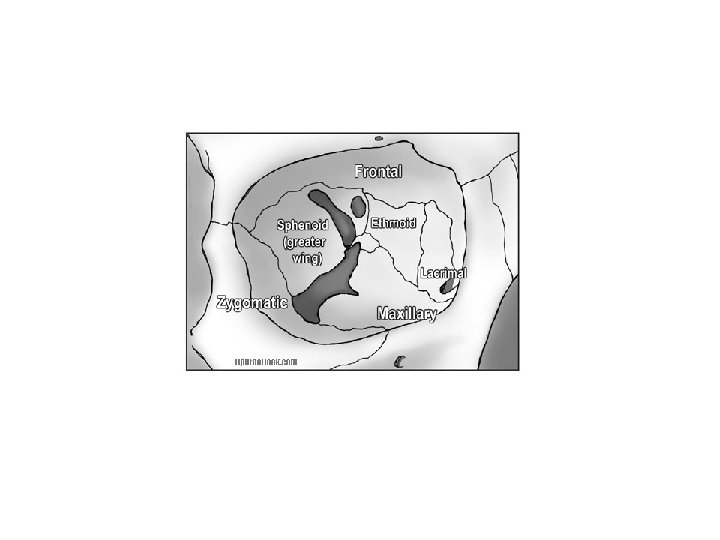
Bones of the orbit • • Frontal bone Zygomatic bone Maxillary bone Sphenoid bone Ethmoid bone Lacrimal bone Palatine bone

The seven bones

The frontal bone

The zygomatic bone

The maxillary bone

The sphenoid bone

The ethmoid bone

The lacrimal bone

The palatine bone

The communications of the orbit

The superior orbital fissure

The optic foramen

The inferior orbital fissure

Contents of the orbit Eyeball Extra-ocular muscles Fascia Vessels Nerves Retro-orbital fat NO LYMPHATICS

Relations of the orbit

Orbital rim

Apex of the orbit • Superior orbital fissure • Optic foramen
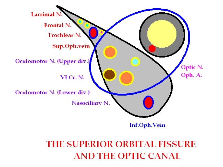
 IV cranial nerve")
• • II cranial nerve III cranial nerve (2 divisions) IV cranial nerve (3 branches of the Ophthalmic division) • VI cranial nerve • Ophthalmic veins (superior and inferior) • Ophthalmic artery

Inferior orbital fissure

Spaces of the orbit • • Sub-periosteal space Peribulbar space Retrobulbar space Sub Tenon’s space
- Slides: 26