Anatomy of the neck By Dr Medhat Abd

Anatomy of the neck By Dr. Medhat Abd El-Haleem

TRIANGLES OF THE NECK The anterolateral part of the neck is quadrilateral area which is divided by sternomastoid muscle into 2 large triangular areas (anterior and posterior). The investing layer of deep fascia roofs both triangles.

Sternomastoid muscle : Origin: - Sternal head: from front of manubrium sterni. - Clavicular head: front and upper aspect of medial ⅓ of clavicle. Insertion: - Mastoid process. - Lateral 1/3 of superior nuchal line.

Nerve supply: - Motor: spinal accessory n. - Sensory: C 1, C 2, C 3. proprioceptive fibers. Action: - One muscle act: rotate head to same side and turn face to opposite side. - Both muscles act: neck flexion.

Omohyoid muscle Sternomastoid muscle

The anterior triangle of the neck

Site: It lies in front of the sternomastoid muscle. It is bounded by : 1 - anterior border of sternomastoid muscle (behind). 2 - anterior midline of the neck ( in front ) 3 - lower border of the mandible (base )

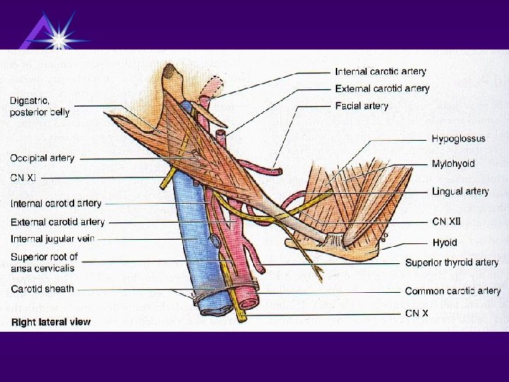
Anterior belly of digastric d Digastric triangle Posterior belly of digastric Mylohyoid muscle forms the floor of the mouth Carotid triangle Muscular triangle

It is Subdivided into 4 triangles by means of the superior belly of omohyoid muscle , the anterior and posterior bellies of digastric muscle as follows : 1 - Digastric triangle 2 - Carotid triangle 3 - Muscular triangle 4 - Submental triangle

Posterior belly of digastric Stylohyoid Anterior belly of digastric muscle Sternohyoid muscle

Internal carotid artery External carotid artery Common carotid artery

The infrahyoid muscles They are strap like muscles occupy each side of the midline of the neck , from the hyoid bone to the manubrium sterni. They consist of 4 muscles arranged into 2 layers : 1 - Superficial layer : A- sternohyoid B- omohyoid 2 - Deep layer: A- sternothyroid B- thyrohyoid

• Nerve supply of The infrahyoid muscles: • All The infrahyoid muscles are supplied by Ansa cervicalis except thyrohyoid muscle supplied by fibres from C 1 through hypoglossal nerve.

Ansa cervicalis Ansa = loop It is a loop of cervical nerve fibres derived from C 1, 2, 3 which lies below the carotid sheath below the middle of the neck. It consists of 2 roots : 1 - descendes hypoglossi C 1: derived from hypoglossal nerve 2 - descendes cervicalis : C 2, 3 : from the cervical plexus

Suprahyoid muscles 1 - digastric muscle 2 - mylohyoid muscle 3 - hyoglossus muscle 4 - geniohyoid muscle 5 - stylohyoid muscle

Geniohyoid muscle Hyoglossus

Digastric muscle It consists of 2 bellies anterior and posterior , that connected together by an intermediate tendon. Origin: has two bellies: - anterior belly: from the digastric fossa of the mandible. - posterior belly: from digastric notch of temporal bone. Insertion: in an intermediate tendon which runs in a fibrous sling attached to hyoid bone.

Digastric muscle Posterior belly Anterior belly

Nerve supply : 1 - anterior belly : from nerve to mylohyoid which is a branch of mandibular nerve. 2 - posterior belly : the facial nerve Action : - When the mandible is fixed , it raises the hyoid bone during swallowing. - When the hyoid bone is fixed , the anterior belly can depress the mandible

mylohyoid muscle : Origin: Mylohyoid line of mandible. Insertion: - anterior ⅔: into median raphe extend from symphysis menti to body of hyoid. - posterior ⅓: into body of hyoid. Nerve supply : nerve to mylohyoid , which is a branch of the mandibular nerve.

Action - - - : elevate hyoid and floor of the mouth during swallowing. depress mandible(open the mouth). support floor of mouth.

Geniohyoid muscle Origin: inferior genial tubercles. Insertion: fibers pass backward and downward to insert on the body of hyoid. Action: elevate hyoid bone, important in swallowing and phonation. Nerve supply: by a branch from hyoglossal nerve.
- Slides: 23