Anatomy of the kidney ureter and bladder David

Anatomy of the kidney, ureter and bladder David Dora Assistant lecturer 2018. april

Anatomy of the kidney - Kidneys are paired parenchymal organs with 130 -140 grams of mass - Situated in the abdominal cavity retroperitoneally - Left kidney: Th 12 -L 2 - Right kidney: L 1 -L 3

Topography of the kidneys I. cardia hepatoduodenal ligament superior mesenteric a. /v. cut edge of parietal peritoneum ascending colon origin of mesocolon transversum cut edge of parietal peritoneum descending colon radix mesenterii

Topography of the kidneys II. left right suprarenal glands liver right colic flexure renal hilum spleen stomach pancreas duodenum colon descendens

POSTERIOR ASPECT Right kidney XI. rib diaphragm psoas major musculus quadratus lumborum musculus transversus abdominis

Topography of the kidneys III. right lung right suprarenal gland right kidney renal fascia posterior layer parietal peritoneum duodenum descending part transverse colon renal fascia anterior layer

Coverings of the kidney renal capsule renal hilum parietal peritonem renal fascia ant. transversalis fascia sul cap m. transversus abdominis sa o dip aa psoas major sinus renalis m. obliquus abdominis int. quadratus lumborum m. obliquus abdominis ext. erector spinae perinephric fat thoracolumbal Fascia renal fascia post. pararenal fat body

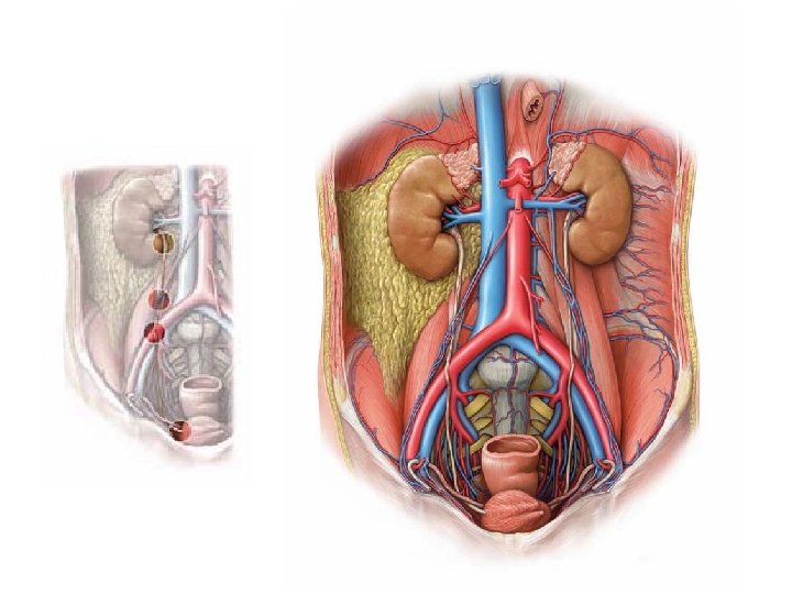
Structure of the kidney I. ANTERIOR hilum renalis superior polus Suprarenal arteries sup. /med. /inf renal artery renal vein fibrous capsule hilum renalis suprarenal gland POSTERIOR renal pelvis Inferior polus ureter Anterior – Posterior order in the hilum: VENA – ARTERIA – URETER VAU Superior – Inferior order in the hilum: ARTERIA – VENA – URETER

Structure of the kidney II. renal pyramid alis renal papilla calyx major renal artery and vein medullary rays vis medulla calyx minor pel cortex ureter renal columnes „Bertini”

right and left kidney left renal artery

right renal vein Left renal vein

ureters hilum renalis

renal cortex Columns of Bertini

~ 30 renal papillae 9 calyces minores 3 calyces majores pelvis renalis ureter

calyces minores calyces majores

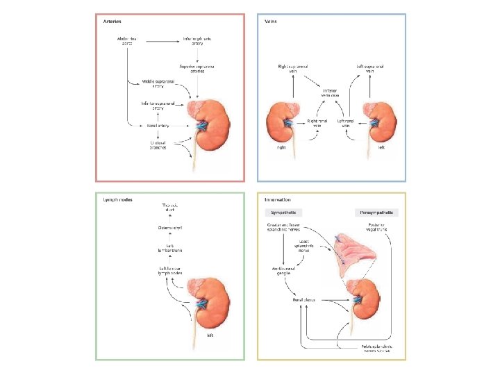
Inferior vena cava right testicular/ovaric vein Left left suprarenal vein rena l ve in aorta left testicular/ovaric vein

Ureter • retroperitoneal, L 2 • lengths: 25 -30 cm • parts: abdominal part, pelvic part, intramural part Crossings: Physiological constrictions: 1. (genitofemoral nerve) – ant. 1. Renal pelvis – ureter transition 2. Gonadal a/v - post. 3. Common iliac a. , -external and –internal ant. 4. Uterine a. / vas defferens - post. / inf. Where’s the ureter from the other? 2. (Crossing with gonadal vessels) 3. Crossing with common iliac artery 4. Intramural part
 (lig. umbilicale")
Urinary bladder - Infraperitoneal - Parts: fundus-, corpus- and vesical vertex (apex) (lig. umbilicale medianum urachus) - Vesical trigone: ureteric ostia, internal urethral orifice - Volume: 300 -350 ml - Blood supply: superior vesical arteries, inferior vesical artery (branches of internal iliac artery)

interureteric fold intramural part of left ureter intramural part of right ureteric orifice vesical trigone detrusor muscles neck internal vesical sphincter internal urethral orifice

m rectu urethra aorta ut s ter ra ureth a vagin m rectu bladder ure rectum prostate

Thank you for your attention! References: Gray’s Anatomy for Students Thieme: Atlas of Anatomy, Neck and Internal Organs Grant’s Atlas of Anatomy 13 th edition studyblue. com
- Slides: 23