Anatomy of the Eye II histology and retinal


 is lost, the “optic nerve” is reduced to")


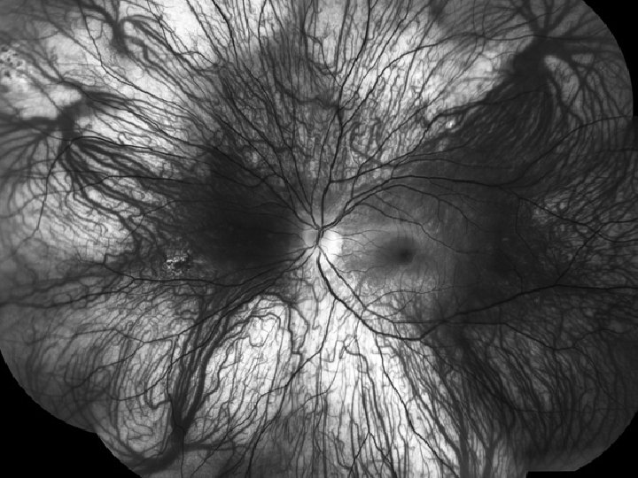
 easily visible (A red-free")


- Slides: 35
Anatomy of the Eye: II histology and retinal landmarks 1. 2. We will discuss retinal histology, the 10 layers of the retina, and how the retina functions We will review the 6 key landmarks to recognize in examination of the retina
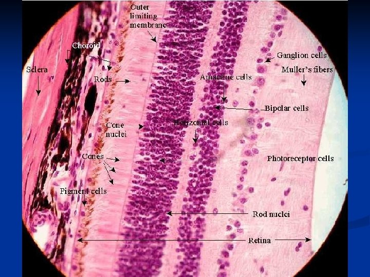
Ten layers of the retinal
How the retina functions Light travels thru the retina and is captured by the photoreceptor cells, the “rods and cones. ” The light impulse is then analyzed and interpreted by the other types of retinal nerve cells and converted into a “message” to send to the brain. The “message” is then transferred up to the Nerve Fiber Layer, at the surface of the retina. The NFL is the “wiring” that transfers the visual message from the retina to the brain. There approximately 1 million nerve fibers that come together to form the Optic nerve, and travel without synapse into the substance of the brain.
1. Clinical relevance of the 10 retinal layers for the AIDS clinic Nerve Fiber Layer (NFL) 1. 2. Oriented horizontally - blood in the NFL has linear orientation Layer 3 -10 1. 2. Oriented vertically & below (deeper to) retinal blood vessels - blood or lesions within these layers appear round An infectious process that begins within these layers may initially appear slightly “yellow”
Retinal landmarks 1. 2. 3. 4. 5. 6. optic disc Nerve fiber layer Superior and inferior temporal vascular arcades Fovea Vortex veins / the equator of the eye Choroidal markings seen thru the normal translucent retina
Landmark # 1: optic disc / optic nerve
Normal Optic Nerve or Optic Disc
1. The nerve fiber layer is oriented horizontally. The other retinal layers are oriented vertically. 2. The nerve fiber layer comes together at the optic disc to form the optic nerve, an extremely “crowded intersection”
If the Nerve Fiber Layer (NFL) is lost, the “optic nerve” is reduced to only an “optic cup” with no “pink” neuroretinal rim
Landmark # 2: Nerve fiber layer
SCHEMATIC ILLUSTRATION OF THE NERVE FIBER LAYER
Nerve Fiber Layer
An area of the nerve fiber layer where the myelin sheath congenitally remains, outlining the NFL
NERVE FIBER LAYER DEFECT
INFERIOR NERVE FIBER LAYER DEFECT CORRESPONDING TO AN AREA OF RETINAL NECROSIS (AND NOW SCARRING) FROM CMV RETINITIS
Landmark # 3: superior and inferior temporal vascular arcades
Superior temporal arcade fovea Optic nerve Inferior temporal arcade
Landmark # 4: fovea
The fovea is slightly darker than the background retina. It is temporal to optic disk by 1. 5 - 2 DD. All fine vision or reading vision is from this area
The fovea is avascular
In people under about 40 years, the surface of the foveal depression acts as a “mirror” and focuses the light from the ophthalmoscope back, to a spot just above the retinal surface, seen as a bright spot of light - the “foveal light reflex. ” If this reflex is present, the fovea is “normal” – the basic anatomy is intact
Landmark #5: vortex veins
A Vortex Vein part of the Choroidal circulation seen thru the translucent retina
vortex veins mark the “equator” of the eye the dividing line between “Zone 2” and “Zone 3” for CMV retinitis
Vortex veins look Bizarre think “mutant octopus” about 7 per eye, at least one each quadrant.
The vortex veins divide Zone 2 from Zone 3 Zone 1: within 1500 microns (1 DD) or the optic nerve, or 3000 microns (2 DD) of the fovea Zone 2: from Zone 1 to the equator (vortex veins) Zone 3: from the equator to the ora serata
Landmark # 6: Choroid / choroidal markings
Choroid n The choroid is the network of blood vessels below the retina. n It has the highest blood flow/unit volume of any tissue in the body. n It acts as the “radiator” of the eye, diffusing the heat created by the light that is focused on the retina by the cornea and the lens. n It can be seen thru the translucent retina as the ribbon-like network of freely anastomosing
Normal Retina with normal choroidal markings visible through the translucent retina.
Example of both retinal vessels and choroidal vessels (choroidal markings) easily visible (A red-free photograph, that makes blood appear “black” is a patient with an extremely thin retina) (also great example of vortex veins!)
TEST
Familiarity with retinal anatomy is critical for indirect ophthalmoscopy Orientation is always confusing because the retinal image is upside down and backwards Another way to imagine the view with the indirect ophthalmoscope: “rotated 180 degrees”
The solution for performing a perfect examination Knowing landmarks !!! n Controlling and knowing the direction of the patient’s gaze n Always following a systematic scheme for conducting the examination n