Anatomy of the Cranial Bones Lesson Objective Describe

Anatomy of the Cranial Bones

Lesson Objective • Describe the anatomy of the cranial bones

ANATOMY OF THE SKULL

Topic Objectives • • • List the skeletal bones within each division of the cranium. Describe the bone classification and composition of the cranium. Identify the sutures of an adult and the fontanels of a newborn skull. Describe the anatomy of the cranium. Identify the basic localization points and planes of the skull. Identify the anatomical features of the cranial bones on an illustration.

Skeletal Divisions of the Cranium • Composed of 22 bones total • 2 Divisions – Cranial (8 bones) – Facial (14 bones)

Cranial Division • 8 bones • Houses and protects the brain • Provides attachment for muscles for chewing and head movements • Forms the cranial cavity that includes – Calvaria (skull cap) – The floor or base

Facial Division • 14 bones total • 2 are classified as single bones – Vomer – Mandible • 6 pairs of bones (12) (Facial division will be covered in separate class)

BONE CLASSIFICATION & COMPOSITION

Topic Objective Describe the bone classification of the cranium
 • Irregular (facial and some cranial")
Bone Classifications • Flat (majority of cranial bones) • Irregular (facial and some cranial bones)

Bone Composition • Composed of 3 layers • Cranial vault composed of 2 plates of compact tissue – Outer plate thicker than inner (mostly) – Both plates are separated by an inner layer of spongy tissue called diploe (varies in thickness)

SUTURES

Topic Objective • Identify the fontanels of a newborn skull on an illustration

Sutures • Join the bones of the cranium and face via fibrous joints • Adults have sutures (rigid immovable articulations) – – Coronal Sagittal Squamosal Lambdoidal

Coronal Suture separates frontal bone from two parietals

Saggital Suture located midline on top of the head, separates 2 parietal bones

Squamosal Suture formed by the junctions of the parietal bones and temporal bones

Lambdoidal Suture separates both parietal bones from the occipital bone

Suture Junctions • • Bregma Lambda Pterions Asterions

Suture Junctions • Bregma – anterior end of the sagittal suture that intersects with the coronal suture • Pterions – right & left located on lateral aspect of the skull where the parietal bone, squamosal suture, and greater wing of the sphenoid meet

Suture Junctions • Lambda – posterior end of the sagittal suture that intersects with the lambdoidal suture • Asterions – located behind the ear at the junction of the occipital bone, parietal bone, and mastoids

Fontanels • Infants have 6 areas of incomplete ossification • Membrane-covered openings or soft spots – – 1 anterior 1 posterior 2 sphenoid (right and left) 2 mastoid (right and left)

Fontanels • Ossification completed: – End of 1 st and 3 rd month for posterior & sphenoidal fontanels – End of 2 nd year for anterior & mastoid fontanels

BONES OF THE CRANIUM

Topic Objectives • • • Describe the bones of the cranium Identify the anatomical features of the cranial bones on an illustration Describe the bones that encompass the orbit cavities
 forms the forehead –")
Frontal Bone • 2 main parts – Vertical portion (Squama) forms the forehead – Horizontal portion (Orbital) forms roofs of orbits & roof of nasal cavity • Articulates with – Right & left parietals – Sphenoid – Ethmoid bones • Structural landmarks – Glabella – Nasion

Frontal Bone Nasion
 • Forms lateral sides of the cranium & part of the")
Parietal Bones (2) • Forms lateral sides of the cranium & part of the posterior cranial roof • Somewhat square in appearance, has convex external surface & concave internal surface

Parietal Bones • Articulate with 5 cranial bones: – – – Frontal Occipital Temporal Sphenoid Opposite parietal bone

Parietal Bones

Parietal Bones

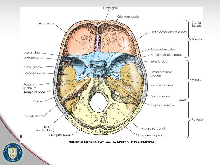
Occipital Bone • Forms posterior half of the cranial base and greater portion of the posterior cranial fossa • 4 main parts – Squama – Occipital condyles (2) – Basilar portion

Occipital Bone • Squama- rounded portion that forms most of the back of the head (inion) • Occipital condyles- oval processes that extend anteriorly on each side of the foramen magnum & articulate with the superior facets of C 1 (atlanto-occipital joint)

Occipital Bone • Basilar portion - curves anteriorly & superiorly to join the body of the sphenoid, resulting in a continuous bone (foramen magnum) • Articulates with 6 bones: – 2 parietals – 2 temporals – Sphenoid – Atlas (C 1)
")
Occipital Bone (External surface)
")
Occipital Bone (Internal surface)

Occipital Bone- Lateroinferior • Basilar Portion
 • Irregularly shaped bones • Located on each side of the")
Temporal Bones (2) • Irregularly shaped bones • Located on each side of the cranial base between the occipital bone & greater wings of the sphenoid bone • Form a large portion of the middle fossa of the cranium & a small part of the posterior fossa

Temporal Bones • Consist of 5 main parts – Squamous portion – Mastoid portion – Petrous portion – Tympanic portion – Styloid process

Temporal Bones
")
Temporal Bones (Lateral)
")
Temporal Bones (Internal)

Temporal Bones Coronal section through mastoid and petrous portions of the temporal bone

Sphenoid Bone • Irregularly wedge-shaped bone situated in the base of the cranium anterior to the temporal bone • Serves as the anchor for all 8 cranial bones

Sphenoid Bone • Consists of 4 primary parts: – Body • Contains 2 sphenoidal sinuses and the sella turcica – 2 Lesser wings • Project laterally from the upper sphenoid bone – 2 Greater wings • Includes 3 pairs of small foramina – 4 Pterygoid processes • Form part of the lateral walls of the nasal cavity

Sphenoid Bone

Sphenoid Bone

Sphenoid Bone

Ethmoid Bone • Small cubed-shaped bone • Lies between the orbits & forms part of the anterior cranial fossa, nasal cavity, bony nasal septum, & orbital walls • Articulates with 2 cranial bones – Frontal – Sphenoid
 •")
Ethmoid Bone • Consists of 4 main parts – Horizontal portion (cribriform plate) • Contains many small foramina that transmit olfactory nerves • Includes a thick conical process called the crista galli – Vertical portion (perpendicular plate) • Forms the bony nasal septum – 2 Lateral labyrinths (masses) • Contain ethmoidal sinuses • Superior & middle nasal conchae
")
Ethmoid Bone (Anterior)
")
Ethmoid Bone (Lateral)
")
Ethmoid Bone (Superior)

Ethmoid/Sphenoid Bones

LOCALIZATION POINTS & PLANES OF THE HEAD

Topic Objective • Identify the localization points & planes of the head

OML Orbitomeatal Line • Located between the outer canthus of the eye & EAM

IOML Infraorbitomeatal Line • Formed by connecting the infraorbital margin to EAM

AML Acanthomeatal line • Line formed between the acanthion & EAM

MML Mentomeatal Line • Formed by connecting a line from the mental point and EAM

GML Glabellomeatal Line • Most superior of the positioning lines between glabella and EAM

IPL Interpupillary Line • Line connecting the pupils or outer canthi of eyes

IOM Infraorbital Margin • Inferior rim of the bony orbit of the eye

SOM Supraorbital Margin • Superior rim of the bony orbit of the eye

Outer Canthus • Lateral junction of the outside of the upper and lower eyelids

Acanthion • Midline point at the junction of the upper lip & nasal septum (where nose & upper lip meet)

Skull Types • Dolichocephalic long from the front to the back, and narrow from side to side.

Skull Types • Mesocephalic - average or most common sized skull

Skull Types • Brachycephalic - short from the front to the back, and wide from side to side

Review • • • Skeletal divisions of the skull Bone classifications Bone composition Sutures and fontanels Bones of the cranium Basic localization points and planes of the head

Review Question How many bones are there in a skull? Answer 22

Review Question Skull is classified as what bone? Answer Flat and irregular bone

Review Question What are the two divisions of skull? Answer Cranium and facial

Review Question What is an inner layer of spongy bone called in a skull? Answer Diploe

Review Question What suture is between the temporal and the parietal bone? Answer Squamosal suture

Review Question What is called an incomplete ossification in newborn cranial bones? Answer Fontanels

Review Question What is known as the site of the antero lateral fontanel? Answer Pterion

QUESTIONS?
- Slides: 79