Anatomy of the coronary arteries Angiographic VISUALIZATION Coronary


 • RCA ostium-3.")


Conus artery/ Infundibular/ Third coronary/ Adipose /Arteria of Vieussens(1 st branch")
SA nodal artery arises from– RCA-55 -65%, LCX -35 -45% •")


 Right ventricular branches - one or more ventricular branches arising")
 Right atrial artery-originates at about the level of acute")
 Acute marginal artery -relatively large and constant vessel -arises")
 AV nodal artery(80%) -slender , straight vessel -appears almost")
 Posterior descending artery -The PDA courses in the inferior interventricular groove")


 length & 3 -6")


 • Based on length and")

 Diagonals - In most cases 1 st branch of LAD is 1")
 Septals • vary in number • Arise in about 90* angle")


 RV branches • gives 1 or more branches to the right ventricle.")
 Terminal branches of LAD type 3 • Apical branches • Usually 2 branches")


 - LCA")


 trial was")


 -LAD 58%")
 • Type")


- Slides: 102
Anatomy of the coronary arteries & Angiographic VISUALIZATION
Coronary arterial anatomy • 1 st anatomical drawings- Leonardo da Vinci • Oblique inverted crown
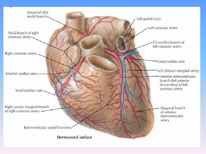
• The coronary arteries and their major branches are sub-epicardially located
The LEIDEN convention • Each artery arises from respective aortic sinuses - Right coronary sinus(anterior) - Left coronary sinus(left posterior) - Non-coronary sinus(right posterior) 1 R 2 LCx pattern
• LCA ostium -4. 7 mm(Range 1. 0 -8. 5) • RCA ostium-3. 7 mm(Range 0. 5 -7. 0) • RCA takes off at right angle and LCA takes off at more acute angles
Right coronary artery • Arises from the right sinus of valsalva , lies inbetween pulmonary conus and right atrium. • Moves sharply towards left of the observer and is directed towards sternum. • Curves down following right AV groove in direction of acute margin of heart and diaphragm. • Curves posteriorly and follows AV groove towards crux cordis.
Right Coronary Artery Proximal RCA Mid RCA Distal RCA PDA LAO cranial Best for visualization of ostial and proximal RCA Distal RCA PDA/P LV RAO cranial Best for visualization of mid RCA and PDA PA Cranial Best for visualization of distal RCA and its bifurcation
Surgical division of the RCA • Proximal - Ostium to 1 st main RV branch • Mid - 1 st RV branch to acute marginal branch • Distal - acute margin to the crux
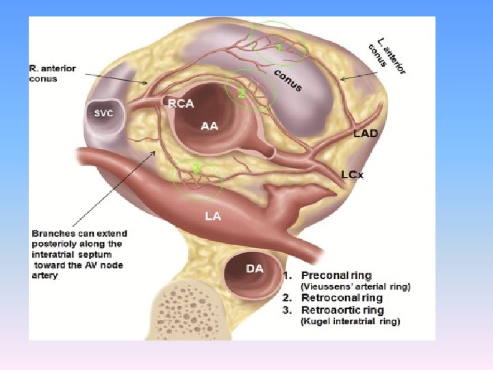
Right coronary artery-branches 1)Conus artery/ Infundibular/ Third coronary/ Adipose /Arteria of Vieussens(1 st branch in 60% cases) - Separate ostium in 23% - 51% - Curves away from main artery and proceeds ventrally encircling the outflow tract of RV at the level of pulmonary valve.
Right coronary artery-branches… 2)SA nodal artery arises from– RCA-55 -65%, LCX -35 -45% • Runs in opposite direction to the conus branch • Runs cranially, dorsally and to the right • Divides into two rami, one recurrent branch which supplies the SA node, the other runs posteriorly a left atrial branch. • Two divisions form a wide Y acquiring shape of ram’s horn.
Ram’s horn
• Left horn-encircles the SVC and supplies the SA node. • Right horn-provides blood supply to superior and posterior walls of left atrium. • When SA nodal a. arises from LCX it arises from proximal portion then ascends to the right beneath the left atrial appendage and behind the aorta crossing the posterior aspect of the LA to reach the IAS and the SVC.
Right coronary artery-branches… 3) Right ventricular branches - one or more ventricular branches arising in the AV groove. - Often reach interventricular sulcus and anastomose with branches of LAD when occluded.
Right coronary artery-branches… • 4) Right atrial artery-originates at about the level of acute marginal artery travels in opposite direction towards right heart border -receives branches from SA nodal artery and bypasses obstruction in proximal portion of the RCA.
Right coronary artery-branches… • 5) Acute marginal artery -relatively large and constant vessel -arises at lower aspect of right atrium just before or at the acute margin of the heart.
Right coronary artery-branches… • 6) AV nodal artery(80%) -slender , straight vessel -appears almost vertical in LAO view -directed towards centre of heart shadow -it arises from RCA where it forms a characteristic inverted U curve as the artery passes in the interventricular sulcus around the interventricular vein.
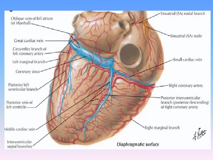
RCA-branches…. • 7) Posterior descending artery -The PDA courses in the inferior interventricular groove gives rise to a number of small inferior septal branches which supply lower part of IVS and interdigitate with superior septal branches from LAD. • 8) Posterolateral branches -After giving rise to PDA , the RCA continues beyond the crux cordis as the right posterior AV branch terminating in one or several posterolateral branches to supply the diaphragmatic surface of the LV.
• At times the posterlateral branches may be larger than the PDA itself. • The entire sweep of RCA resembles a sickle, the blade formed by main stem of RCA and handle formed by PDA and posterior LV branches.
“Dominance” • giving rise to PDA, at least 1 postero lateral branch - 85% right dominant -8% left dominant -7% co-dominant • Left dominance is 25 -30% in Bi-Ao. V
Surgical standpoint of Rt. dominance • Whenever the RCA is dominant in patients subjected to CABG , any clinically significant obstruction should be bypassed with graft placed downstream to most distal lesion. • If RCA is not dominant any right sided lesion is considered surgically insignificant.
Shepherd’s-crook RCA • ~5% • Acute superiorly angled take-off of the RCA from the aorta. • Difficult RCA lesion angioplasty Ethan Halpern. Cardiac CT. Functional anatomy.
Left coronary artery LMCA - 10 -15 mm(upto 30 mm) length & 3 -6 mm(upto 10 mm diameter), diameter is inversely proportional to its length, - Trifurcates in 1/3 rd : Ramus intermedius/ median artery/straight LV artery/left diagonal artery.
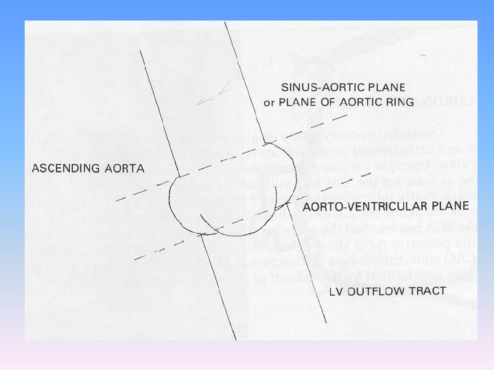
LMCA Largest and shortest coronary artery Ostium of LCA 1 cm higher than that of RCA Axis of ostium is 30 -40* posterior to frontal plane Vessel is directed anteriorly, left and downward between the pulmonary trunk and left atrium. • Ostium is thus located posterior to axis of artery. • Hence catheterization is best in LAO 30*but course of artery is best outlined in shallow view. • •
LMCA –area perfused • Entire LV, LA except the posterior portion of IVS and adjacent area when PDA is branch of RCA
LMCA variations • When LM is absent the coronary ostium appears like shallow funnel with two separate openings. • When LM is short visualization may be difficult and catheter tip appears to be in direct contact with bifurcation. • In patients with high diaphragm with horizontal hearts a weeping willow view LAO 60* cranial 30* may improve visualisation of LM and its branches.
Absent LMCA Topaz et al. Absent left main coronary artery: angiographic findings in 83 patients with separate ostia of the left anterior descending and circumflex arteries at the left aortic sinus. Am Heart J. 1991 Aug; 122(2): 447 -52.
LAD –angiographic classification • By D. B. Effler(personal communication) • Based on length and amount of myocardium it perfuses • Type 1 -small caliber vessel reaches only 2/3 rd of way from base of heart to apex, more prevalent in women • Type 2 -larger caliber reaches the apex of LV • Type 3 -extends from base to apex wraps around the diaphragmatic surface of LV where it augments the perfusion pattern of PDA.
LAD • For practical purposes is a continuation of the LMCA • Passes to the left of pulmonary trunk and travels into the upper portion of IV sulcus. • As it turns around the pulmonary artery and begins its downward course the LAD forms a 90* angle often highlighted by origin of 2 nd diagonal. • Surgical importance of this location is that it is the point where LAD is amenable to bypass.
LAD-branches 1) Diagonals - In most cases 1 st branch of LAD is 1 st diagonal branch Arises close to bifurcation of LM 2 -9 in number Supplies whole of LV free wall
LAD-branches. . 2) Septals • vary in number • Arise in about 90* angle • Runs along septum front to back and in caudal direction • The anterior septal perforators mechanically immobilize the LAD, fixing it to the heart, limiting its motion, and preventing buckling of the artery during systole.
SEPTALS-territory supplied • 2/3 rds of upper portion of septum and almost entire septum in inferior 3 rd. • When PDA arises from LCX the LCA is the sole source of supply for entire IVS.
SEPTALS-ANGIOGRAPHIC CHARACTERISTICS • More cranial septal branches are better demonstrated than lower septal branches because of greater length and caliber • Characteristic branching of 90*from LAD and straight course, lacking slight tortuosity of other branches • Another characteristic feature is relative lack of motion(evident when LCA is viewed against the background of LCX in RAO view)
LAD-branches… 3) RV branches • gives 1 or more branches to the right ventricle. • Highest of these rise to the level of pulmonary valve forming vieussen s anastomosis. • The other branches run obliquely over the RV surface anastomosing with similar branches of the RCA. • Form importanat cllateral channel.
4) Terminal branches of LAD type 3 • Apical branches • Usually 2 branches can be seen • Recurrent posterior-supplies diaphragmatic portion • Recurrent lateral-supplies lateral aspect of apex. • Referred to as pitch fork, mustache or whale’s tail
Myocardial bridging • short segments of the LAD can travel within the myocardium (covered by a so-called myocardial bridge) resulting systolic luminal narrowing is probably benign in the vast majority of people. • 0. 5 to 1. 6% in general population, 28% in children, 3050% in HOCM. • associated with a poor prognosis (higher incidence of myocardial ischemia and sudden death) in patients with hypertrophic cardiomyopathy
Surgical division of the LAD • Proximal - Ostium to 1 st major septal perforator or 1 st diagonal artery whichever is first • Mid - 1 st perforator to D 2 (90 degree angle) • Distal - D 2 to end
Left circumflex artery - Departs at a sharp angle from LM to run posteriorly along the AV groove towards the crux cordis. Reaches crux only in 16% cases Course nearly mirrors that of RCA.
LCX-branches • Soon after origin divides into 2 parallel branches • Lower and larger gives origin to ventricular branch • Upper gives atrial circumflex to atrial wall • Largest and constant branch is obtuse marginal • Runs along the ventricular wall posteriorly and in direction of apex
• The proximal portion of the obtuse marginal, LAD and diagonal branches may be closely superimposed on one another when the origin of the obtuse marginal is along the initial segment of the circumflex • Bunching up of branches, difficult to recognize minor lesions in RAO and LAO view • RAO 30*cranial 20* may be necessary.
LCX-branches • After the obtuse marginal the LCX travels all way around the left AV groove and gives rise to posterolateral branches. • Because of its close relation to the AV groove (and mitral valve ring) this portion of artery moves widely with systole (towards apex) and diastole(away from apex). • Reaches crux and enters the posterior IV groove as PDA. Regardless of origin PDA follows same course in posterior IV groove. • Its origin from circumflex is best seen in LAO view.
LCX-territory supplied • Supplies lateral wall of LV above the diaphragmatic surface
Surgical division of the LCX • Proximal - Ostium to 1 st major obtuse marginal branch • Mid - OM 1 to OM 2 • Distal - OM 2 to end
Optimal angiographic views for coronary segments Carlo Di Mario, Nilesh Sutaria. CORONARY ANGIOGRAPHY IN THE ANGIOPLASTY ERA: PROJECTIONS WITH A MEANING Heart 2005; 91: 968– 976.
RAO- RCA
Optimal angiographic views for coronary segments Carlo Di Mario, Nilesh Sutaria. CORONARY ANGIOGRAPHY IN THE ANGIOPLASTY ERA: PROJECTIONS WITH A MEANING Heart 2005; 91: 968– 976.
PA 5 -10* cranial 35 -45*- LCA
PA Cranial view -RCA
RAO cranial - RCA
Optimal angiographic views for coronary segments Carlo Di Mario, Nilesh Sutaria. CORONARY ANGIOGRAPHY IN THE ANGIOPLASTY ERA: PROJECTIONS WITH A MEANING Heart 2005; 91: 968– 976.
RAO 30 -45* caudal 30 -40* - LCA
Optimal angiographic views for coronary segments Carlo Di Mario, Nilesh Sutaria. CORONARY ANGIOGRAPHY IN THE ANGIOPLASTY ERA: PROJECTIONS WITH A MEANING Heart 2005; 91: 968– 976.
AP 5 -10* caudal 25 -35*- LCA
Optimal angiographic views for coronary segments Carlo Di Mario, Nilesh Sutaria. CORONARY ANGIOGRAPHY IN THE ANGIOPLASTY ERA: PROJECTIONS WITH A MEANING Heart 2005; 91: 968– 976.
LAO 30 -45* cranial 25 -35*- LCA
LAO cranial-RCA
Optimal angiographic views for coronary segments Carlo Di Mario, Nilesh Sutaria. CORONARY ANGIOGRAPHY IN THE ANGIOPLASTY ERA: PROJECTIONS WITH A MEANING Heart 2005; 91: 968– 976.
LAO 40 -50* caudal 25 -40* (Spider view) - LCA
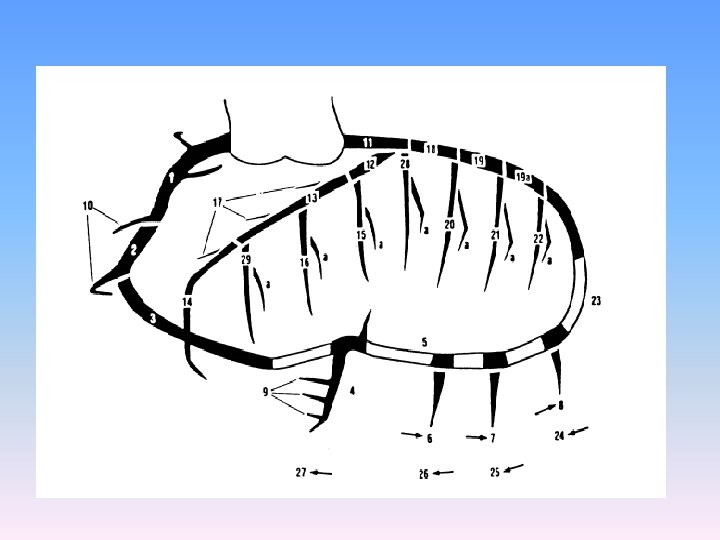
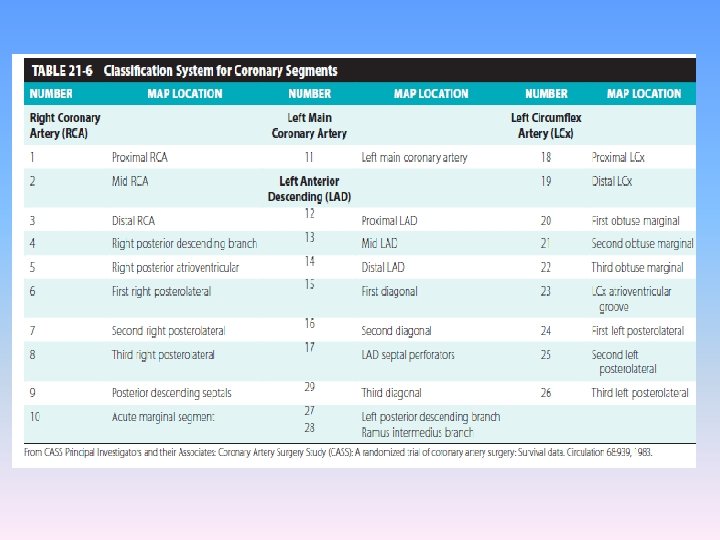
Coronary segment classification system • CASS investigators – 27 segments • BARI – 29 segments ( ramus intermedius and 3 rd diagonal branch)
Thank you
SYNTAX SCORE • An angiographic tool grading the complexity of coronary artery disease • A semiquantitative visual score that will help us to be aware of the anatomical complexity and to anticipate procedural difficulties
• One drawback in these comparisons is that there is heterogencity in the complexity of CAD of the patients enrolled. • Absence of grading of severity of CAD and lack of comparison of lesion complexity between various groups severely limits the interpretation of results.
• For example pts with distal LM trifurcation disease with occluded RCA is pooled together as TVD with patients with 3 focal lesions in midportion of the 3 coronary arteries. • The first has a greater therapeutic challenge for PCI and both have completely different prognosis regardless of revascularisation.
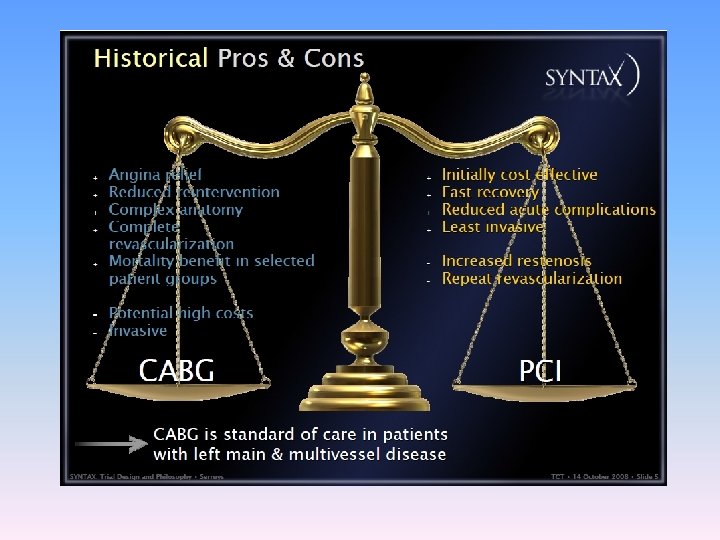
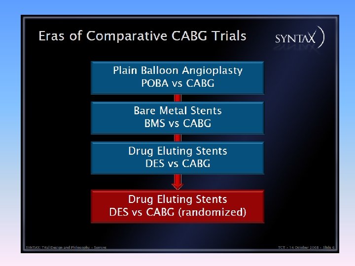
• SYNTAX (Synergy between PCI with TAXUS stent and cardiac surgery) trial was organised for patients with significant lesion in LM and /or TVD. • The syntax score has been used in this study to categorize the coronary vasculature with respect to the number of lesions their functional impact, location and complexity.
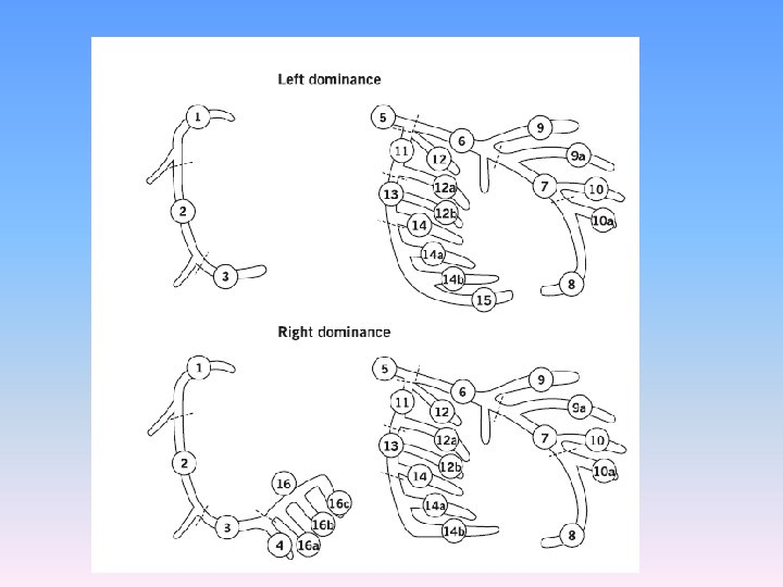
• The SYNTAX score has been developed based on the following: • 1. The AHA classification of the coronary tree segments modified for the ARTS study • 2. The Leaman score • 3. The ACC/AHA lesions classification system • 4. The total occlusion classification system • 5. The Duke and ICPS classification systems for bifurcation lesions • 6. Consultation of experts
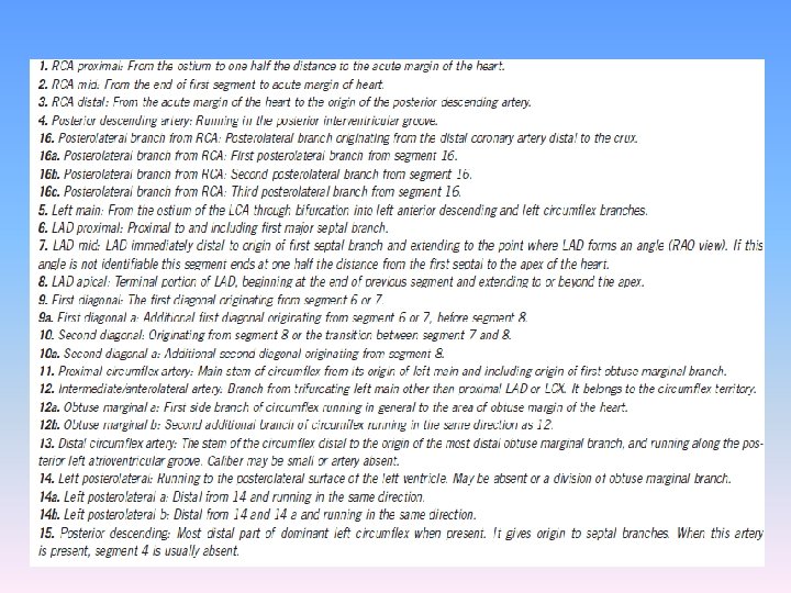
AHA Definition of coronary tree segments • Arterial tree is divided into 16 segments • This system has been adopted for the syntax scoring.
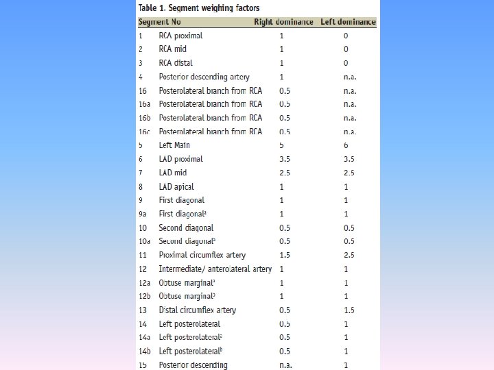
LEAMAN SCORE • Based on severity of luminal diameter narrowing • Weighed according to usual blood flow to LV by each vessel
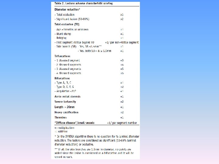
Severity of luminal diameter narrowing • significant lesion-50% reduction in lumen diameter by visual assessment in vessels >1. 5 mm in diameter. • Less severe lesions not included • Percent diameter stenosis is not included • Only occlusive lesions (100% stenosis)-MF 5 • And non occlusive lesions (50 -99% stenosis)MF 2
• In right dominant system -RCA supplies 16% -LCA supplies 84% of flow to LV • Of the 84%, 66% is by LAD and 33% by LCX. • The LM supplies approximately 5 times, the LAD app. 3. 5 times and LCX app. 1. 5 times blood as the RCA to the LV.
• For left dominant system -LM supplies 100%(hence multiplication factor 6) -LAD 58% (MF-3. 5) -LCX 42% (MF-2. 5) • The contribution is used as a multiplication factor -
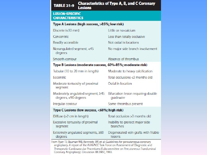
ACC/AHA lesion classification system • Type A (high success , low risk) • Type B (mod success , mod risk) • Type C (low success , high risk)
Total occlusion classification system • No antegrade flow is visible distal to lesion • Distal segments may be filled via bridging , ipsilateral or contralateral collaterals. • Parameters included are -Age of occlusion -blunt stump -presence of bridging collaterals -presence of side branch -occlusion length
DUKE and ICPS bifurcation lesion classification • Defined as junction of main vessel and a side branch (1. 5 mm) • Not involving ostium(A, B, C) • Involving ostium(D, E, F, G)
DUKE and ICPS bifurcation lesion classification
Bifurcation segments • • 5/6/11 6/7/9 7/8/10 11/13/12 a 13/14/14 a 3/4/16 13/14/15
Trifurcation segments • • • 3/4/16/16 a 5/6/11/12 a/12 b/13 6/7/9/9 a 7/8/10/10 a
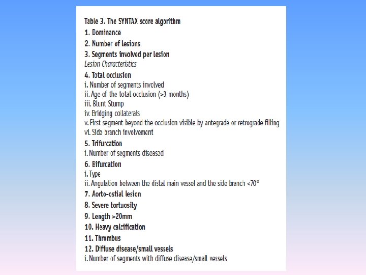
SCORING method • Started from RCA to qualify the dominance • Visually score each lesion starting from RCA • Only lesions >= 50% in a vessel >=1. 5 mm in diameter should be scored • Tandem lesions should be considered as single lesion • Points calculated for each lesion and then added together.
LIMITATION • Does not entail any clinical variable • Comorbidities are known to impact early outcomes of patients undergoing revascularisation. • Hence limited use in guiding decision making between CABG and PCI. • Relies on pure visual interpretation of lesion severity and subjective variables hence poor reproducibility.
Functional syntax score • Incorporates ischemia producing lesions as determined by FFR.
Clinical syntax score • • Syntax score + Age Creatinine EF
SYNTAX score II • • Anatomical syntax score Age Creatinine clearance LVEF ULMCA Peripheral Vascular Disease Female sex COPD
• Aids in decision making between CABG and PCI • Interaction for particular baseline characteristic was defined by the hazard ratio. • Hazard ratio is defined as HR of mortality of that characteristic undergoing PCI to the HR of mortality in those undergoing CABG.
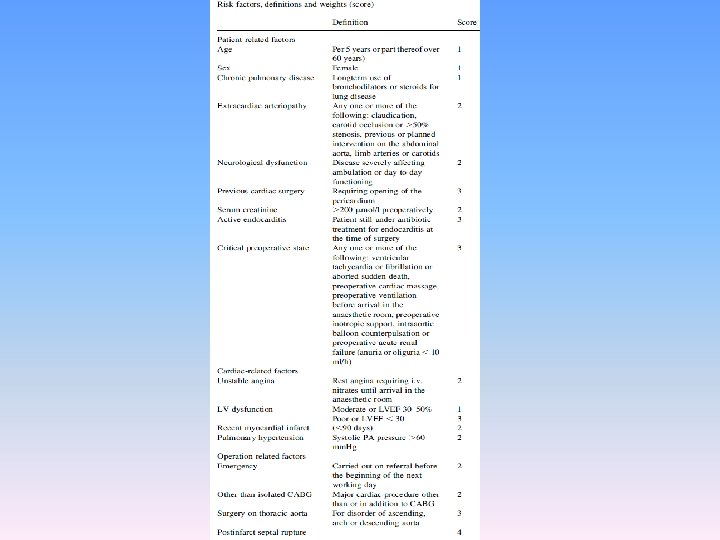
EURO SCORE-European System for cardiac operative risk evaluation
MACCE to 5 years by Syntax Score Tercile Left Main Disease PCI or CABG
Thank You
References • • Hurst’s The Heart 13 th Edition Braunwalds Heart Disease 2 nd edition Grey’s Anatomy Kern’s Handbook of Interventional Catheterization Grossman’s Textbook of Cardiac Catheterization Carlo Di Mario, Nilesh Sutaria. CORONARY ANGIOGRAPHY IN THE ANGIOPLASTY ERA: PROJECTIONS WITH A MEANING Heart 2005; 91: 968– 976 David M Fiss. Normal coronary anatomy and anatomic variations. Applied Radiology, Jan 2007. Horia Muresian. Coronary arterial anomalies and variations. MAEDICA. A journal of clinical Medicine, 1(1), 2006.