
ANATOMY OF SKIN Skin v Specialize Volume 8
- Slides: 27
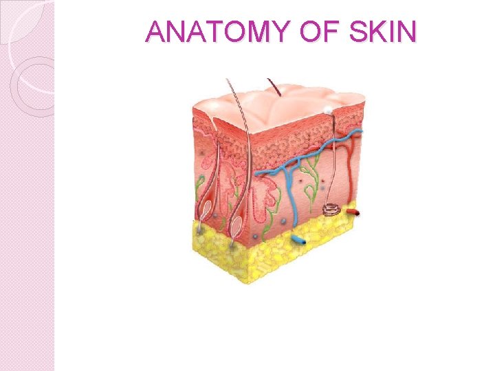
ANATOMY OF SKIN
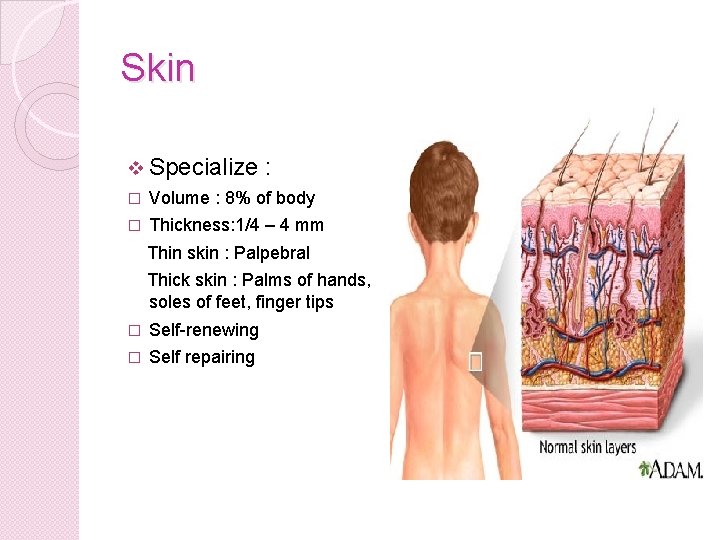
Skin v Specialize : � Volume : 8% of body � Thickness: 1/4 – 4 mm Thin skin : Palpebral Thick skin : Palms of hands, soles of feet, finger tips � Self-renewing � Self repairing
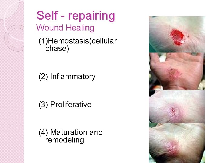
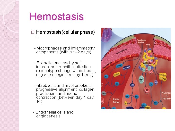
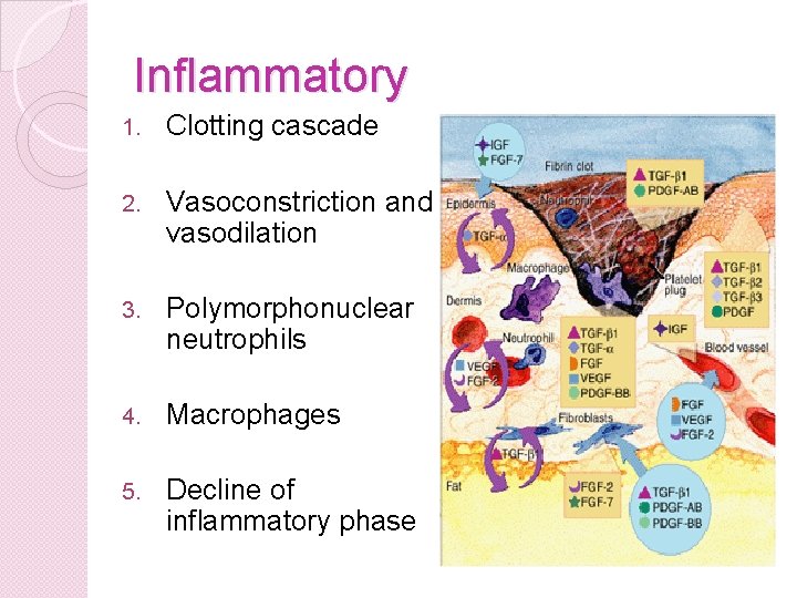
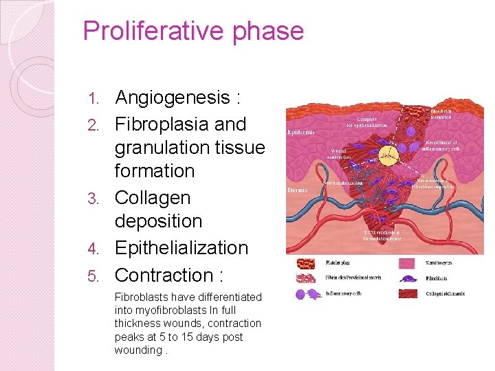
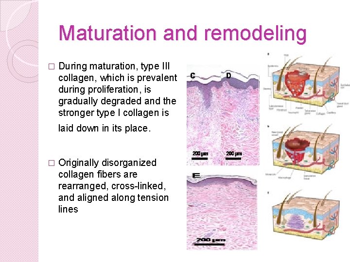
Self - repairing Wound Healing (1)Hemostasis(cellular phase) (2) Inflammatory (3) Proliferative (4) Maturation and remodeling
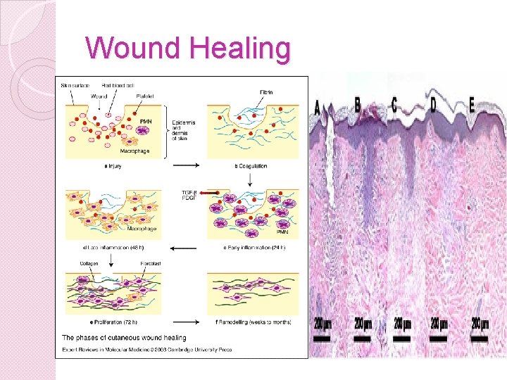
Wound Healing
Hemostasis � Hemostasis(cellular phase) : - Macrophages and inflammatory components (within 1– 2 days) - Epithelial-mesenchymal interaction: re-epithelialization (phenotype change within hours, migration begins on day 1 or 2) -Fibroblasts and myofibroblasts: progressive alignment, collagen production, and matrix contraction (between day 4 day 14) - Endothelial cells and angiogenesis
Inflammatory 1. Clotting cascade 2. Vasoconstriction and vasodilation 3. Polymorphonuclear neutrophils 4. Macrophages 5. Decline of inflammatory phase
Proliferative phase 1. 2. 3. 4. 5. Angiogenesis : Fibroplasia and granulation tissue formation Collagen deposition Epithelialization Contraction : Fibroblasts have differentiated into myofibroblasts In full thickness wounds, contraction peaks at 5 to 15 days post wounding.
Maturation and remodeling � During maturation, type III collagen, which is prevalent during proliferation, is gradually degraded and the stronger type I collagen is laid down in its place. � Originally disorganized collagen fibers are rearranged, cross-linked, and aligned along tension lines
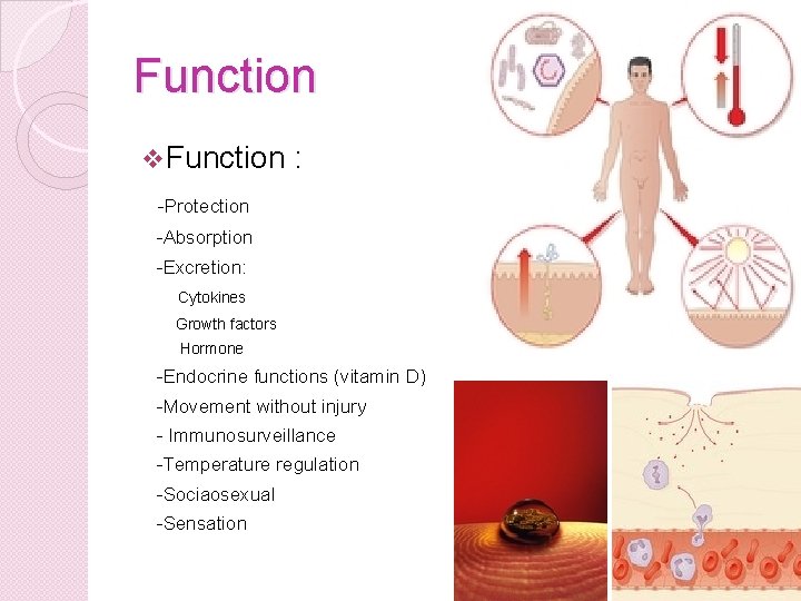
Function v Function : -Protection -Absorption -Excretion: Cytokines Growth factors Hormone -Endocrine functions (vitamin D) -Movement without injury - Immunosurveillance -Temperature regulation -Sociaosexual -Sensation
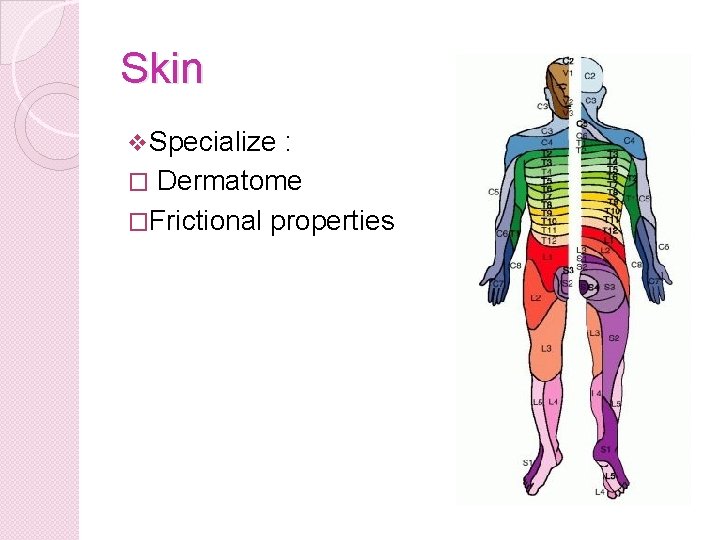
Skin v Specialize : � Dermatome �Frictional properties
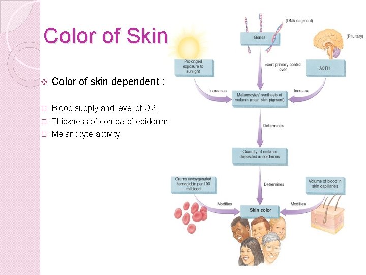
Color of Skin v Color of skin dependent : � Blood supply and level of O 2 � Thickness of cornea of epidermal � Melanocyte activity
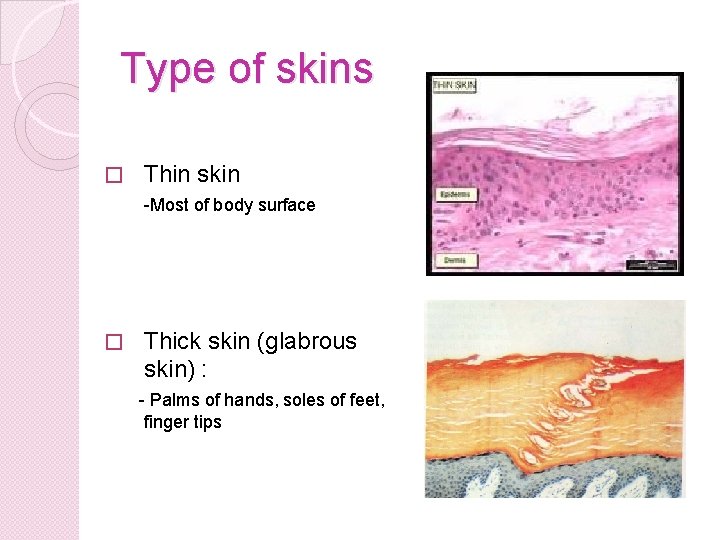
Type of skins � Thin skin -Most of body surface � Thick skin (glabrous skin) : - Palms of hands, soles of feet, finger tips
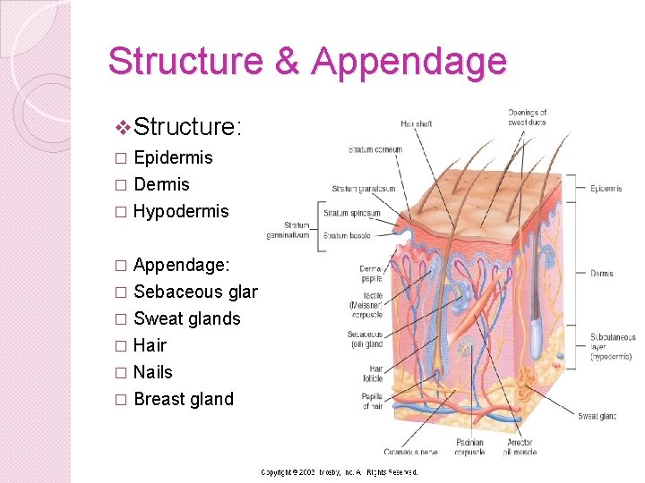
Structure & Appendage v Structure: � Epidermis Dermis � Hypodermis � Appendage: � Sebaceous glands � Sweat glands � Hair � Nails � Breast gland �
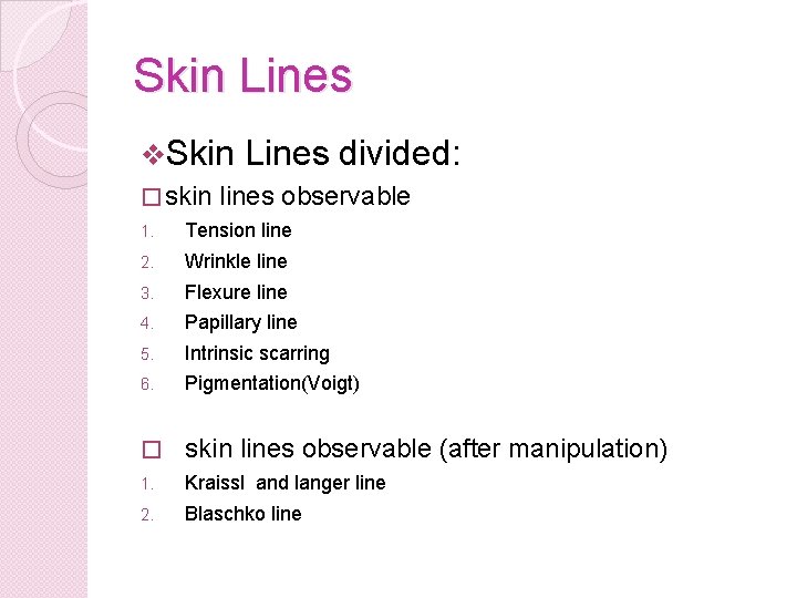
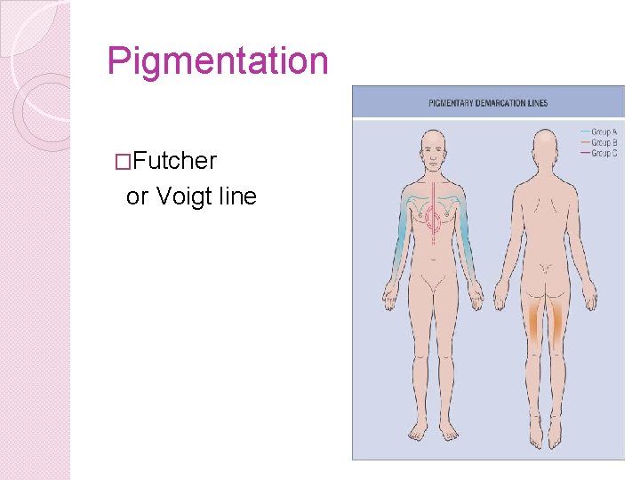
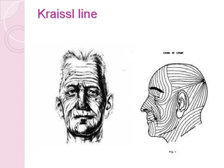
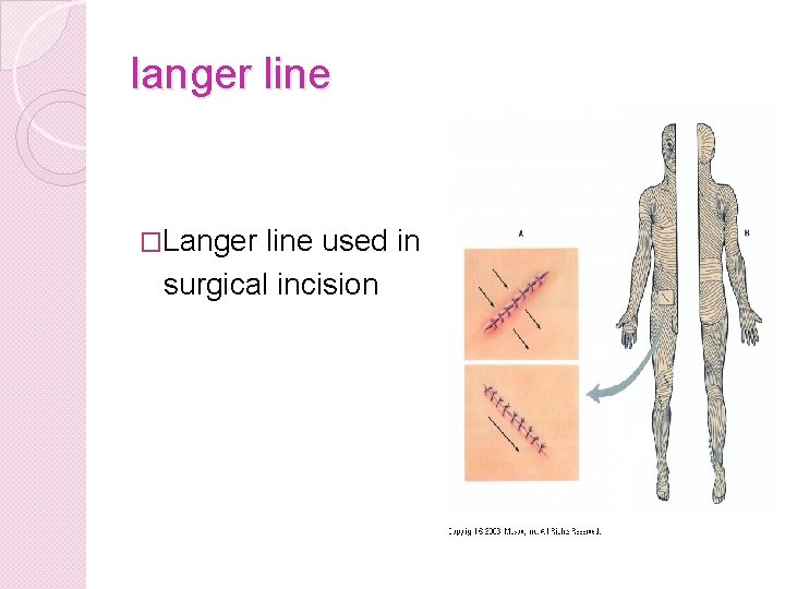
Skin Lines v. Skin � skin Lines divided: lines observable 1. Tension line 2. Wrinkle line 3. Flexure line 4. Papillary line 5. Intrinsic scarring 6. Pigmentation(Voigt) � skin lines observable (after manipulation) 1. Kraissl and langer line 2. Blaschko line
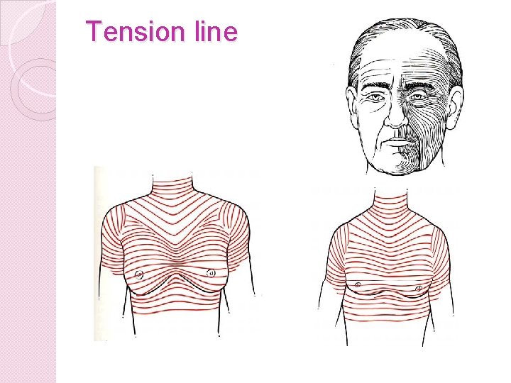
Tension line
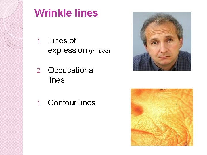
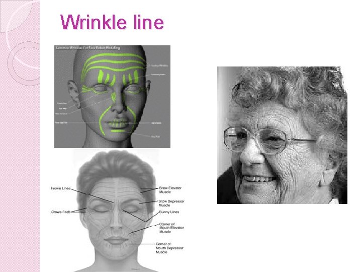
Wrinkle lines 1. Lines of expression (in face) 2. Occupational lines 1. Contour lines
Wrinkle line
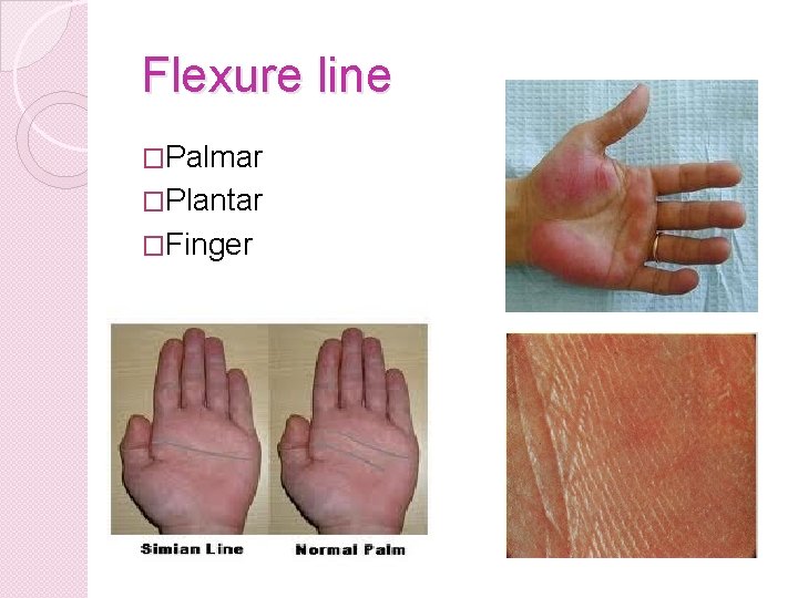
Flexure line �Palmar �Plantar �Finger
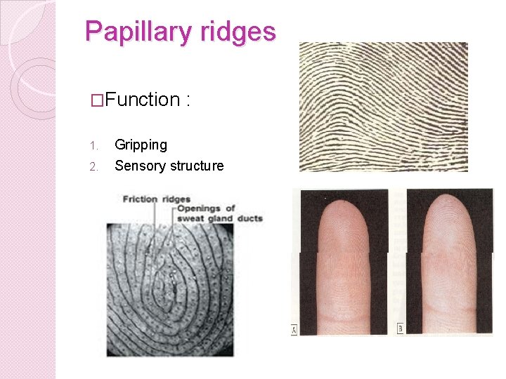
Papillary ridges �Function 1. 2. : Gripping Sensory structure
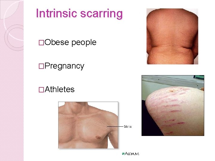
Intrinsic scarring �Obese people �Pregnancy �Athletes
Pigmentation �Futcher or Voigt line
Kraissl line
langer line �Langer line used in surgical incision
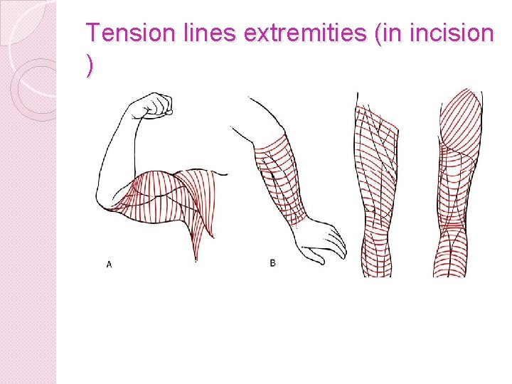
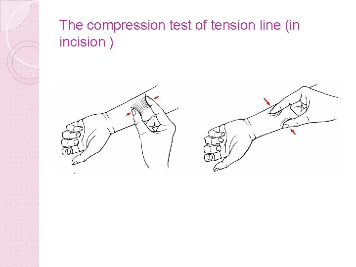
Tension lines extremities (in incision )
The compression test of tension line (in incision )
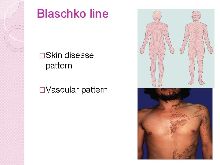
Blaschko line �Skin disease pattern �Vascular pattern
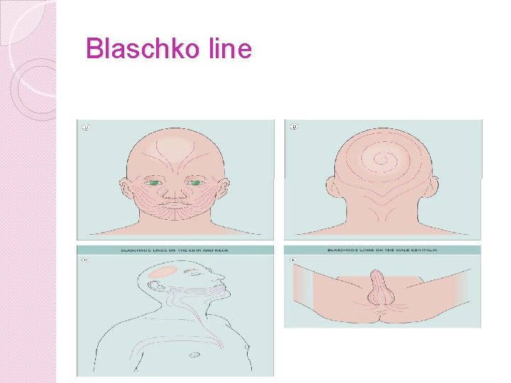
Blaschko line