ANATOMY OF PANCREAS BY Dr Manjula Vastrad Asst

ANATOMY OF PANCREAS BY : Dr Manjula Vastrad Asst Prof SMVVS RKM AMC Vijayapura

DEFNIt is a soft, compound, recemose transversely situated retroperitoneal gland having endocrine & exocrine function. EXOCRINE SECRETION- Pancreatic juice ENDOCRINE SECRETION- Insulin & glucagon EXTENTFrom the concavity of the duodenum to the hilum of spleen which crosses transversely with slightly upwards to the left through the post. abd. wall. CONSISTENCYSoft COLOR-Grayish pink SHAPE-Retort flask shaped MEASURMENTLength-12 to 15 cm Breadth-3 to 4 cm Thickness- 1. 5 to 2 cm Weight-average-85 gm Situation-Epigastric & left hypochondric region.

SURFACE ANATOMY

PARTS 1. Head 2. Neck 3. Body 4. Tail

HEAD It is the rt. Enlarged portion of the pancreas lodged in the duodenal curve. � Situationin the curvature of 1 st, 2 nd & 3 rd part of duodenum. � Vertebral level. At the leval of L 1 to L 2 veretebra. � FEATURES 3 Borders 1. Superior b. /upper b. Relation-1 st part of duodenum 2. Inferior b. /lower b. Relation-Uncinate process 3 rd part of duodenum 3. Right b. Relation-2 nd part of duodenum Anastomosis betn sup. & inf. Pancraticoduodenal vessels.

Surfaces 1. Ant. s. Direction-Forwards, upwards & left. Continuation-with the neck. Relation-Ant. presents a groove for gastroduodenal artery. Transverse colon Loose connective tissue Coils of jejunum Peritoneal relation-Upper part is nonperitoneal up to the transverse colon, below that it is peritoneal. 2. Post. s. Inf. vena cava Terminal parts renal veins Rt. crus of the diaphargm Bile duct it may embeded into the pancreatic substance Peritoneal reln. : Non perotoneal �
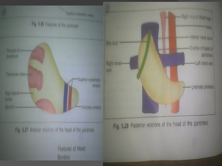

Arterial supply. Sup. & Inf. pancreticoduenal artery.

UNCINATE PROCESS The head consist of a process named as uncinate process. It projects from the lower part of the head directed upwards & medially. Relation. Ant. -Sup. mesenteric vessels. Post. -Abd. aorta.

NECK Length- 2 cm Direction-Forwards, Upwards & to the left where the neck meet with body FEATURESBorders 1. Sup. b. Relation-1 s. T part of duodenum celiac plexus 2. Inf. b. Relation-Attachment of the root of the transverse mesocolon.

SURFACE 1. Ant. s. Relation-peritoneum divistion of gastroduodenal artery pylorus of stomach 2. Post. s. -Relation-Superior mesenteric vein Beginning of portal vein. Arterial supply. Sup. & inf. pancreaticoduodenal artery.

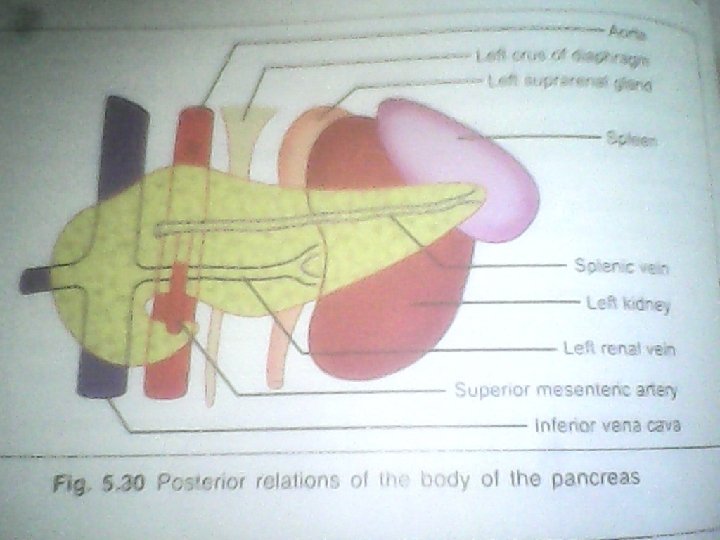
BODY Intro 1. The body is elongated & the largest part of the pancreas. 2. This extended from the front of aorta to the front of the left kidney 3. This passes towards the left with slight upwards & backwards inclination. � Shape- Prism shaped � Features. Borders 1. Sup. b. Relation-celiac artery Splenic artery Commom hepatic artery At the rt. End of this border, there is a tuberosity called tuber omentale. �

2. Inf. b. a. This border separates the post. surface from the inf. surface b. The sup. mesenteric vessels emerge through its rt. extremity 3. Ant. b. It is the differentiating border betn the ant. & inf. surface. Attachment. It provides attachment of the 2 layer of transverse mesocolon. � SURFACE 1. Ant. s. Direction-Anterosuperiorly Relation-Peritoneum Posteroinferior surface of stomach separated by the omental bursa 2. Post. s. Relation. Peritineal relation-Nonperitoneal Other relation-Abd. aorta.

Sup. mesenteric artery Left crus of the diaphragm Left suprarenal gland Left kidney Left renal vessels Splenic vein Inf. surface. Relation. Peritoneum Duodenojejunal flexure Coils of jejunum Left colic flexure � Arterial supply. Arteria pancreatica magna branch of splenic artery.

TAIL Intro 1. It Is the narrow left end of the pancreas 2. It passes betn. The layers of lienorenal lig. & extend up to the hilum of the spleen. 3. It is the most mobile part of the gland. � Situation. In betn the 2 layers of the lienorenal lig. along with the splenic vessels, to reaches the spleen. Relation. Anteriorly. Peritoneum of the lesser sac separates from the stomach. Posteriorly. Spleen & splenic vessels Inferiorly. Left colic flexure � Arterial supplybranch from splenic artery. �

Venous Drainage. Splenic, sup. mesenteric & portal vein Lymphatic Drainage. Into the pancreaticosplienic lymph nodes & efferent vessels drained into celiac lymph nodes. Nerve supply. Sympathetic. From the celiac & sup. mesenteric plexuses. Parasympathetic. Rt. & lf. vagus nerves.

DUCTS OF PANCREAS � Main pancreatic duct Beginning. It commences from the tail of the pancreas by the union of numerous smaller ducts, which are open at rt. Angles & regular resembling herring bone pattern. End. At the posteromedial wall of the 2 nd part of duodenum where it unites with the bile duct to form ampulla of vater, 10 cm distal from the pyloric end of stomach at the summit of the major duodenal papilla.

Accessory pancreatic duct- 1. It receives the secretion from the uncinate process & the lower part of the head. 2. It ends by piercing the medial wall of the 2 nd part of duodenum, at the summit of minor duodenal papilla, 2 cm proximal from the ampulla of vater.

DEVELOPMENT It arises from the caudal end of the foregut by 2 sources 1. Dorsal diverticulum. Arises from the dorsal wall of the primitive duodenum cephalic to the hepatic bud. From dorsal diverticulum develops tail, body & upper part of the head. 2. Ventral diverticulum. It develops from the stalk of hepatic diverticulum. From ventral diverticulum develops rest part of head & uncinate process.

FUNCTIONS 1. Digestive. Pancreatic juice contains many digestive enzymes of which the important, Trypsin breaks down proteins to lower peptides. Amylase hydrolyses starch & glycogen to disaccharides. Lipase breaks down fat into fatty acids & glycerol. � 2. Endocrine. Carbohydrates are the immediate source of energy. Insulin helps in utilizations of sugar in the cells. Deficiency of insulin results in hyperglycaemia. The disease is called diabetes mellitus. � 3. Pancreatic juice. It provides appropriate alkaline medium(p. H 8) for the activity of the pancreatic enzymes. �

HISTOLOGY It is made up of small clusters of glandular epithelial cells. About 99% of the clusters, called acini, constitute the exocrine portion of the organ. The cells within acini secrete a mixture of fluid & digestive enzymes called pancreatic juice. The remaining 1 % of the clusters, called pancreatic islets(islets of Langerhans), form the endocrine portion of the pancreas. These cells secrete the hormones glucagon, insulin, somatostatic &pancreatic polypeptide.

Exocrine function Acinar cells - exocrine cells of the pancreas that produce and transport digestive enzymes Amylase, lipase, phospholipase, proteases (trypsinogen, chymotrypsinogen)

Endocrine function Islets of Langerhans - endocrine cells of the pancreas that produce and secrete hormones into the bloodstream Glucagon - Alpha cells (A cells) - raises the level of glucose (sugar) in the blood Insulin - Beta cells (B cells) - stimulates cells to use glucose Somatostatin - Delta cells (D cells) - regulate the secretion of glucagons and insulin

Pancreatic Hormones, Insulin and Glucagon, Regulate Metabolism

Pancreas

APPLIED ANATOMY

CYSTS IN PANCREAS It may develop in the pancreas, which may press upon the duodenum & bile duct.

TRAUMA IN PANCREAS Trauma in pancreas may be by the sudden blow to the abdomen, etc. can be compressed & tear the pancreas against the vertebral column.

RUPTURE OF THE PANCREAS Pancreatic injury can result from sudden, severe, forceful compression of the abd. � Such as the force of a seat belt in an automobile accident. � Because the pancreas lies transeversaly, the vertebral column acts like an anvil & traumatic force may rupture the pancreas. � It frequently tears the pancreatic duct which causing digestion of the pancreatic parechyma & adjacent tissues due to release of pancreatic juice contain highly potent enzymes. �

DAMAGE OF THE PANCREATIC TAIL During splenectomy may cause serious result because tail of pancreas is rich in islets of langerhans cells.

ACUTE PANCREATITIS It is Graves disease. It may be a complication of mumps.

PANCREATITIS If pancreatic duct is blocked the pancreas may be inflamed this condition called………. CAUSES 1. Reflex of the bile from the ampulla of vater 2. Swelling of the head of the pancreas due to inflammation. 3. Spasm of the hepatopancreatic spincter present around the ampulla of vater.

ACCESSORY PANCREATIC TISSUE 1. It is not unusual for pancreatic tissue to develop in the stomach, duodenum, & ileum or Meckels diverticulum. The most commen site is stomach & duodenum. 2. The accessary pancreatic tissue may contain islets of Langerhans cells that produce insulin & glucagon.

BLOCKAGE OF THE AMPULLA OF VATER Because the main pancreatic duct joins the bile duct to from the hepatopancreatic ampulla & pierces the duodenal wall, a gallstone passing along the extrahepatic bile passage may lodge in the constricted distal end of the ampulla, where it opens at the summit of the major duodenal papilla. In this case, both the biliary & pancreatic duct systems are blocked & neither bile nor pancreatic juice can enter the duodenum. However, bile may block up & enter the pancreatic duct. Asimilar refllux of the bile sometimes results from spasms of the hepatopancreatic sphincter. Normally, the sphincter of the pancreatic duct prevents reflux of bile into pancreatic duct, however, if the hepatopancreatic ampulla is obstructed, the weak pancreatic duct sphincter may be unable to withstand the excessive pressure of the bile in the hepatopancreatic ampulla.

DIABETES MELLITUS Deficiency of insulin causes the disease……. .

CARCINOMA It is commen head in pancreas. Pressure over the bile duct leads to persistent obstructive jaundice. It may press upon the portal vein, causing ascites or may involve the stomach, causing pyloric obstruction.

PANCREATECTOMIE S 1. In some cases of chronic pancreatitis most of the pancreas is removed. � 2. But arrangement of the blood supply between the head of the pancreas, bile duct & duodenum in such a pattern that is impossible to remove whole of the head of the pancreas. � 3. Therefore, a rim of the head of the pancreas is retained along the medial border of the 2 nd part of the duodenum to maintain the duodenal blood supply. �

THANK YOU
- Slides: 39