Anatomy of Nose and PNS DR ABHISHEK BHARDWAJ

Anatomy of Nose and PNS DR. ABHISHEK BHARDWAJ

EMBRYOLOGY • Nose develops from frontonasal process which grows between primitive forebrain and the stomodaeum. • Frontonasal process gets divided into median nasal process and two lateral process.

• Olfactory placodes on the frontonasal process become depressed to form olfactory pits which later form nasal cavity.

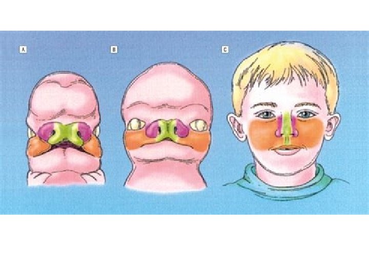

• Primitive nasal cavities are closed at their posterior ends by bucconasal membrane which ruptures and forms choanae. • Clinical significance: choanal atresia

• • • Maxillary sinus First sinus to appear Completely develops by 17 -18 years of age Sphenoid sinus , Frontal sinus Poorly developed at birth

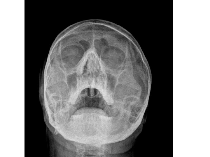
EXTERNAL NOSE • External nose is shaped like a pyramid with its root up and base directed downwards. • Consists of osteocartilagenous framework covered by muscle and skin.

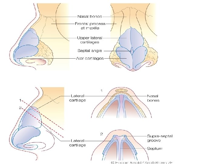

• • Osteocartilagenous framework: Upper 1/3 rd - bony Lower 2/3 rd – cartilagenous Bony framework a) Nasal bones b) Nasal processes of frontal bone c) Frontal processes of maxilla
 Upper lateral cartilages b) Lower lateral cartilages")
• • • Cartilagenous framework a) Upper lateral cartilages b) Lower lateral cartilages (alar cartilages) c) Lesser cartilages (sesamoid cartilages) d) Septal cartilage
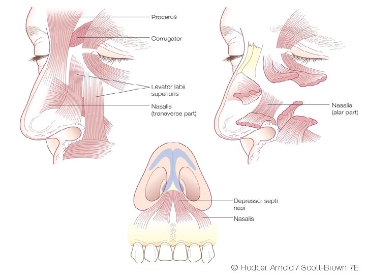
 Procerus b) Nasalis (transverse and alar part)")
• • • Nasal musculature: a) Procerus b) Nasalis (transverse and alar part) c) Levator labi superioris alaque nasi d) Anterior and posterior dialator naris e) Depressor septi

• Nasal skin: • Over nasal bone and upper lateral cartilage is thin and freely mobile • On alar cartilages is thick and adherent and contains sebaceous glan

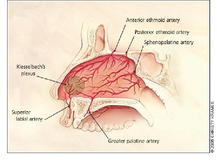
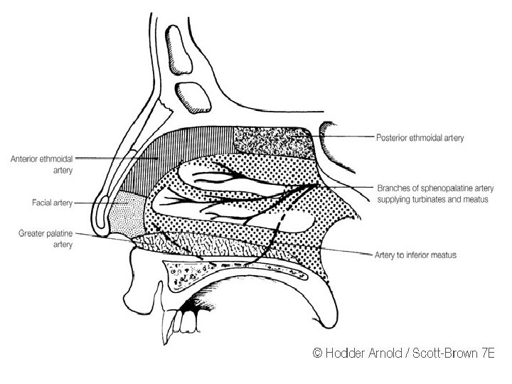
• Blood supply: facial and ophthalmic arteries and veins • Lymphatic drainage: preauricular and submandibular lymph nodes

INTERNAL NOSE • Divided into right and left nasal cavities by nasal septum. • Nasal cavity proper: bounded by lateral wall, medial wall, roof and a floor.
 Palatine process of maxilla (anterior 3/4 th")
• Floor: Formed by • a) Palatine process of maxilla (anterior 3/4 th ) • b) Horizontal process of palatine bone (posterior 1/4 th )
 Nasal bones b) Frontal bone c)")
• • • Roof: formed by a) Nasal bones b) Frontal bone c) body of sphenoid d) cribriform plate of ethmoid
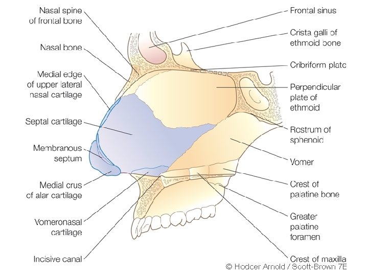
 Columellar septum • b)")
• Nasal septum consists of three parts • a) Columellar septum • b) Membranous septum (lies between columella and caudal border of septal cartilage) • c) Septum proper: consists of osteocartilagenous framework covered with nasal mucous membrane

Lateral wall of nose

• Lateral wall is marked by three bony projections called turbinates or conchae • superior (part of ethmoid), • middle (part of ethmoid), • inferior (separate bone).

• Sometimes a fourth turbinate concha suprema may also be present. • Below and lateral to each turbinate is a corresponding meatus

• Inferior meatus- nasolacrimal duct opens in its anterior part. • Middle meatus- consists of bulla ethmoidalis, hiatus semilunaris, infundibulum. • Frontal, maxillary and anterior ethmoidal sinuses open into middle meatus.

• Superior meatus- posterior ethmoidal sinuses open into it. • Sphenoethmoidal recess- triangular fossa above the superior meatus. Sphenoidal sinus opens into it.
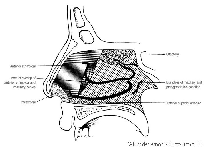

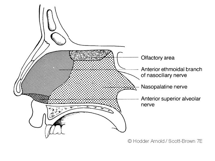
• Trigeminal nerve carries the common sensation via ophthalmic and maxillary divisions. • Special sensory (smell) carried via olfactory nerves.

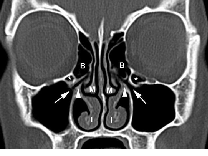
• OSTEOMEATAL COMPLEX • The middle meatus is the space below and lateral to the middle turbinate, and is often functionally referred to as the osteomeatal complex. • It contains the drainage pathways for the anterior ethmoids, the maxillary and the frontal sinuses.

• The middle meatus is the area that is most commonly involved in the pathophysiology of chronic rhinosinusitis.

• Bulla ethmoidalis • one of the most constant and largest of the anterior ethmoid air cells. • Hiatus semilunaris- hiatus semilunaris is a crescent shaped gap between the posterior free margin of the uncinate process and the anterior wall of the ethmoid bulla

• Ethmoidal infundibulum – • funnel-shaped passage through which the secretions from various anterior ethmoid cells, the maxillary sinus, and, in some cases, the frontal sinus are transported or channeled into the middle meatus.
 Largest of the paranasal sinuses Pyramidal")
• • Maxillary sinus (Antrum of Highmore) Largest of the paranasal sinuses Pyramidal in shape with base towards lateral wall of nose and apex directed into zygomatic process

• Capacity- 10 -20 ML boundaries

THANK YOU
- Slides: 40