ANATOMY OF LARYNX AND TRACHEOBRONCHIAL TREE Dr Mukesh

ANATOMY OF LARYNX AND TRACHEOBRONCHIAL TREE Dr Mukesh Singla

STRIDOR Harsh, high-pitched musical sound Produced by turbulence of airflow through a partial obstruction in the airway passage

ANATOMY OF LARYNX Situated in midline of neck from the level of C-3 to C-6 vertebrae lying in front of laryngopharynx.

LARYNX • Musculocartilagenous structure, • Lined with mucous membrane • Connected to pharynx and superior part of trachea • Acts as essential sphincter guarding entrance into trachea • Secondarily function as organ of voice • Formed of nine cartilages connected by ligaments and muscles

Topography of the extrinsic musculature of the larynx

ADAM’S APPLE At puberty male larynx increases in size rapidly, and the thyroid cartilage projects to form the Adam’s apple.
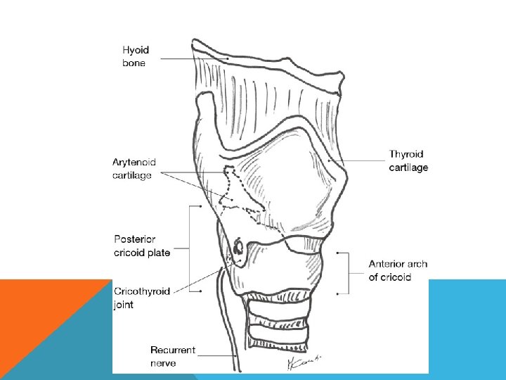

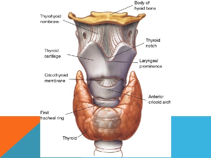
CACARTILAGEGESSOFOFR L Y N X A L Y R X N

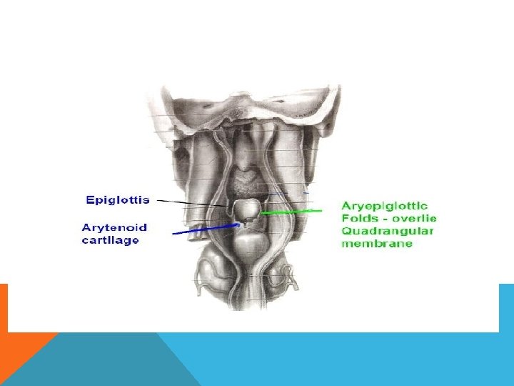
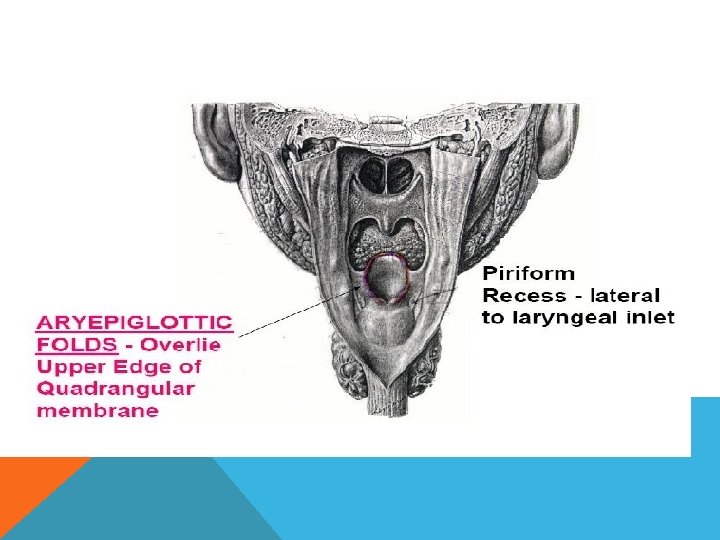
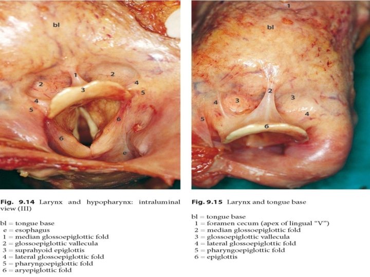
CAVITY OF LARYNX EXTENT- from laryngeal inlet above to lower border of cricoid cartilage. Inlet of larynx communicates with laryngopharynx.

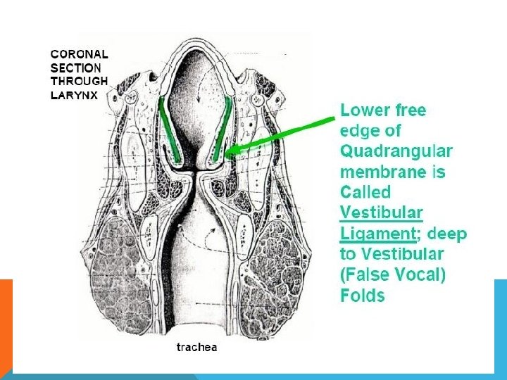
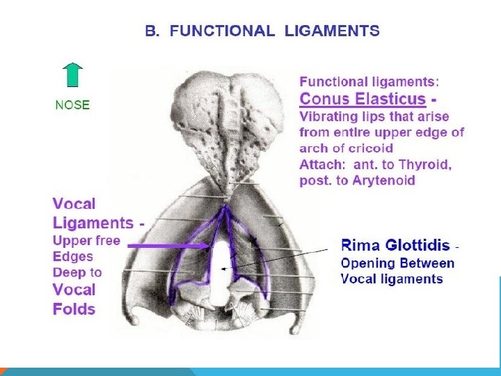
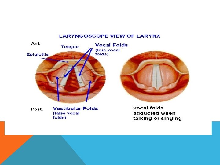
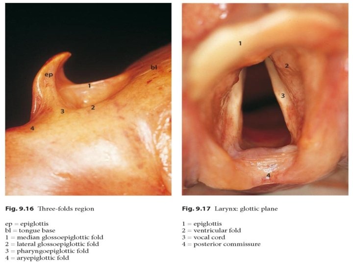
CAVITY OF LARYNX Divided into three parts by 2 folds of mucus membrane: False cords and True vocal cords Parts are Vestibule, ventricle and subglottic space Glottis is the opening between vocal folds


MUSCLES OF LARYNX- INTRINSIC

HISTOLOGY OF LARYNX Mucous membrane lines the entire larynx. Stratified squamous epithelium lines true vocal cords and upper parts of vestibule. Pseudostratified Columnar ciliated epithelium lines rest of the cavity. Mucous glands preset in all parts except on free edges of vocal cords

LYMPHATIC DRAINAGE SUPRAGLOTTIS-pre-epiglottic and upper deep cervical nodes. GLOTTIS-Lymphatics are practically absent SUBGLOTTIS-prelaryngeal and pretracheal nodes also lower deep cervical nodes

LARYNX OF AN INFANT DIFFERENT FROM ADULT Infant's larynx is positioned high in the neck opposite C 3 or C 4 (vocal cord level ) at rest and reaches C 1 or C 2 during swallowing. This high position allows the epiglottis to meet soft palate and make a nasopharyngeal channel for nasal breathing during suckling. The milk feed passes separately over the dorsum of tongue and the side of epiglottis, thus allowing breathing and feeding to go on simultaneously.

LARYNX OF AN INFANT

LARYNX OF AN INFANT DIFFERENT FROM ADULT Laryngeal cartilages are soft and collapse easily. Epiglottis is omega-shaped and arytenoids relatively large covering significant portion of the posterior glottis.

LARYNX OF AN INFANT DIFFERENT FROM ADULT Thyroid cartilage in an infant is flat. It also overlaps the cricoid cartilage and is in turn overlapped by the hyoid bone. Thus cricothyroid and thyrohyoid spaces are narrow and not easily discernible as landmarks when performing tracheostomy.

LARYNX OF AN INFANT DIFFERENT FROM ADULT Infant's larynx is small and conical. The diameter of cricoid cartilage is smaller than the size of glottis, making subglottis the narrowest part. It has a bearing in the selection of paediatric endotracheal tube. In adults, subglottic-glottic dimensions are approximately same and larynx is cylindrical.

LARYNX OF AN INFANT DIFFERENT FROM ADULT Submucosal tissues of infant's larynx are comparatively loose and easily undergo oedematous change with trauma or inflammation leading to obstruction. Infant's larynx shows two spurts in growth. In the first three years of life larynx grows in width and length, and thus obviates the need for any airway surgery in certain congenital anomalies.

LARYNX OF AN INFANT DIFFERENT FROM ADULT The second spurt in growth occurs during adolescence when the thyroid angle develops. The length of vocal cords then increases leading to voice changes associated with puberty. With growth of the neck, larynx gradually descends to adult position opposite Cs (vocal cord level).

LARYNX OF AN INFANT DIFFERENT FROM ADULT In childhood, vocal cord is 6 mm in females and 8 mm in males. It increases to 15 -19 mm in adult females and 17 -23 in adult male. Adult male voices are usually lowerpitched and have larger folds.

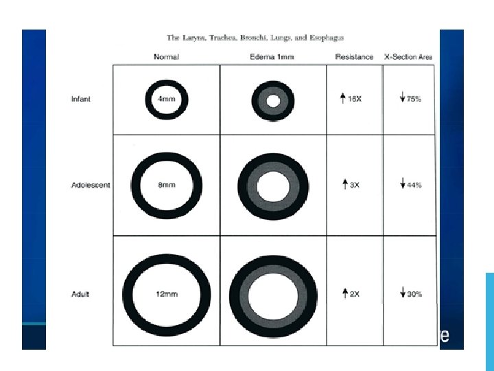
INFANT AIRWAY The subglottic diameter measures approximately 4. 5 A diameter of less then 3. 5 mm suggests a marginal subglottic airway and is consistent with subglottic stenosis

INFANT AIRWAY • Circumferential mucosal edema of 1 mm within the larynx of an infant causes a glottis to narrow by over 60% to 75%.

LOCATION OF OBSTRUCTION Stridor can be localized to discrete areas of the airway according to the nature of the sound in relationship to the phase of breathing

LOCATION OF OBSTRUCTION These discrete regions can be divided into three zones Supraglottic and supralaryngeal zone which includes the pharynx Extrathoracic tracheal zone including both glottis subglottis and cervical trachea Intrathoracic tracheal zone which includes primary and secondary bronchi

LOCATION OF OBSTRUCTION Supraglottis or Pharynx - Inspiratory and highpitched. Glottis and Subglottis (extrathoracic tracheal zone)- Biphasic of intermediate pitch Intrathoracic tracheal/bronchial zone- Expiratory often confused with wheezing

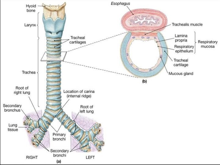
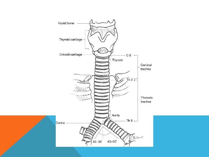
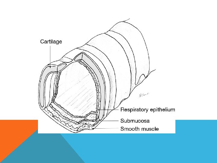
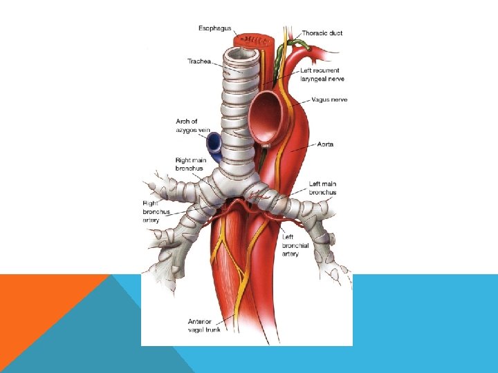
ANATOMY OF TRACHEA Tube made up of cartilage and membrane and measures approx 10 -11 cm in adult Extends from C-6 to T-5 There are 16 -20 incomplete cartilaginous rings. In neck 6 -7 rings are present.
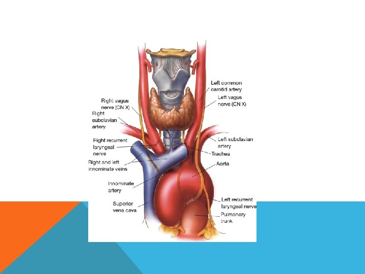

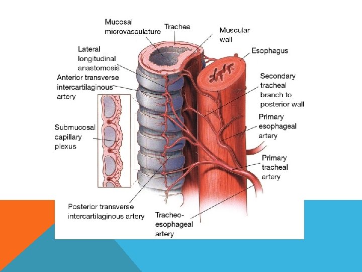
BLOOD SUPPLY The inferior thyroid vessels and their tracheoesophageal branches provide blood supply to the proximal trachea while the bronchial arteries vascularize the distal trachea, carina, and main bronchi The trachea is also supplied by small branches originating from the subclavian artery, internal mammary artery, and innominate artery.

VENOUS AND LYMPHATIC DRAINAGE Venous drainage is through the azygos and hemi azygos systems while lymphatic drainage is through the low and high paratracheal nodal chains eventually reaching the deep cervical nodes.


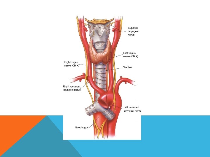
NERVE SUPPLY Vagus, recurrent laryngeal nerve and sympathetic trunk

LYMPHATICS Pretracheal and paratracheal lymph nodes
- Slides: 49