Anatomy of JEJUNUM ILEUM By Dr Manjula Vastrad

Anatomy of JEJUNUM & ILEUM By: Dr Manjula Vastrad Asst Prof Dept of Rachana Shareera SMVVS RKM AMC Vijayapura.
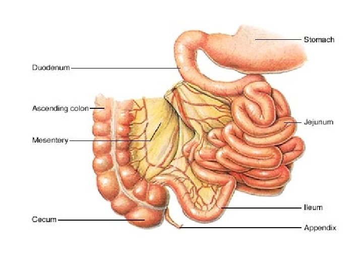

• The jejunum begins at the duodenojejunal flexure. • Together, the jejunum and ileum are 5 to 7 metres long. (avg 6 m) • The jejunum and ileum are the greatly coiled parts of the small intestine. • They are covered to varying extents by the greater omentum. • Although there is no clear line of demarcation, the character of the intestine gradually changes.

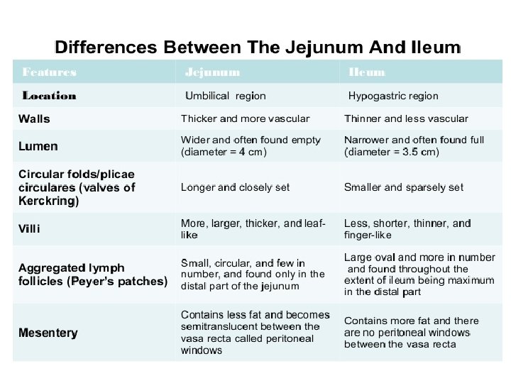
The jejunum • The jejunum is middle part of the small intestine in humans and most higher vertebrates. • It lies between duodenumand the ileum. • The jejunum arises from the duodenojejunal flexure.

Ileum • Part of the small intestine beyond the jejunum and up to to the ileocecal valve i. e. before the colon(large intestine). • The wall itself is made up of folds, each of which has many tiny finger-like projections known as villi on its surface. – The function of the ileum is mainly to absorb vitamin B 12 and bile salts and whatever products of digestion were not absorbed by the jejunum.

STRUCTURE OF SMALL INTESTINE: • The wall structure of the small intestine includes four layers. – From inside to outside, these are: • Mucosa • Submucosa • Muscle layer • Serosa

The jejunum and ileum are attached to the posterior abdominal wall by the root of the mesentery (about 15 cm long) is directed obliquely. • It extends from the duodenojejunal junction on the left side of vertebra L 2 to the ileocolic junction and the right sacroiliac joint. • Between these two points, the root of the mesentery crosses: 1) The horizontal (3 rd & 4 rh ) parts of the duodenum 2) Aorta 3) Inferior vena cava 4) Psoas major muscle 5) Right ureter 6) Right testicular (or ovarian) vessels

Blood Supply of the Jejunum and Ileum • The arterial supply to the jejunoileum is from the superior mesenteric artery. • The superior mesenteric artery arises from the aorta at the level of the L 1 vertebrae, immediately inferior to the coeliac trunk. • It moves in between layers of mesentery, splitting into approximately 20 branches. • These branches anastomose to form loops, called arcades. From the arcades, long and straight arteries arise, called vasa recta.

Venous Drainage of the Jejunum and Ileum • The superior mesenteric vein drains the jejunum and ileum. • This accompanies the superior mesenteric artery, lying anterior and to its right in the root of the mesentery. • This vein unites with the splenic vein to form the portal vein

Lymphatic Drainage of the Jejunum and Ileum • The lacteals in the intestinal villi empty their milk-like fluid (L. lactis, milk) into a plexus of lymph vessels in the walls of the jejunum and ileum.

Innervation of the Jejunum and Ileum • The innervation is through the superior mesenteric plexus extensions along the arteries. • The sympathetic supply is from the greater splanchnic and lesser splanchnic nerves. • The parasympathetic supply is from the posterior vagal trunk via the coeliac plexus.

CLINICAL ANATOMY : vpeptic ulcer v. Trauma to the Jejunum and Ileum v. Tumors and Cysts of the Mesentery of the Small Intestine v. Mesenteric Arterial Occlusion v. Mesenteric Vein Thrombosis v. Strangulation of a coil of small intestine in an inguinal hernia

- Slides: 14