Anatomy of Inner Ear Audition Cochlea detects sound


")


, head acceleration (vestibular) Stereocilia")
![Endolymph of inner ear high [K+], positively charged fluid endolymph Fox Figure 10. 13](https://slidetodoc.com/presentation_image_h2/963dd6ac5c5c07d2ec32db4834ab5532/image-13.jpg "Endolymph of inner ear high [K+], positively charged fluid endolymph Fox Figure 10. 13")


![Endolymph of inner ear has high [K+] K+ in hair cells works like Na+](https://slidetodoc.com/presentation_image_h2/963dd6ac5c5c07d2ec32db4834ab5532/image-18.jpg "Endolymph of inner ear has high [K+] K+ in hair cells works like Na+")


![Basilar membrane vibration bends hair cells cochlear duct with Endolymph [K+] Perilymph Vibrates see](https://slidetodoc.com/presentation_image_h2/963dd6ac5c5c07d2ec32db4834ab5532/image-26.jpg "Basilar membrane vibration bends hair cells cochlear duct with Endolymph [K+] Perilymph Vibrates see")


")


- Slides: 50
Anatomy of Inner Ear Audition Cochlea detects sound waves Hair Cells Receptor cells for audition and vestibular system. Inner ear connected by ducts filled with endolymph, a high [K+] fluid T
Figure 10. 12 Vestibulocochlear cranial nerve 8
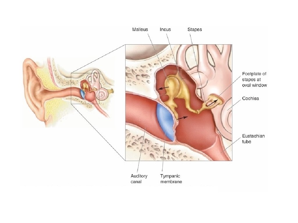
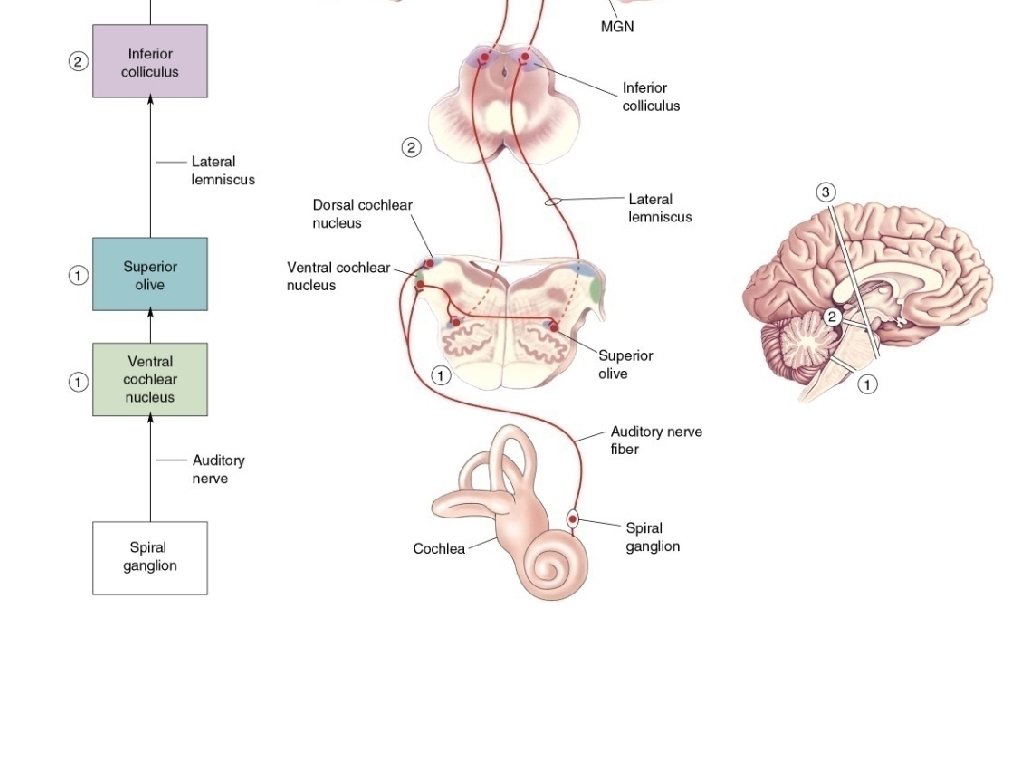
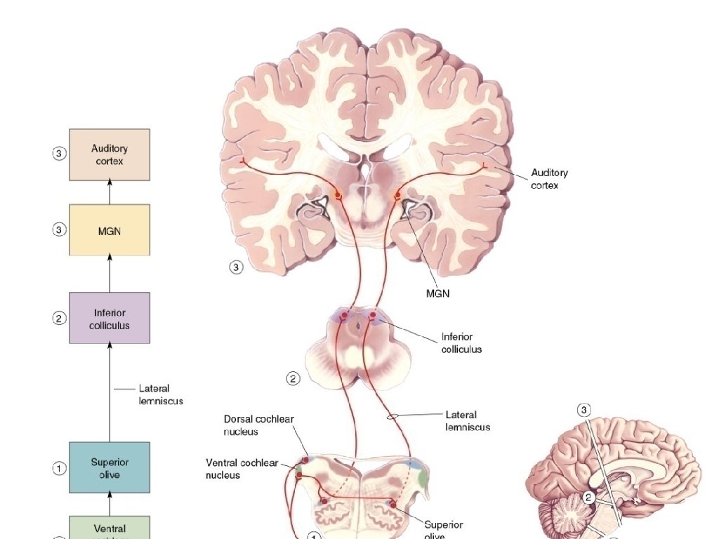
Properties of Auditory System 1. Capture sound using outer ear (auricle, external auditory meatus) to the middle ear via tympanic membrane and middle ear bones (malleus, incus, stapes): transfer & amplify vibration to the oval window 2. Transmit to receptors: vibration of basilar membrane that runs length of cochlea. Response of basilar membrane varies across its length. Low frequency sound vibrates apex of cochlea. High frequency sound vibrates base of cochlea. 3. Transduction of sound frequency into spatial location (tonotopy) to stimulate auditory nerve fibers (Cranial Nerve 8: vestibulocochlear nerve). 4. Receptive Field of Auditory Neuron: tuned to characteristic frequency. Neuron’s response (rate of action potentials) reflects intensity of sound at characteristic frequency. 5. Cranial Nerve 8 projects to cochlear nucleus in the brainstem. T
The Nature of Sound • Audible variations in air pressure – Sound frequency: Number of cycles per second expressed in units called hertz (Hz) – Cycle: Distance between successive compressed patches
The Nature of Sound – Range: 20 Hz to 20, 000 Hz – Pitch: High pitch = high frequency; low frequency = low pitch – Intensity: High intensity louder than low intensity
6
Figure 10. 19 8
Cochlea & Sound Detection Vestibulocochlear Cranial Nerve 8 = “snail” Fox Figure 10. 12
Figure 10. 20 10
Basilar membrane to hair cells
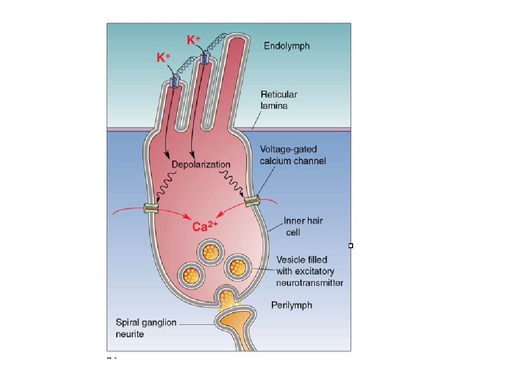
Hair Cells of inner ear Mechanoreceptors that detect vibration (audition), head acceleration (vestibular) Stereocilia -- tufted projections that stick into endolymph and gelatinous tectoral membrane and bend with vibration. Bending of stereocilia causes change in membrane potential, and regulates release of neurotransmitter onto afferent nerve depolarization -> more transmitter release -> more Action Potentials hyperpolarization -> less transmitter release -> fewer Action Potentials Bending of stereocilia opens K+ channels. Because endolymph is high in K+, K+ rushes into hair cell to cause depolarization. T
Endolymph of inner ear high [K+], positively charged fluid endolymph Fox Figure 10. 13
Hair cell Stereocilia -- tufted projections that stick into gelatinous tectoral membrane and bend with vibration
Figure 10. 14
Vibration bends hair cells
Deflection of stereocilia opens K+ channels K+ rushes into hair cell, causing depolarization
Endolymph of inner ear has high [K+] K+ in hair cells works like Na+ in neurons [K+] = 157 m. M [Na+] = 1. 3 m. M
Bending of stereocilia causes change in membrane potential K+ channels opened more transmitter release onto sensory nerve K+ channels closed less transmitter release onto sensory nerve
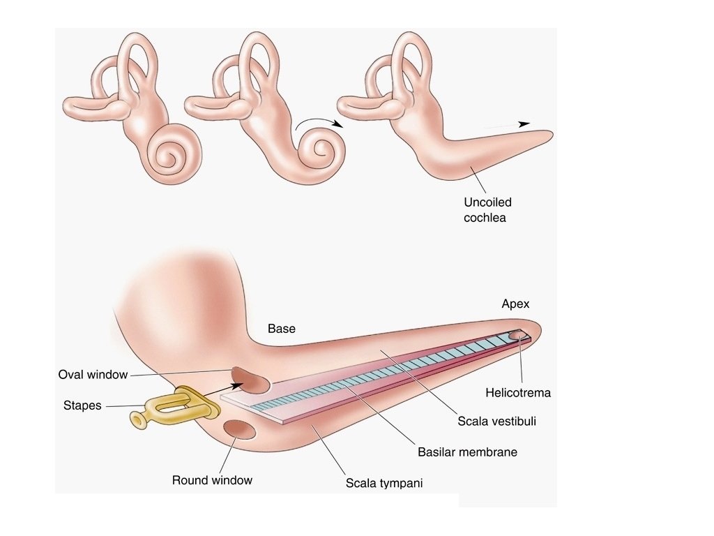
Vibration of Basilar Membrane Vibrations of oval window -> vibrations in endolymph -> vibration of basilar membrane. Response of basilar membrane varies across its length. Low frequency sound vibrates apex of cochlea (basilar membrane thicker). High frequency sound vibrates base of cochlea (basilar membrane thinner). Transduce sound frequency into spatial location.
Figure 10. 20 22
s e t a r b i v , k c i th : x e y p c a n e u q e r f w at lo t a s e t a r ib v , n i h t : base y c n e u q e high fr
Figure 10. 21 25
Basilar membrane vibration bends hair cells cochlear duct with Endolymph [K+] Perilymph Vibrates see Fox Figure 10. 22
Hair cells
Deflection of stereocilia opens K+ channels
Bending of stereocilia causes change in membrane potential more transmitter release onto auditory nerve less transmitter release onto auditory nerve
Bending of stereocilia causes change in membrane potential
Hair cells are tuned by position in cochlea base apex
Frequencies Map on to Extent of Cochlea: High frequencies at base, low frequencies at apex. Fox Figure 10. 23
• Stimulus Frequency – Tonotopic maps on the basilar membrane – 20 - 20, 000 Hz range, 0. 3% discrimination outside of skull in brainstem
Somatosensory receptive fields of spinal sensory nerves
• Stimulus Frequency – Tonotopic maps on the basilar membrane – 20 - 20, 000 Hz range, 0. 3% discrimination outside of skull in brainstem
Each Auditory Neuron responds best to characteristic frequency (position of hair cells in cochlea = receptive field of neuron)
Auditory Nerve encodes intensity of sound (increased rate of action potentials = louder sound) Number of spikes per sec
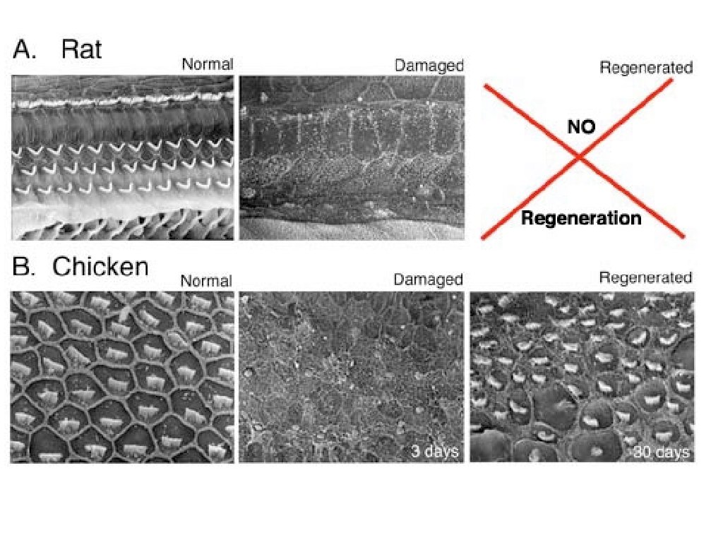
Damage to Inner Ear Hair Cells In mammals, hair cells die with old age (e. g. hi frequency hair cells) or after damage due to high intensity noise. Hair cells do not regenerate (although they can regenerate in lower animals). Cochlear Implants reproduce function of basilar membrane and hair cells: stimulate auditory nerve endings at appropriate point in cochlea to reproduce tonotopic mapping of missing hair cells. Example: sound of voice, music if only a small number of frequencies are restored. T
Map frequencies to electrode location in cochlea
http: //graemeclarkfoundation. org/bionic_ear/CI%20 Function. ht m
Cochlear implant to cure deafness by directly stimulating auditory nerves cochlear implant -> 32 different frequencies (normal inner ear -> 300+ different frequencies http: //graemeclarkfoundation. org/bionic_ear/CI%20 Function. ht m
Central Representation of Audition Auditory input projects to mostly contralateral auditory cortex and some ipsilateral auditory cortex. Tonotopy is preserved in auditory cortex: cortical neurons respond to characteristic frequencies, with mapping from low to high frequency across the auditory cortex. Auditory cortex neurons send projections to higher levels of cortex that extract features from sound: Wernicke’s Area -- extracts meaning from words, integrate with vision Broca’s Area -- generates speech via projections to motor cortex (to move the tongue lips, & throat). Mc. Gurk Effect: example of sound integration with visual information to change perception of syllable. T
Mechanisms of Sound Localization • Techniques for Sound Localization – Horizontal: Left-right, Vertical: Up-down • Localization of Sound in Horizontal Plane – Interaural time delay: Time taken for sound to reach from ear to ear – Interaural intensity difference: Sound at high frequency from one side of ear – Duplex theory of sound localization: • Interaural time delay: 20 -2000 Hz • Interaural intensity difference: 2000 -20000 Hz
• Interaural time delay and interaural intensity difference 47
Auditory Localization 1. Compare delay times from each ear 2. Calculate doppler and echo (bats)
Auditory Cortex Inner surface of Temporal Lobe Fox Figure 10. 25
Feature Extraction and Integration of Auditory Input Integration with vision, word extraction, speech generation Fox Figure 8. 14