ANATOMY OF HIP BONE Dr Luqman Asghar Lecturer

ANATOMY OF HIP BONE Dr. Luqman Asghar Lecturer Anatomy

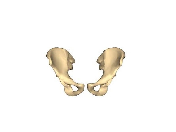
SIDE DETERMINATION • The upper and lower parts of the bone are expanded while the middle portion is constricted • The expanded portion with foramen is inferior • The constriction is marked by a deep hollow(acetabulum) which faces laterally

NORMAL ANATOMICAL POSITION • Pubic tubercle and anterior superior iliac spine lie in the same coronal plane • The symphyseal surface of the pubis lie anteriorly in the median plane • Upper border of the pubic symphysis and ischial spine lie in the same horizontal plane

PARTS OF HIP BONE • ilium • ischium • pubis

Two ends • upper • lower Three borders • anterior • Posterior • Medial Three surfaces • Gluteal • Pelvic • sacropelvic ILIUM

UPPER END • Known as iliac crest • convex upwards in vertical plane • concave inwards anteriorly and convex inwards posteriorly in horizontal plane • highest point of iliac crest is at the level of interval between 3 rd and 4 th lumbar spines • anterior superior iliac spine is anterior end of iliac crest • posterior superior iliac spine is posterior end of iliac crest

Morphologic division of the iliac crest Iliac crest is divided into a long ventral segment and a short dorsal segment. The ventral segment: • anterior two-thirds of the crest. • outer lip • an inner lip • intermediate area • tubercle of the iliac crest

Dorsal segment The dorsal segment forms less than the posterior one-third of the crest. • lateral slope • medial slope • intermediate ridge

BORDERS OF ILIUM Anterior border: • Starts at the anterior superior iliac spine(felt in living) and runs downwards to the acetabulum • The upper part of the border presents a notch • Lower part shows an elevated area called the anterior inferior iliac spine.

posterior border • Extends from the posterior superior iliac spine to the upper end of the posterior border of the ischium • Few centimeters below the posterior superior iliac spine it presents another prominence called the posterior inferior iliac spine • Still lower down the posterior border is marked by a large deep notch called the greater sciatic notch

Medial border • It extends on the inner or pelvic surface of the ilium from the iliac crest to the iliopubic eminence • It separates the iliac fossa from the sacropelvic surface • Its lower rounded part forms the iliac parts of the arcuate line or inlet of pelvis.

SURFACES OF ILIUM Gluteal surface outer surface of the ilium, which is convex in front and concave behind, like the iliac crest It is divided into four areas by three gluteal lines • posterior gluteal line (shortest) begins 5 cm in front of the posterior superior spine, and ends just in front of the posterior inferior spine • The anterior gluteal line (longest) begins about 2. 5 cm behind the anterior superior spine, runs backwards and downwards to end at the middle of the upper border of the greater sciatic notch • The inferior gluteal line (most ill-defined) begins a little above and behind the anterior inferior spine, runs backwards and downwards to end near the apex of the greater sciatic notch.
 Large concave area on the inner surface of the ilium Situated")
Iliac surface(iliac fossa) Large concave area on the inner surface of the ilium Situated in front of its medial border Forms the lateral wall of the false pelvis Sacropelvic surface Divided into • iliac tuberosity • Auricular surface • Pelvic surface

Iliac tuberosity • Upper, large, roughened area, lying just below the dorsal segment of the iliac crest • Raised in the middle and depressed both above and below Auricular surface • Auricular surface is articular • It lies anteroinferior to the iliac tuberosity • It articulates with the sacrum to form the sacroiliac joint Pelvic surface • Smooth and lies anteroinferior to the auricular surface • Forms a part of the lateral wall of the true pelvis • Along the upper border of the greater sciatic notch, this surface is marked by the preauricular sulcus(females=deeper)
 Anterior superior iliac spine: • lateral end of the inguinal")
ATTACHMENTS ILIAC CREST(ventral segment) Anterior superior iliac spine: • lateral end of the inguinal ligament • sartorius muscle Outer lip: origins • tensor fasciae latae in front of the tubercle • latis-simus dorsi just behind the highest point of the crest. Insertions External oblique in its anterior two-thirds Attachments fascia lata in its whole extent

inner lip Origins • transversus abdominis in its anterior two-thirds • quadratus lumborum in its posterior one-third Attachments • fascia transversalis in its anterior two-thirds • fascia iliaca in its anterior two-thirds • thoracolumbar fascia around quadratus lumborum intermediate area Origin internal oblique muscle in its anterior two-thirds
 Lateral slope Origin • gluteus maximus medial slope Origin • erector")
ILIAC CREST(dorsal segment) Lateral slope Origin • gluteus maximus medial slope Origin • erector spinae Attachments • interosseous sacroiliac ligament • dorsal sacroiliac ligaments (these ligamnts are deep to attachmentof the erector spinae)

Attachments Of The Iliac Crest
 Attachments")
Anterior inferior iliac spine Origin • straight head of the rectus femoris(upper half) Attachments • Ilio-femoral ligament(rough lower part) posterior border Origin • few fibres of the piriformis from the upper margin of the greater sciatic notch attachments • to the upper fibres of the sacrotuberous ligament above the greater sciatic notch

Gluteal surface behind the posterior gluteal line Origin • upper fibres of the gluteus maximus area between anterior and posterior gluteal lines Origin • Gluteus medius area between the anterior and inferior gluteal lines Origin • Gluteus minimus Below the inferior gluteal line origin • Reflected head of the rectus femoris

Attachments Of The Gluteal Region

ILIAC FOSSA upper two-thirds Origin • Iliacus lower grooved part • covered by the iliac bursa

SACRO-PELVIC SURFACE iliac tuberosity Attachments • the interosseous sacroiliac ligament in its greater part • the dorsal sacroiliac ligament posteriorly • the iliolumbar ligament superiorly auricular surface Attachments • convex margin gives attachment to the ventral sacroiliac ligament

Pelvic surface preauricular sulcus attachment lower fibres of the ventral sacroiliac ligament. lateral to preauricular sulcus Origin few fibres of the piriformis Rest of pelvic surface Origin upper half of the obturator internus

Attachments Of Ligaments

ISCHIUM • The ischium forms the posteroinferior part of the hip bone, and the adjoining two-fifths of the acetabulum. • It forms the posterior boundary of the obturator foramen. PARTS • Body • Ramus

Body of the Ischium This is a thick and massive mass of bone that lies below and behind the acetabulum Two ends, • Upper(forms postero-inferior two fifth of the acetabulum) • Lower(forms ischial tuberosity) Three borders • Anterior • posterior • Lateral • Three surfaces • Femoral • Dorsal • pelvic

BORDERS OF ISCHIAL BODY anterior border • forms the posterior margin of the obturator foramen. posterior border • continuous above with the posterior border of the ilium • Below, it ends at the upper end of the ischial tuberosity • It forms part of the lower border of ilium • It also forms part of the lower border of the greater sciatic notch • Below the greater sciatic notch the posterior margin shows a projection called the ischial spine • Below the spine the posterior border shows a concavity called the lesser sciatic notch. lateral border forms the lateral margin of the ischial tuberosity, except at the upper end where it is rounded.

SURFACES OF ISCHIAL BODY Femoral surface • lies between the anterior and lateral borders. Dorsal surface • continuous above with the gluteal surface of the ilium Three parts • Upper convex surface adjoining the acetabulum • Middle wide shallow groove • Lower the upper part of the ischial tuberosity Pubic surface • Forms lateral boundary of the lower part of true pelvis

ISCHIAL TUBEROSITY The ischial tuberosity is divided by a transverse ridge into an upper and a lower area Upper area subdivided by an oblique ridge into a • superolateral area • inferomedial area Lower area subdivided by a longitudinal ridge into an • Outer area • inner area
")
Ischial tuberosity(left hip bone)

Conjoined Ischiopubic Rami The inferior ramus of the pubis unites with the ramus of the ischium on the medial side of the obturator foramen The site of union may be marked by a localized thickening. The conjoined rami have Two borders • Upper forms part of the margin of the obturator foramen • Lower Forms part of the pubic arch Two surfaces • outer • inner. The inner surface is convex and smooth (It is divided into three areas, upper, middle and lower, by two ridges)

ATTACHMENTS OF ISCHIUM ischial spine Origin • posterior fibres of the levator ani from its pelvic surface Attachments • sacrospinous ligament along its margins

Lesser Sciatic Notch occupied by the tendon of the obturator internus There is a bursa deep to the tendon The notch is lined by hyaline cartilage. Two margins Upper margin Origin • Gamellus superior Lower margin Origin • Gamellus inferior

Femoral surface Origins • obturator externus along the margin of the obturator foramen • quadratus femoris along the lateral border of the upper part of the ischial tuberosity
 superolateral area Origin • Semimembranosus Inferomedial area Origin • Semitendinosus •")
Gluteal tuberosity(Ischial tuberosity) superolateral area Origin • Semimembranosus Inferomedial area Origin • Semitendinosus • Long head of the biceps femoris outer lower area Origins • adductor magnus • inner lower area covered with fibrofatty tissue(supports body weight in the sitting position) Attachments of ischial tuberosity • The sharp medial margin of the tuberosity gives attachment to the sacrotuberous ligament. • The lateral border of the ischial tuberosity provides attachment to the ischiofemoral ligament, just below the acetabulum.

Attachments of ischial tuberosity
 upper border Attachment • obturator membrane. Lower")
pelvic surface Origin • obturator internus(greater part) upper border Attachment • obturator membrane. Lower border Attachment • Fascia lata • Membranous layer of superficial fascia or Colles' fascia of the perineum

ISCHIO-PUBIC RAMI upper border Attachment • obturator membrane. Lower border Attachment • Fascia lata • Membranous layer of superficial fascia or Colles' fascia of the perineum

outer surface origins • obturator externus, near the obturator margin of both rami • adductor brevis, chiefly from the pubic ramus • gracilis, chiefly from the pubic ramus • adductor magnus, chiefly from the ischial ramus

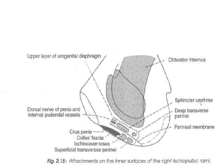
Inner surface upper ridge Attachments • upper layer of the urogenital diaphragm lower ridge Attachments • perineal membrane Upper area Origin • obturator internus Middle area Origin • Sphincter urethrae • Deep transverse perinei

Lower area Origins • Ischiacavernosus • Superficial transverse perinei

PUBIS • It forms the anteroinferior part of the hip bone and the anterior one-fifth of the acetabulum • It forms the anterior boundary of the obturator foramen Parts • a body anteriorly • superior ramus supero-laterally • inferior ramus infero-laterally

Body of Pubis This is flattened from before backwards Parts • Superior border called the pubic crest • Pubic tubercle at the lateral end of the pubic crest(important landmark) • Three surfaces(anterior, posterior and medial) Anterior: directed downwards, forwards and laterally, rough supero-medially and smooth elsewhere. Posterior: directed upwards backwards, smooth, anterior wall of true pelvis Medial: symphyseal surface

Superior Ramus It extends from the body of the pubis to the acetabulum, above the obturator foramen Borders • Superior border(pectineal line or pecten pubis) Sharp crest extending from just behind the pubic tubercle to the posterior part of the iliopubic eminence With the pubic crest it forms the pubic part of the arcuate line. • Anterior border(obturator crest) Rounded ridge, extending from the pubic tubercle to the acetabular notch • Inferior border Upper margin of the obturator foramen

Surfaces Pectineal surface Triangular area between the anterior and superior borders, extending from the pubic tubercle to the iliopubic eminence. Pelvic surface Between the superior and inferior borders. It is smooth and is continuous with the pelvic surface of the body of the pubis. Obturator surface Between the anterior and inferior borders. It presents the obturator groove.

Inferior Ramus • It extends from the body of the pubis to the ramus of the ischium, medial to the obturator foramen • It unites with the ramus of the ischium to form the conjoined ischiopubic rami.

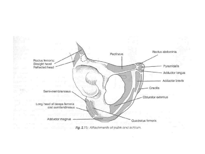
Attachments Of Pubis Pubic tubercle • Attachment to the medial end of the inguinal ligament • Ascending loops of the cremaster muscle. Pubic crest Origins • Lateral head of the rectus abdominis • Pyramidalis

BODY Anterior surface Origins • Adductor longus • Gracilis • Adductor brevis • obturator externus Attachment • anterior pubic ligament medially

Attachments

Posterior surface Origins • Levator ani • Obturator internus Attachments • Puboprostatic ligaments
 Attachments • Conjoint tendon •")
Pectineal line Origins • Pectineus • Psoas minor(when present) Attachments • Conjoint tendon • Lacunar ligament • Pectinate ligament(along whole length)

Pectineal surface Origin pectineus Pelvic surface The pelvic surface is crossed by the ductus deferens in males, and the round ligament of the uterus in females. Obturator groove Transmits the obturator vessels and nerve

THANK YOU
- Slides: 57