
Anatomy of female genital tract SUPPORTS OF UTERUS



- Slides: 99
Anatomy of female genital tract
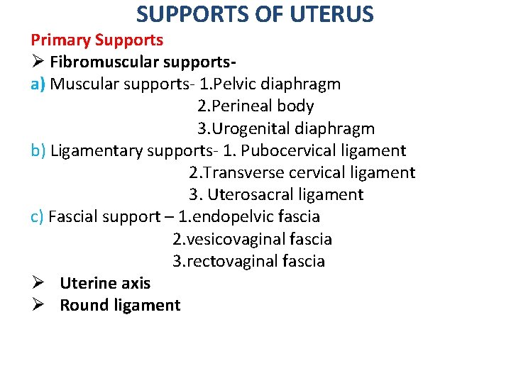
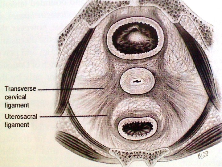
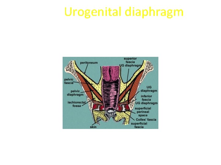
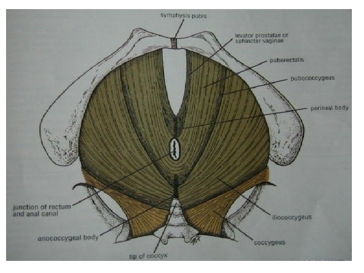
SUPPORTS OF UTERUS Primary Supports Ø Fibromuscular supportsa) Muscular supports- 1. Pelvic diaphragm 2. Perineal body 3. Urogenital diaphragm b) Ligamentary supports- 1. Pubocervical ligament 2. Transverse cervical ligament 3. Uterosacral ligament c) Fascial support – 1. endopelvic fascia 2. vesicovaginal fascia 3. rectovaginal fascia Ø Uterine axis Ø Round ligament
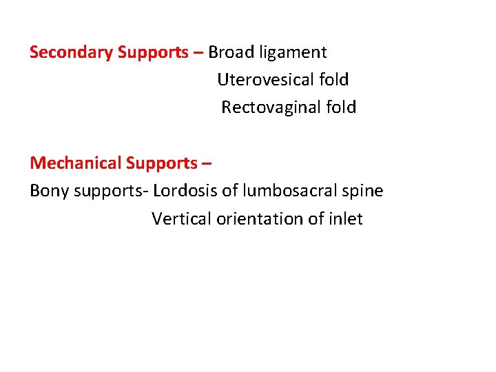
Secondary Supports – Broad ligament Uterovesical fold Rectovaginal fold Mechanical Supports – Bony supports- Lordosis of lumbosacral spine Vertical orientation of inlet
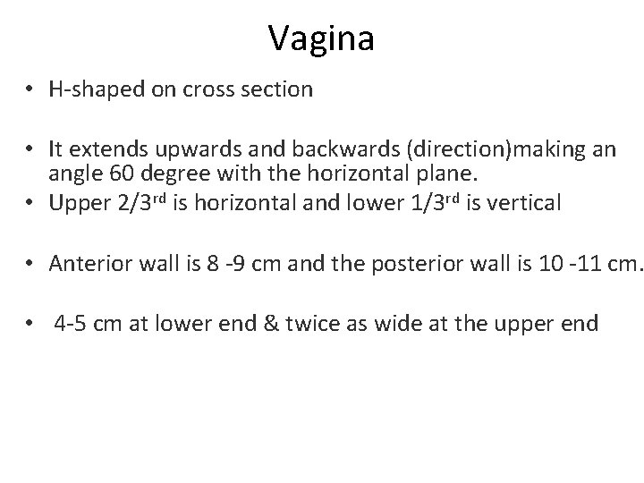
Vagina • H-shaped on cross section • It extends upwards and backwards (direction)making an angle 60 degree with the horizontal plane. • Upper 2/3 rd is horizontal and lower 1/3 rd is vertical • Anterior wall is 8 -9 cm and the posterior wall is 10 -11 cm. • 4 -5 cm at lower end & twice as wide at the upper end
Pericervical ring
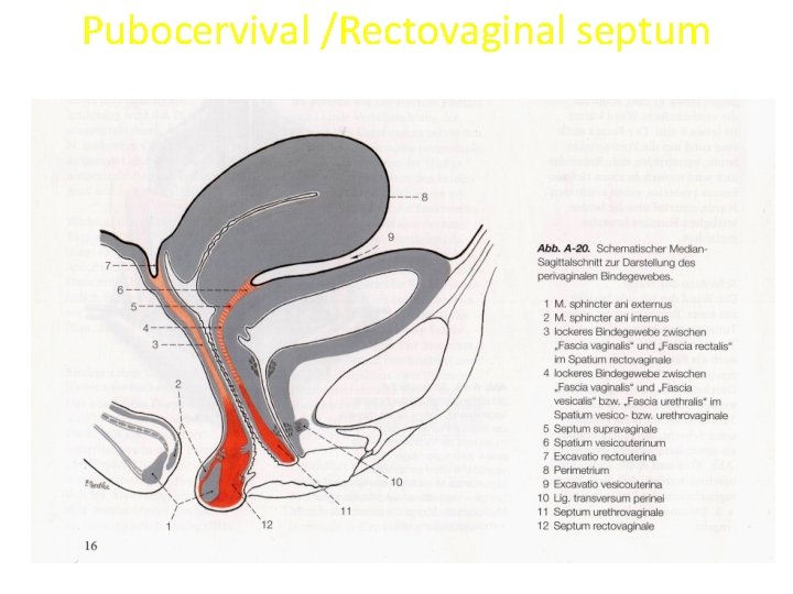
Pubocervival /Rectovaginal septum
Urogenital diaphragm
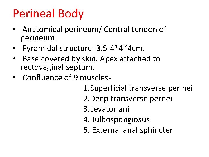
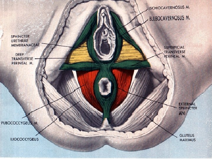
Perineal Body • Anatomical perineum/ Central tendon of perineum. • Pyramidal structure. 3. 5 -4*4*4 cm. • Base covered by skin. Apex attached to rectovaginal septum. • Confluence of 9 muscles 1. Superficial transverse perinei 2. Deep transverse pernei 3. Levator ani 4. Bulbospongiosus 5. External anal sphincter
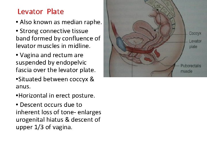
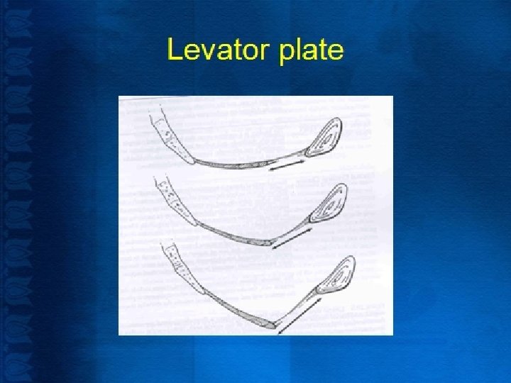
Levator Plate • Also known as median raphe. • Strong connective tissue band formed by confluence of levator muscles in midline. • Vagina and rectum are suspended by endopelvic fascia over the levator plate. • Situated between coccyx & anus. • Horizontal in erect posture. • Descent occurs due to inherent loss of tone- enlarges urogenital hiatus & descent of upper 1/3 of vagina.
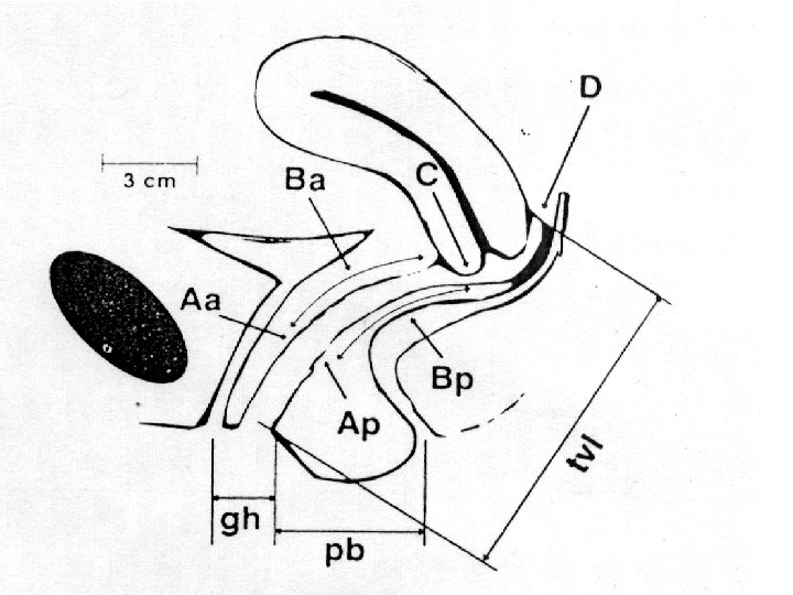
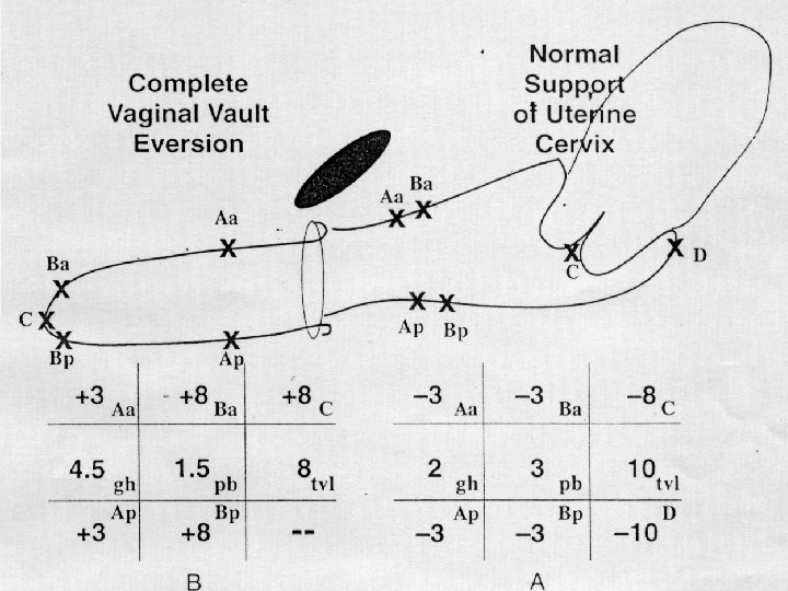
POP -Q • Approved by the International Continence Society, the American Urogynecologic Society, and the Society of Gynecologic Surgeons for the description of female pelvic organ prolapse • Ordinal staging system created to make comparative analyses and clinical communications more practical
• Reduce the prolapse and mark Aa (3 cm from external urinary meatus) and Ap (3 cm from hymen on the posterior vaginal wall) points, measure TVL(forchette to posterior fornix) , GH (urethra to forchette) and PB (forchette to midpoint of anus) • At maximal excursion mark Ba, Bp, C and D points, now measure all 6 points from hymen
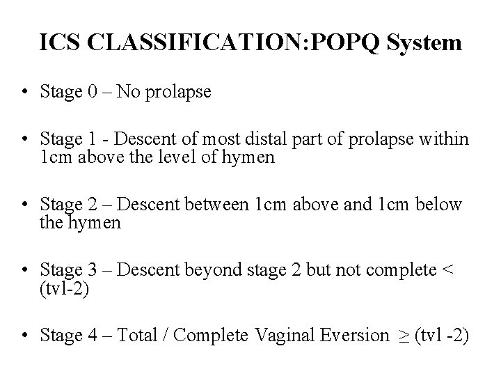
ICS CLASSIFICATION: POPQ System • Stage 0 – No prolapse • Stage 1 - Descent of most distal part of prolapse within 1 cm above the level of hymen • Stage 2 – Descent between 1 cm above and 1 cm below the hymen • Stage 3 – Descent beyond stage 2 but not complete < (tvl-2) • Stage 4 – Total / Complete Vaginal Eversion ≥ (tvl -2)
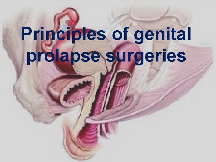
Principles of genital prolapse surgeries
General prolapse • All three supports are weak – • Ligaments – sling/mesh/SSF • Fascia – native tissue repair, attachment, mesh • Muscular - perineorrhaphy
UV prolapse • • 1 st and 2 nd degree – Ligaments are strong Manchester operation 3 rd and 4 th degree – Ligaments are also weak Sling/mesh/SSF
Congenital/Nulliparous prolapse • Severe weakness of connective tissue – congenital prolapse • Moderate weakness – Nulliparous prolapse • Mild weakness – Nulliparous prolapse after easy child birth
Case 1 • Nulliparous prolapse • 28 yr old P 1 L 1 comes with mass per vagina • O/E – III Uterine descent without the descent of vaginal wall
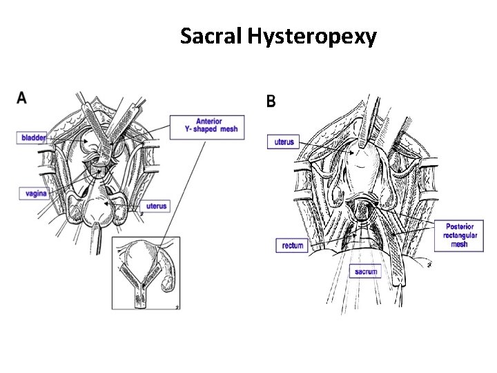
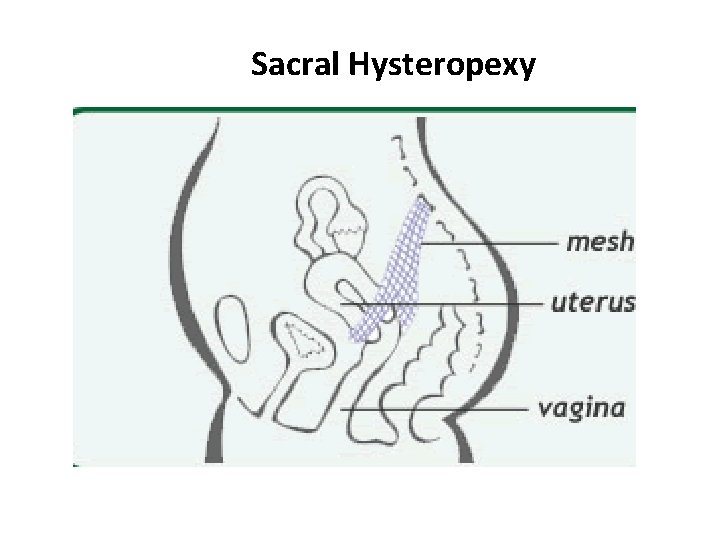
Defect – Ligamentary support is weak Choice of surgery – • Sacrohysteropexy – fixing the uterus to the anterior longitudinal ligament at S 2 S 3 level (simulating Uterosacrals)
Sacral Hysteropexy
Sacral Hysteropexy
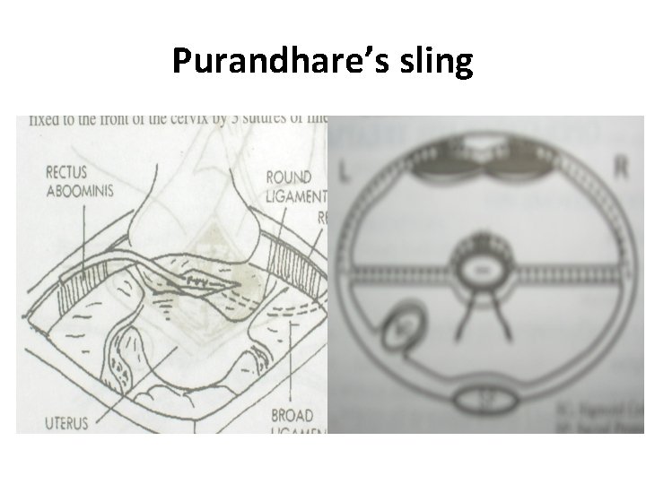
Sling surgeries • Modified Purandare – fixing the uterus to the anterior rectus sheath – dynamic sling – works only if tone of the rectus muscle is good
Purandhare’s sling
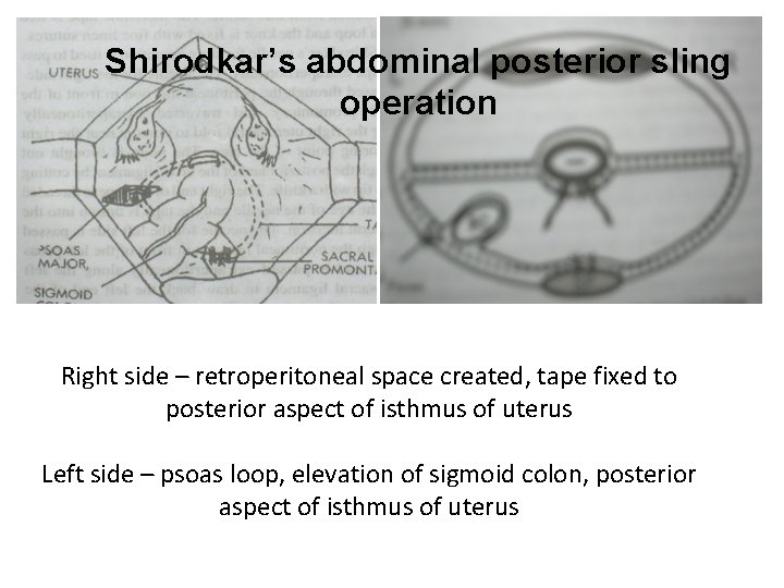
• Shirodkar – uterus fixed to anterior longitudinal ligament at S 2 S 3 level (simulating Uterosacrals) (static sling)
Shirodkar’s abdominal posterior sling operation Right side – retroperitoneal space created, tape fixed to posterior aspect of isthmus of uterus Left side – psoas loop, elevation of sigmoid colon, posterior aspect of isthmus of uterus
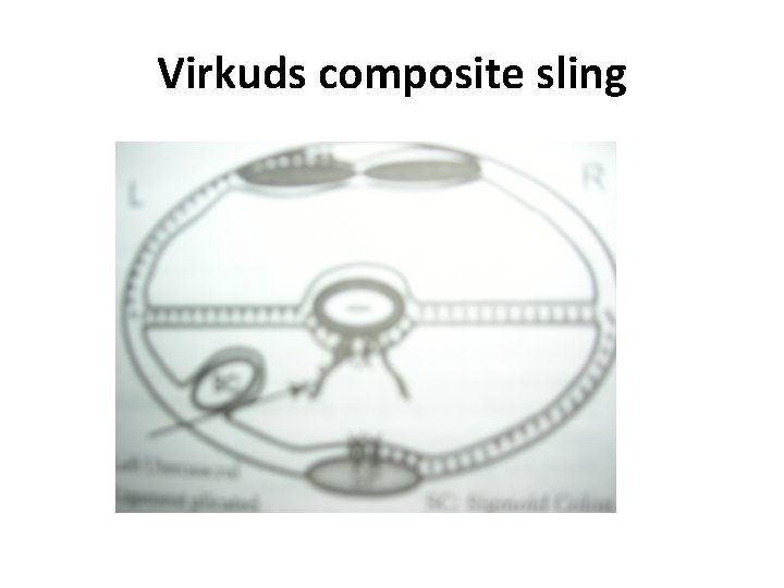
• Virkud - uterus fixed to anterior longitudinal ligament at S 2 S 3 level on right side and to the anterior rectus sheath on left side (static and dynamic sling) • Khanna – Uterus fixed to anterior superior iliac spine (static sling)
Virkuds composite sling
Case 2 • General prolapse • 23 yr old nulliparous lady comes with mass per vagina • O/E – III degree uterine descent without supravaginal elongation and cystocele, enterocele, rectocele
Defect – ligamentary and fascial support weakness Surgery – • Sling surgeries with site specific repair of fascial defects • Extended Manchester – cervix not amputated , uterus is anteflexed by fixing the Mackenrodts and uterosacrals anteriorly, conventional anterior and posterior colporrhaphy
Case 3 • Uterovaginal prolapse • A 28 yr old P 2 L 2 comes with mass per vagina • O/E – III degree uterine descent with supravaginal elongation, cystocele enterocele and deficient perineum
Defect – All three supports ligamentary, fascial and muscular support Surgery – • Modified Fothergill’s - cervix amputated , uterus is anteflexed by fixing the Mackenrodts and uterosacrals anteriorly, conventional anterior and posterior colpoperineorrhaphy • Sling surgeries with site specific repair of fascial defects
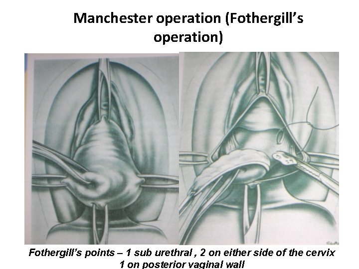
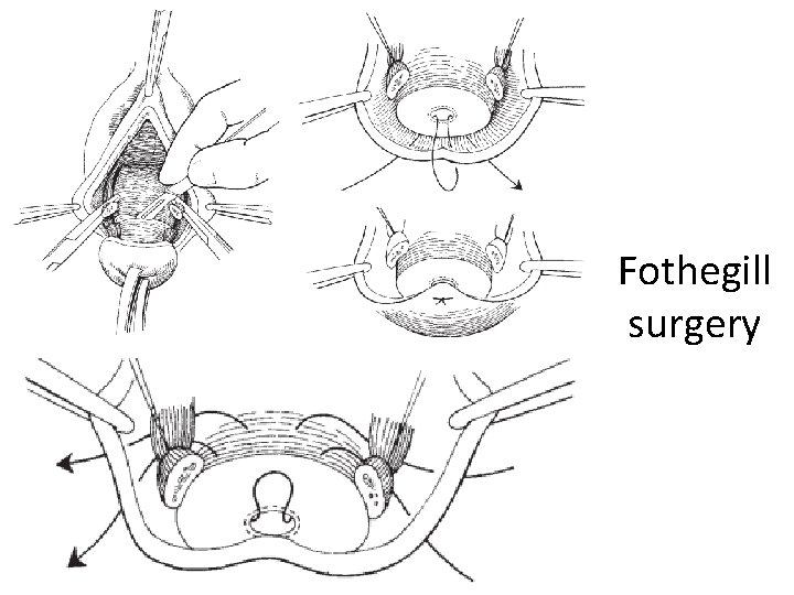
Manchester operation (Fothergill’s operation) Fothergill’s points – 1 sub urethral , 2 on either side of the cervix 1 on posterior vaginal wall
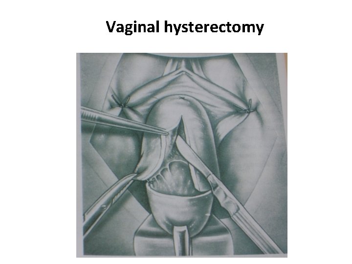
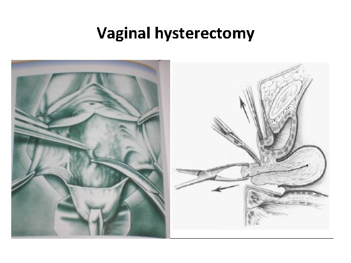
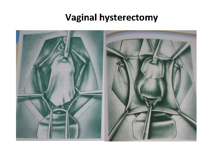
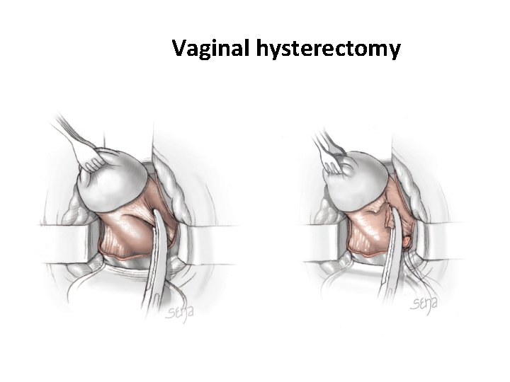
Case 4 • Uterovaginal prolapse in peri and postmenopausal age group Defect – All three supports ligamentary, fascial and muscular support Surgery – Vaginal hysterectomy with site specific repair of fascial defects, perineorrhaphy and SSF
Site specific repair • Reconstruction of pericervical ring • Anchor vesicovaginal fascia and rectovaginal fascia to the pericervical ring • plication of vesicovaginal fascia will detach the fascia from its lateral attachment
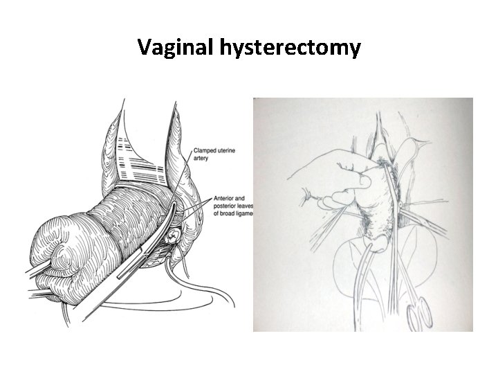
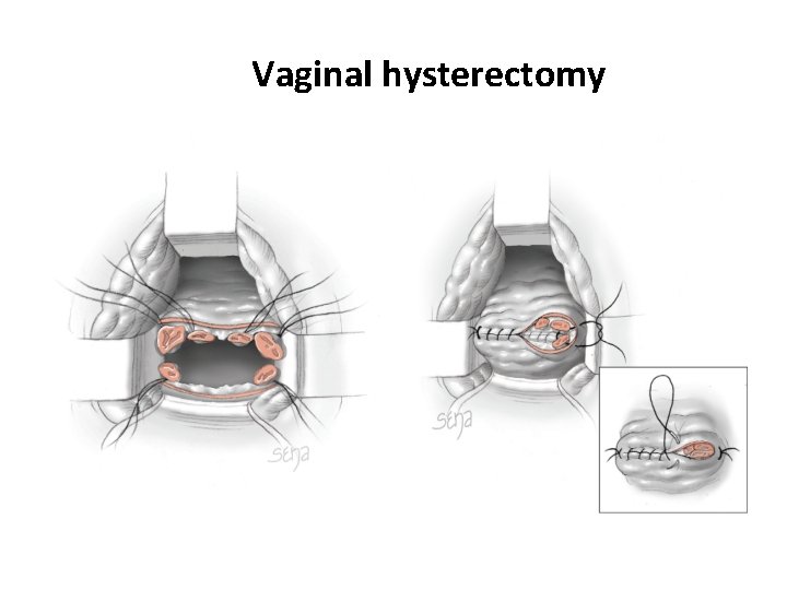
Vaginal hysterectomy
Vaginal hysterectomy
Vaginal hysterectomy
Vaginal hysterectomy
Vaginal hysterectomy
Vaginal hysterectomy
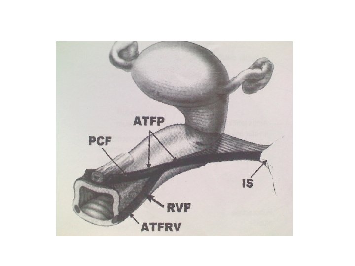
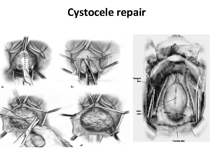
Cystocele repair – - Central – defects in the fascia are closed and vesicovaginal fascia is attached to uterosacrals - Lateral – vesicovaginal fascia is attached laterally to ATFP(Arcus tendinous fascia pelvis) and proximally to uterosacrals
Cystocele repair
• Enterocele repair – rectovaginal fascia is fixed proximally to the uterosacrals
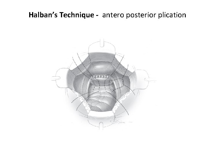
Enterocele correction • Abdominal Ø Moschowitz procedure Ø Halban’s • Vaginal Ø Mc. Caul culdoplasty
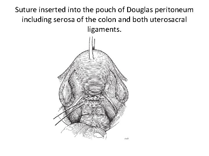
Suture inserted into the pouch of Douglas peritoneum including serosa of the colon and both uterosacral ligaments.
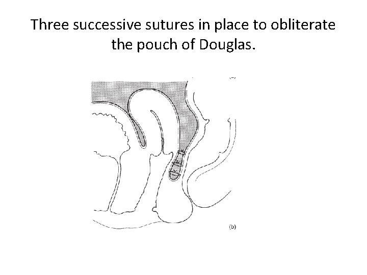
Three successive sutures in place to obliterate the pouch of Douglas.
Halban’s Technique - antero posterior plication
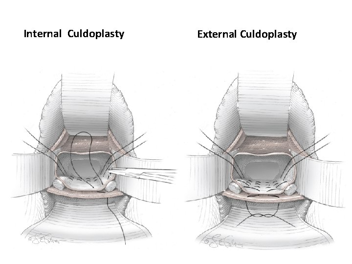
Internal Culdoplasty External Culdoplasty
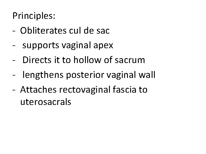
Principles: - Obliterates cul de sac - supports vaginal apex - Directs it to hollow of sacrum - lengthens posterior vaginal wall - Attaches rectovaginal fascia to uterosacrals
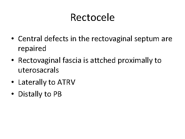
Rectocele • Central defects in the rectovaginal septum are repaired • Rectovaginal fascia is attched proximally to uterosacrals • Laterally to ATRV • Distally to PB
Rectocele repair
Rectocele repair
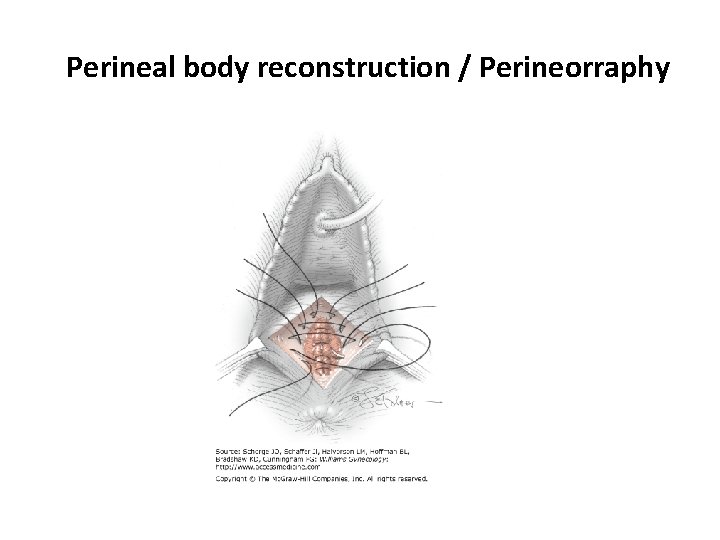
• Perineorrhaphy – approximation of bulbospongiosus and transverse perinnei muscles (distal attachment of rectovaginal septum, narrowing of genital hiatus and horizontal orientation of levator plate)
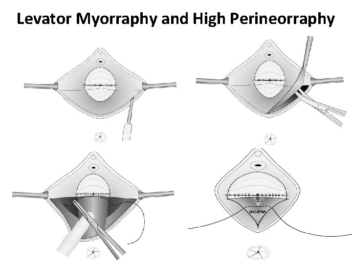
Perineorrhaphy • Essential because it prevents vault prolapse by Ø Anchoring rectovaginal fascia to PB Ø Making levator plate horizontal Ø Narrowing the genital hiatus • Levator myorrhaphy in selected cases Ø Dumble shaped vagina
Perineal body reconstruction / Perineorraphy
Levator Myorraphy and High Perineorraphy
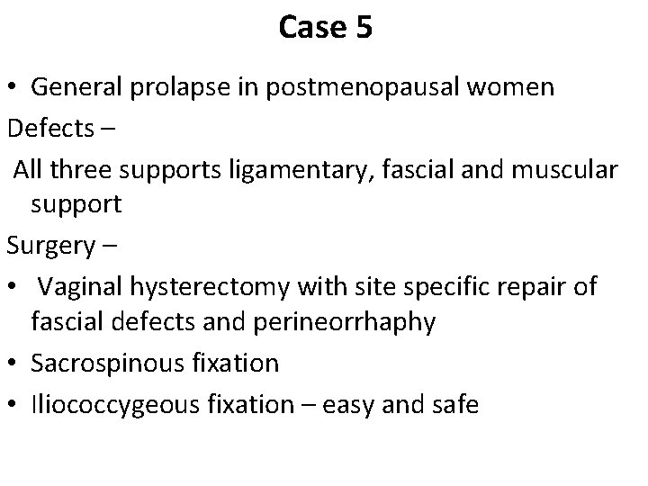
Case 5 • General prolapse in postmenopausal women Defects – All three supports ligamentary, fascial and muscular support Surgery – • Vaginal hysterectomy with site specific repair of fascial defects and perineorrhaphy • Sacrospinous fixation • Iliococcygeous fixation – easy and safe
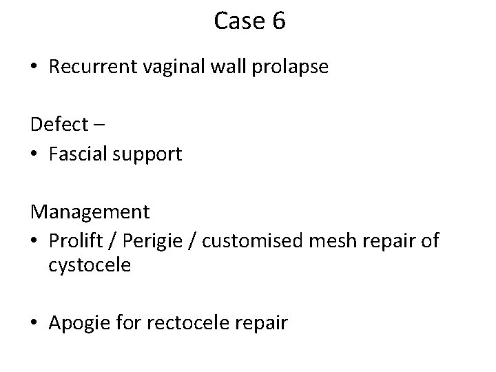
Case 6 • Recurrent vaginal wall prolapse Defect – • Fascial support Management • Prolift / Perigie / customised mesh repair of cystocele • Apogie for rectocele repair
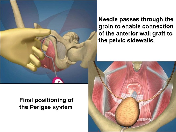
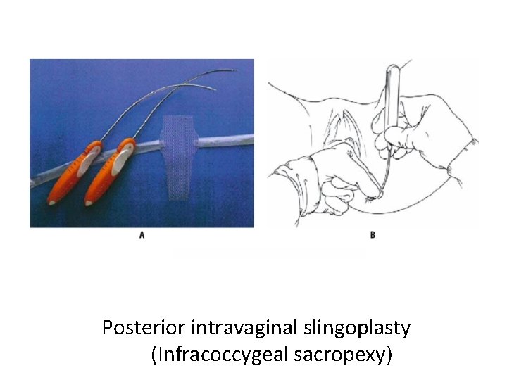
Needle passes through the groin to enable connection of the anterior wall graft to the pelvic sidewalls. Final positioning of the Perigee system
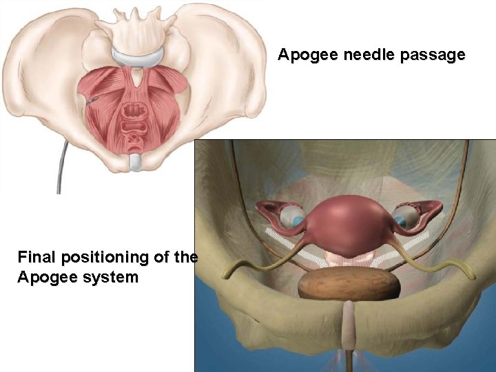
Apogee needle passage Final positioning of the Apogee system
Posterior intravaginal slingoplasty (Infracoccygeal sacropexy)
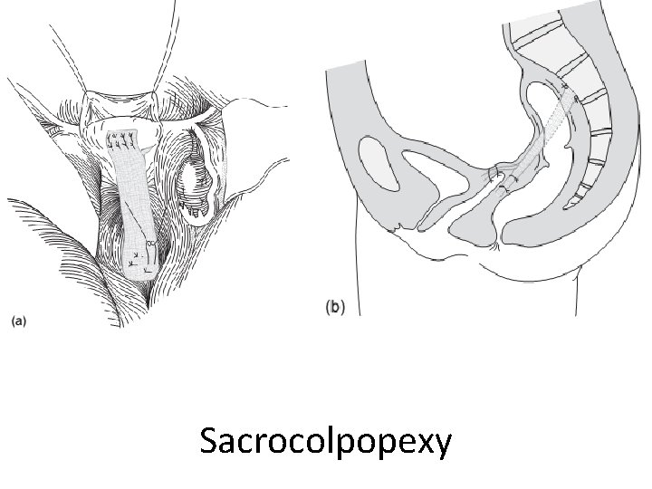
Case 7 • Vault prolapse Defect – Ligamentary support Surgery – • Abdominal sacrocolpopexy • Vaginal sacrospinous / iliococcygeous fixation of the vault
Vault Prolapse • Sacrocolpopexy • Sacrospinous fixation Green-top Guideline No. 46 RCOG/BSUG Joint Guideline | July 2015
Surgery? Who? • Surgical treatment should be offered to women with symptomatic PHVP after appropriate counselling. • PHVP surgery should be performed by an urogynaecologist or gynaecologists who can demonstrate an equivalent level of training or experience.
Postop result • Pelvic Organ Prolapse Quantifiation (POP-Q) stage of I or 0 in the apical compartment seems to be acceptable and widely used as the optimum postoperative result.
Which surgery? • Tailored to the individual patient’s circumstances. • Both ASC and SSF are effective treatments for primary PHVP. ASC is associated with signifiantly lower rates of recurrent vault prolapse, dyspareunia and postoperative stress urinary incontinence (SUI) when compared with SSF.
ASC Vs SSF • However, reoperation rates or higher patient satisfaction remain the same. • SSF is associated with earlier recovery compared with ASC. • SSF may not be appropriate in women with short vaginal length and should be carefully considered in women with pre-existing dyspareunia.
Lap Vs Abdominal • LSC can be equally effective as ASC in selected women with primary PHVP. • LSC can include mesh extension or be combined with other vaginal procedures to correct other compartment prolapse. • There is limited evidence on the effectiveness of RSC.
High uterosacral ligament suspension (HUSLS) • HUSLS - risk of ureteric injury, especially in the laparoscopic approach.
Transvaginal mesh (TVM) kits/grafts? • The limited evidence on TVM kits does not support their use as fist-line treatment of PHVP.
When should colpocleisis be used? • Colpocleisis is a safe and effective procedure that can be considered for frail women and/or women who do not wish to retain sexual function.
Concomitant surgery for occult SUI? • Colposuspension performed at the time of sacrocolpopexy is an effective measure to reduce postoperative symptomatic SUI in previously continent women.
Concomitant surgery for PHVP and overt SUI? • Colposuspension at the time of ASC does not appear to be effective treatment for SUI. • Concomitant mid-urethral sling surgery may be considered when vaginal surgical approaches are used for the treatment of PHVP.
Sacrocolpopexy
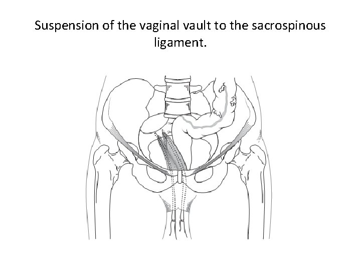
Suspension of the vaginal vault to the sacrospinous ligament.
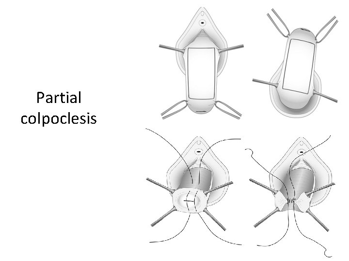
Case 10 • A 85 yr old C/o mass per vagina. K/C/o HTN, old IHD. ECHO – LVEF – 48% • O/E: General prolapse with grade 3 cystocele, enterocele, deficient perineum Management – If muscular support is good - pessary Pt not fit for hysterectomy – Le Fort’s repair
Partial colpoclesis
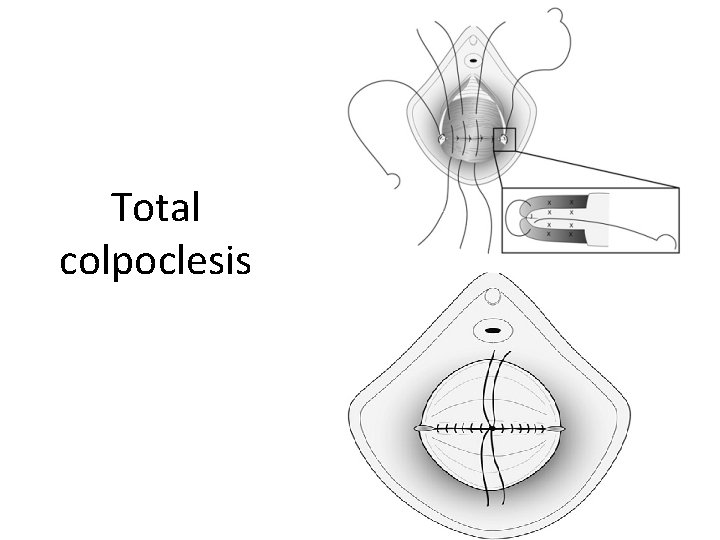
Total colpoclesis
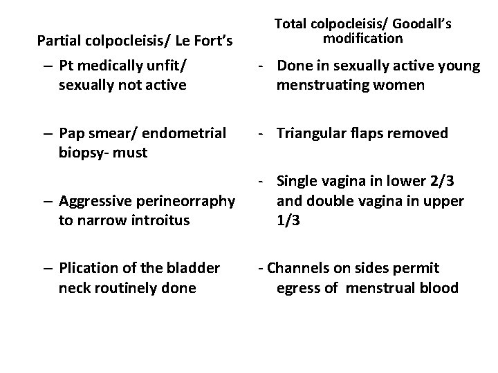
Partial colpocleisis/ Le Fort’s – Pt medically unfit/ sexually not active – Pap smear/ endometrial biopsy- must Total colpocleisis/ Goodall’s modification - Done in sexually active young menstruating women - Triangular flaps removed – Aggressive perineorraphy to narrow introitus - Single vagina in lower 2/3 and double vagina in upper 1/3 – Plication of the bladder neck routinely done - Channels on sides permit egress of menstrual blood
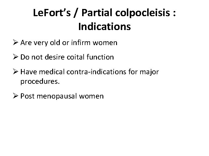
Le. Fort’s / Partial colpocleisis : Indications Ø Are very old or infirm women Ø Do not desire coital function Ø Have medical contra-indications for major procedures. Ø Post menopausal women
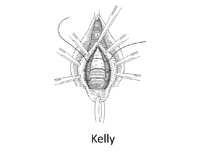
Introital tightening / Dani’s stitch : ØBased on principle of thiersch stitch for rectal prolapse. ØAlternative for Le. Fort’s ØTechnique : -Cerclage of the introitus
Kelly
Fothegill surgery
Wards modification (cystocele, enterocele repair): Ø Purse string suture is passed through UV fold of peritoneum, upper pedicle, Mackenrodt’s uterosacral complex, & highest point on posterior peritoneum Ø United uterosacral and cardinal ligaments are tied
Drawbacks Ø Broad ligaments are drawn into distorted position Ø Vagina is shortened Ø Due to interposition all sutures are under tension
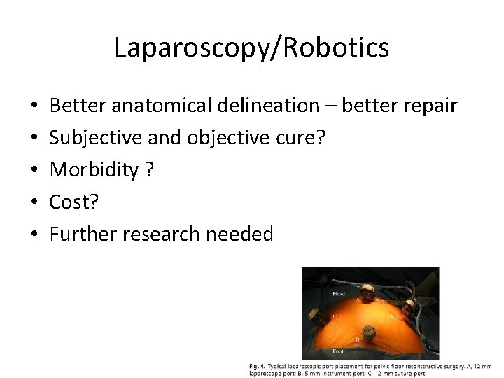
Laparoscopy/Robotics • • • Better anatomical delineation – better repair Subjective and objective cure? Morbidity ? Cost? Further research needed
Case 8 • A 24 yr old G 3 P 2 L 2 with 12 wks gestation with mass per vagina • O/E: III uterine descent + • Treatment Ring pessary, used until 16 weeks of gestation.
Treatment of edematous and congested prolapsed cervix with pregnancy Foot end elevation atleast by 25 cms Cover the prolapsed cervix by soaked guage with glycerine Mg. SO 4. All these measures continued till 18 weeks of gestation. Once replaced patient is allowed is ambulate.
Management of incarcerated pregnant uterus Once it is diagnosed , pregnancy has to terminated, irrespective of period of gestation During labour: A close watch on cervical dilatation needed Generally most of delivery go on spontaneously, if cervical dilatation fails then, At <4 cms inspite of good contraction Consider Em LSCS
At>7 cm with good contraction DUHRSSEN INCISION (2’ 0 and 10’ 0 clock) ↓ Delivery by vacuum and forceps. Hyaluronidase injected at multiple points on the cervical rim helps in cervical dilatation.
Case 9 Post Natal Day 3. P 2 L 2 had FTVD. C/o mass per vagina O/E- III uterine descent Management Consider physiotherapy Use pessaries till corrective repair surgery done Corrective repair surgery after 3 to 6 month
Challenge • Challenges in the management of pelvic organ prolapse still remains…… – High recurrence rates – Lack of randomized control trials – Poorly defined success and failure rates