ANATOMY OF CORONARY SINUS AND CLINICAL APPLICATION Dr


 (26 weeks)—the right horn of the sinus")


, a defect")


 projection showing a near occlusive")


- Slides: 45
ANATOMY OF CORONARY SINUS AND CLINICAL APPLICATION Dr Gaurav Chaudhary MD, DM Cardiology Assistant Professsor Department of Cardiology
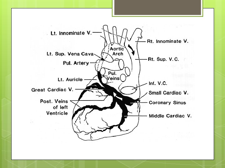
VENOUS DRAINAGE OF HEART Coronary Anterior sinus cardiac vein Thebesian vein
60 % of venous blood of heart drains into right atrium via coronary sinus. 40 % remaining blood drains via anterior cardiac vein
Coronary sinus conveys blood from left coronary territory. Anterior cardiac vein drains most of blood from right coronary artery
CORONARY SINUS Great cardiac vein Oblique vein of left atrium [ vein of marshall ] Posterior Middle Small vein of LV cardiac vein
Coronary Venous System CORONARY SINUS TRIBUTARIES
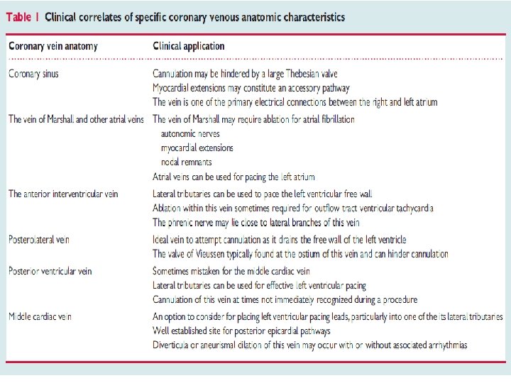
� Great cardiac vein - anterior interventricular sulcus � Oblique vein of LA - post surface of LA � Post . vein of LV - runs on diaphragmatic surface � Middle cardiac vein - posterior IV groove. � Small cardiac vein - accompanies RCA
Developmental anatomy of the coronary sinus (CS) (26 weeks)—the right horn of the sinus venosus remains as the venous portion of the right atrium between the vena cava (light blue). H
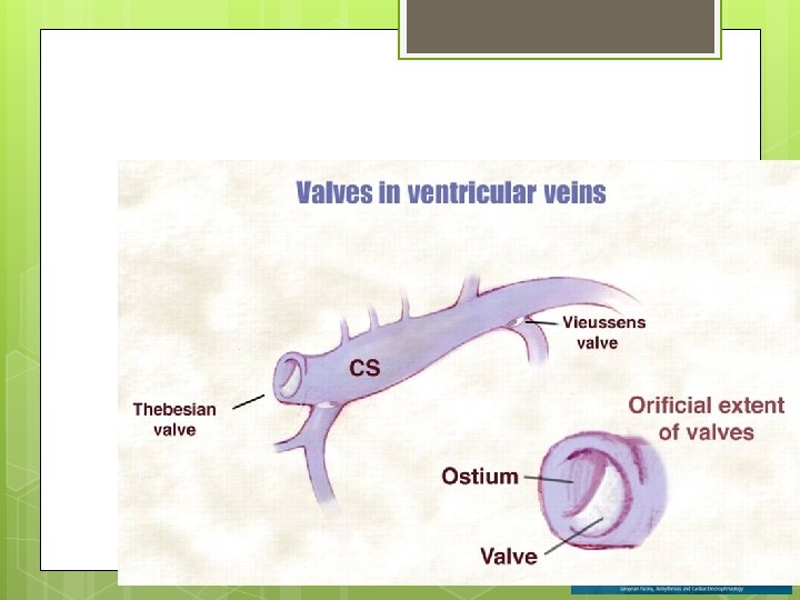
The CS has been dissected open along its long axis, CS musculature is seen in the proximal portion of the CS up to the orifice of the vein of Marshall. In this patient multiple posterior and posterolateral veins are also seen draining into the CS.
The CS has been dissected open along its long axis
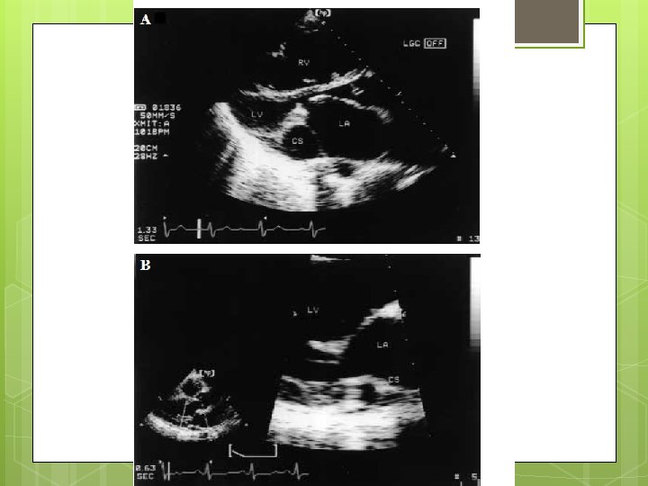
2 -D ECHOCARDIOGRAPHIC IMAGING OF CORONARY SINUS
CORONARY SINUS -2 D ECHO
ANAMOLIES OF CORONARY SINUS Absent thebesian valve. Membranous Absent tributaries of coronary sinus. Obstucted Dilated thebesian valve. coronary sinus ostia coronary sinus
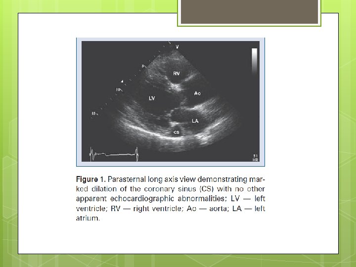
DILATED CORONARY SINUS
ABNORMAL CORONARY SINUS DRAINAGE
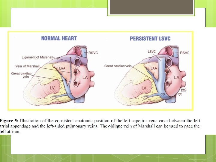
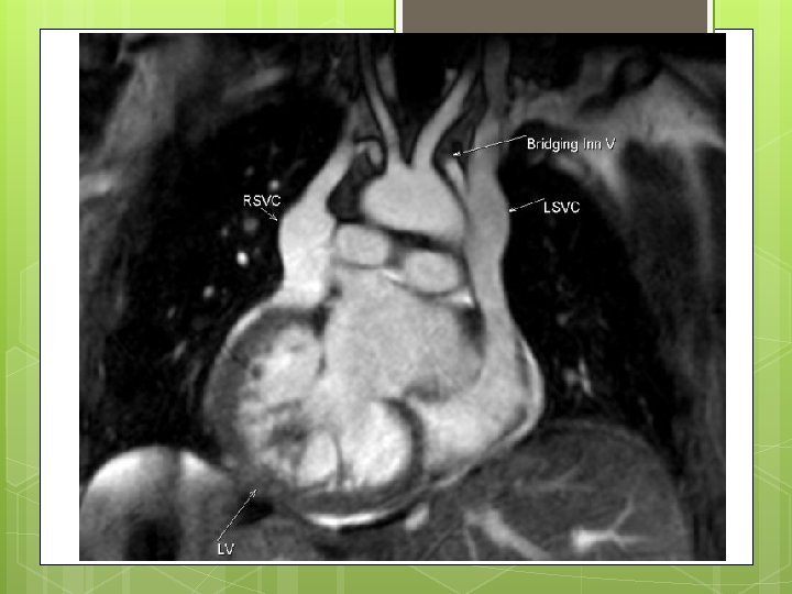
PERSISTENT LSVC
Figure 1. Transesophageal echocardiography revealed both atrial and right ventricular enlargement (left), a defect of the partial coronary sinus (middle), and shunt of the left atrium to the dilated coronary sinus (right) at the near longitudinal plane. Huang X Circulation 2007; 116: e 373 -e 373 Copyright © American Heart Association
FLOUROSCOPIC IMAGING OF CORONARY SINUS
Figure 2. A partial coronary sinus defect beyond the range of the interatrial septum and an intact flap valve of the oval fossa with its muscular rims were revealed simultaneously at ≈140° section by transesophageal echocardiography (left). Huang X Circulation 2007; 116: e 373 -e 373 Copyright © American Heart Association
AP Venogram
The MCV is a very consistent tributary of the CS present in nearly all hearts.
Coronary venous angiogram in the left anterior oblique (LAO) projection showing a near occlusive valve (arrow) in the region of the posterolateral vein (Vieussen's valve).
CT ANGIO IMAGING OF CORONARY SINUS
Coronary Venous Anatomy and Relation Between Coronary Sinus and Mitral Annulus Tops, L. F. et al. J Am Coll Cardiol Img 2008; 1: 94 -106 Copyright © 2008 American College of Cardiology Foundation. Restrictions may apply.
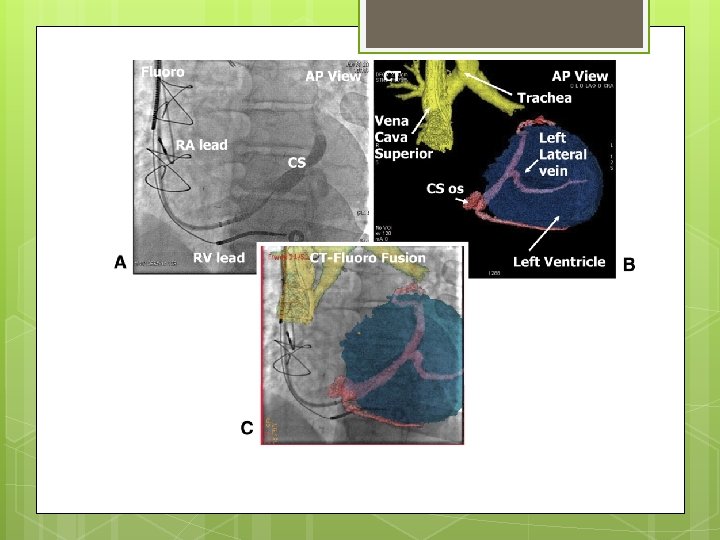
Reconstructed computed tomography image showing the coronary veins
X-ray view of the phantom and fusion of CT reconstruction.
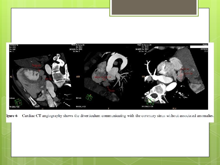
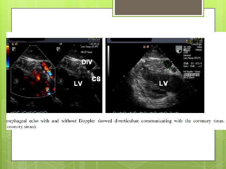
Coronary sinus diverticulum Should be suspected in patient having WPW syndrome , refractory to ablation Angiography reveals coronary diverticulum Associated with refractory posteroseptal pathway
Autopsy heart specimen depicting an absent Thebesian valve.
Examples of membranous Thebesian valves with fenestrations.
Examples of Thebesian valves that are fibromuscular, non-fenestrated, and occlusive.