ANATOMY OF AIRWAY AND INTUBATION NOUR GHNAIMAT Objectives

. ANATOMY OF AIRWAY AND INTUBATION. NOUR GHNAIMAT.

Objectives: � 1. The anatomical structures seen in region of intubation � 2. Different classifications of airway structures � 3. The technique of tracheal intubation � 4. Assessment of patients airway including 1 -2 -3 -Test � 5. Laryngoscopes and type of blades � 6. Tracheal tubes: size and types of tubes Shape of tube and specialized tubes � 7. the laryngeal mask � 8. Other apparatus including oro- and nasopharyngeal airways. � 9. Potential complications of intubation.

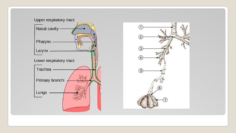
ANATOMY OF RESPIRATORY TRACT �Anatomically into upper and lower tract in relation to vocal cord �Or according to its function into conducting zone and respiratory zone. ◦ Conducting zone : �Nose, pharynx, trachea, bronchioles , terminal bronchioles �Function: filter, warm and moisten air and conduct air to and from the respiratory zone ◦ Respiratory zone : �Respiratory bronchioles , alveolar ducts, alveolar sacs, alveoli �Function : gas exchange

ORAL CAVITY ANATOMY :

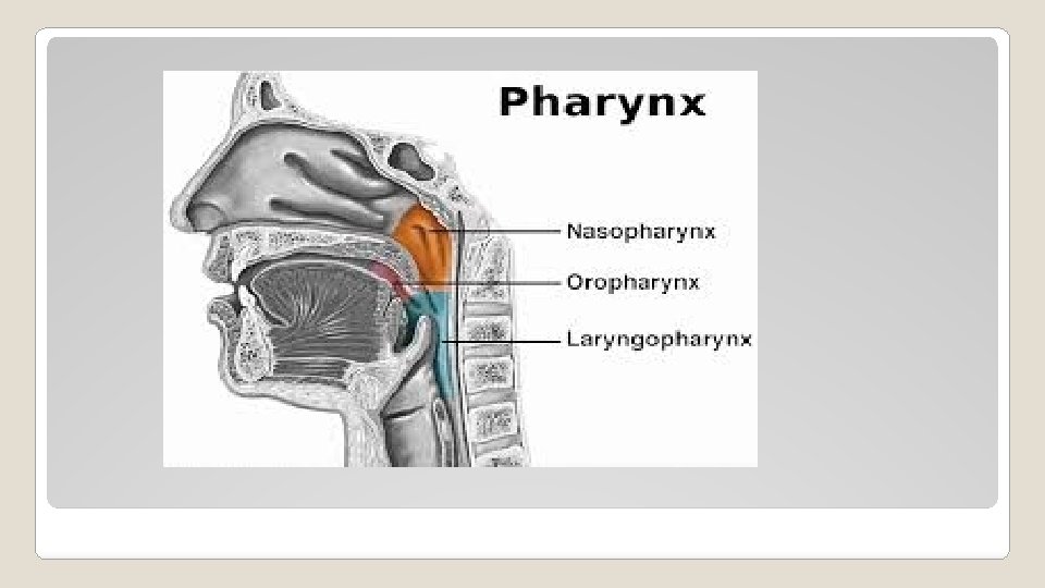
pharynx � fibromuscular structure that extends from the base of the skull to the cricoid cartilage at the entrance to the esophagus. �Parts ? ? � At the base of the tongue, the epiglottis functionally separates the oropharynx from the laryngopharynx (or hypopharynx). epiglottis prevents aspiration by covering the glottis—the opening of the larynx during swallowing.

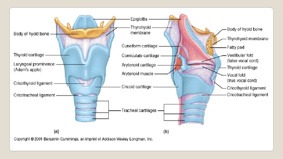
LARYNX � cartilaginous skeleton � ligaments and muscle � Located held together by below the tongue and hyoid bone, between the great vessels of neck. � Level of C 4 -C 6 � 44 mm in males and 36 mm in females � 9 cartilages of larynx ◦ ◦ ◦ Thyroid Cricoid 2 arytenoid 2 corniculate 2 cuneiform Epiglottis

INTRINSIC MUSCLES OF LARYNX

EXTRINSIC MUSCLES OF LARYNX �Sternothyroid muscles depress the larynx. �Omohyoid muscles depress the larynx. �Sternohyoid muscles depress the larynx. �Inferior constrictor muscles �Thyrohyoid muscles elevates the larynx. �Digastric elevates the larynx. �Stylohyoid elevates the larynx. �Mylohyoid elevates the larynx. �Geniohyoid elevates the larynx. �Hyoglossus elevates the larynx. �Genioglossus elevates the larynx

�LARYNGEAL FOLDS ◦ Vestibular fold : fixed , covers vestibular ligament , vascular pink in color. ( false vocal cord ) ◦ Vocal fold : mobile fold , voice production , covers focal ligaments , avascular. ( true vocal cord ) ◦ glottis : gap between the vocal folds, the narrowest part of the larynx

Sensory innervation of airway : � The sensory supply to the upper airway is derived from the cranial nerves.
 ◦ Above the vocal cords by the internal laryngeal")
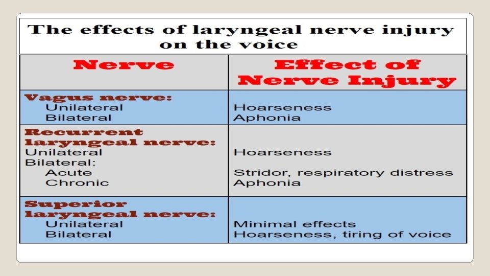
INNERVATION OF LARYNX �SENSORY (mucosa) ◦ Above the vocal cords by the internal laryngeal br of the superior laryngeal br of vagus ◦ Below the vocal cords by the recurrent laryngeal N. �MOTOR ◦ All intrinsic ms of larynx are supplied by the recurrent laryngeal N except the cricothyroid ms ( by external laryngeal branch)

TRACHEA �A cartilaginous and membranous tube � Begins as a continuation of the larynx at the lower border of cricoid cartilage at the level of C 6, and terminates at the carina, at the level of T 5. � Adults – 10 -16 cm long and 2. 5 cm in diameter � Infants – 4 -5 cm long and may be as small as 3 mm in diameter � Kept patent by the presence of C-shaped cartilaginous rings

Thank You
- Slides: 17