ANATOMY III MUDr Ivana Hradilov Svensk Ph D


 arises the neural:")


 (BM) Somatosensory")
 pass through the skull")


, cornu posterius (columna posterior), cornu laterale (columna")


 motoneurons of the")


- Slides: 38
ANATOMY III MUDr. Ivana Hradilová Svíženská, Ph. D.
Lectures 1. Seminars Anatomy of the nervous system, Spinal nerve Gross anatomy and structure of the spinal cord Structure of the spinal cord 2. Structure of the brain stem 3. Structure of the cerebellum and diencephalon 4. Structure of the telencephalon Gross anatomy and structure of the telencephalon Meninges, ventricles and vascular system of the CNS Cranial nerves 1 Cranial nerves 2, Cervical plexus Cranial nerves 2, Cervical plexus Intercostal nerves, Dorsal rami 7. Autonomic nervous system 8. Visual system 9. Regional anatomy of the head and neck Demonstration of topographical regions (head and neck) 5. 6. 10. Gross anatomy and structure of the brain stem Gross anatomy and structure of the cerebellum and diencephalon Dissection course (head, neck) 11. Auditory system 12. Regional anatomy of the body (except limbs) Demonstration of the topographical regions (except limbs) 13. Regional anatomy of the body (except limbs) Demonstration of the topographical regions (except limbs) 14. RTG anatomy 15. Spare lectures
Recommended literature: Liebgott, Bernard. The anatomical basis of dentistry. 3 rd ed. Mosby, ISBN 0 -323 -06807 -3 Dubový, Petr. Gross anatomy and structure of the human nervous system. Part I. Surface anatomy and structural arrangement of the central nervous system. 3 rd ed. Brno: Masarykova univerzita, 2016. 92 s. ISBN 978 -80 -210 -6125 -5 Dubový, Petr. Instructions for anatomical dissection course. 3 rd ed. Brno: Masaryk University, 2013. 71 s. ISBN 9788021062023 Stingl, J. et al. Regional anatomy. 1 st ed. Galén-Karolinum, 2012. 123 s. ISBN 9788072628797 Atlas of Anatomy
Equipment: lab coat dressing forceps gloves
Nervous system
Nervous system ■ is a complex, sophisticated system that regulates and coordinates activities of the body ■ regulates the body's responses to internal and external stimuli ■ has three main functions, sensory input, integration of data and motor output ■ is composed of excitable nerve cells ■ conducts nerve impulses ■ is divided into two categories: the central nervous system- CNS and the peripheral nervous system PNS ■ the basic structural and functional unit - neuron ■ cells providing support and protection for neurons – glial cells
Neuron ■ receives stimuli ■ transforms stimuli to nerve impulses ■ conducts nerve impulses ■ processes information ■ transmits the electrochemical signal across a synapse
TYPES OF NEURONS
Myelinization – node of Ranvier, internodal segment Saltatory conduction oligodendroglia unmyelinated axon Schwann cell Ranvier node Schwann cell nucleus axon
Basic function of NS - reflex Sensor CNS Effector
Interneuron Muscle Skin
Development of NS ■ from ectoderm (under influence of the notochord) arises the neural: plate groove 3. w i. u. tube + neural crest
Parts derived from the neural tube brain spinal cord Parts derived from the neural crest cranial nerve ganglia dorsal root and autonomic ganglia medulla of the suprarenal gland some bones, cartilage and connective tissue of the head pigment cells. . .
Cerebral vesicles from the rostral part of the neural tube 3. 2. 1. Spinal cord medulla spinalis 3. rhombencephalon (hindbrain) 2. mesencephalon (midbrain) 1. prosencephalon (forebrain)
Secondary vesicles 3. 2. 1. myelencephalon medulla oblongata 3. metencephalon pons, cerebellum 2. mesencephalon midbrain diencephalon 1. telencephalon diencephalon telencephalon
dorsal root DRG A B ventral root muscle sulcus limitans Spinal nerve skin
SENSOR Surface of the body Exteroreceptors Organs of motions Proprioreceptors Viscera Interoreceptors Senses EFFECTOR Skeletal muscles Afferent CNS PNS Efferent Smooth muscles + myocardium Glands
Functional types of axons EFFECTOR SENSOR Exteroreceptors Proprioreceptors Somato + branchiomotor (SM) (BM) Somatosensory (SS) Interoreceptors Viscerosensory (VS) Senses Sensory (S) Skeletal muscles CNS Visceromotor (VM) Smooth muscles and myocardium Glands
PNS Cranial nerves III. - XII. (I. - XII. ) pass through the skull base Spinal nerves - 31 pairs pass through the intervertebral foramina
CNS I. Brain ■ medulla oblongata ■ pons ■ mesencephalon ■ cerebellum ■ diencephalon ■ telencephalon II. Spinal cord
Structure of the CNS Gray matter - nuclei White matter – nerve tracts: - tractus - fasciculus (lemniscus)
Spinal cord ■ transmission of neural signals between the brain and the periphery ■ contains neural circuits that can control numerous reflexes and central pattern generators independently on the cortex
MEDULLA SPINALIS
Cauda equina Fila radicularia
Intumescentia cervicalis C 5 – T 1 segments C 4 – T 1 vertebrae Intumescentia lumbalis L 2 – S 1 segments T 9 – T 12 vertebrae
Segment C 1 Segment C 5 Segment C 8 Segment Th 2 Segment Th 10 Segment L 4 Segment L 1 Segment L 4 Segment S 4
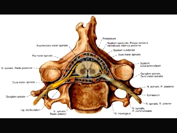
SUBSTANTIA GRISEA – cornu anterius (columna anterior), cornu posterius (columna posterior), cornu laterale (columna lateralis), substantia intermedia, canalis centralis
SUBSTANTIA ALBA – funiculus anterior, lateralis, posterior fissura mediana ant. , sulcus medianus post. , septum medianum posterius, sulcus anterolateralis, posterolateralis
Dorsal horn Ventral horn p. cervicalis p. thoracica p. sacralis
Dorsal root + o i r p o r tors p + ep o r rec e t ex roe t n i Spinal nerve ske leta l + mus cles smo + g oth land Ventral root s
Extero+ proprio - receptors Functional zones of the spinal cord Alar plate Basal plate Intero - receptors GSA GVE GSE Skeletal muscles Sulcus limitans Smoot muscles, glands
Gray matter DORSAL HORN – afferent neurons SUBST. INTERMEDIA (lateral horn) motoneurons of the ANS VENTRAL HORN - motoneurons CC FR Th 1 - L 2 White matter Funiculus post. (fasc. gracilis et cuneatus) Funiculus ant. Funiculus lat. F. anterolateralis
Functional zones in the spinal cord GSA GVE GSE GVE zone T 1 - L 2 - preganglionic sympathetic neurons below L 2 - preganglionic parasympathetic neurons
tr. co-sp lat -sp u r tr. te-sp Multipolar cells p -s tr o c. α, p s e r. r t t tr. ve-sp n a tr. ol-sp Ncl. intermedio-lat. Ncll. motorii Ventral root
Pseudounipol. neurons of the DRG Tr. spino- Fasc. gracilis bulbaris Fasc. cuneatus Radix dorsalis Ncl. posteromarginalis + Subst. gelatinosa Rolandi Ncl. proprius Ncl. thoracicus (Stilling-Clark. ) Tr. spinocerebellaris post. Ncl. intermediomedialis Tr. spinocerebellaris ant. Tr. spino-thalamicus, -reticularis, -tectalis Tr. spino-olivaris
dorsal root GSA GVE GSE tr. sp-bulb ncl apicalis subst gel Rolandi tr. sp -th , te c, tr. s p-c ret er a nt , po st tr. co-sp lat -sp u r tr. e-sp tr. te-sp tr. sp-sp tr. r t tr. ve-sp n a p -s tr. co ncl proprius ncl thoracicus ncl intermed-med ncl intermedio-lat ncll motorii tr. ol-sp ventral root
Illustrations and photographs were copied from: Atlas der Anatomie des Menschen/Sobotta. Putz, R. , und Pabst, R. 20. Auflage. München: Urban & Schwarzenberg, 1993)