
Anatomy for Health Care Professionals NUR 469 Lecture



- Slides: 48
Anatomy for Health Care Professionals NUR 469: Lecture 2 September 14, 2009 Curtis L. Whitehair, MD Georgetown University School of Nursing & Health Studies Graduate Program
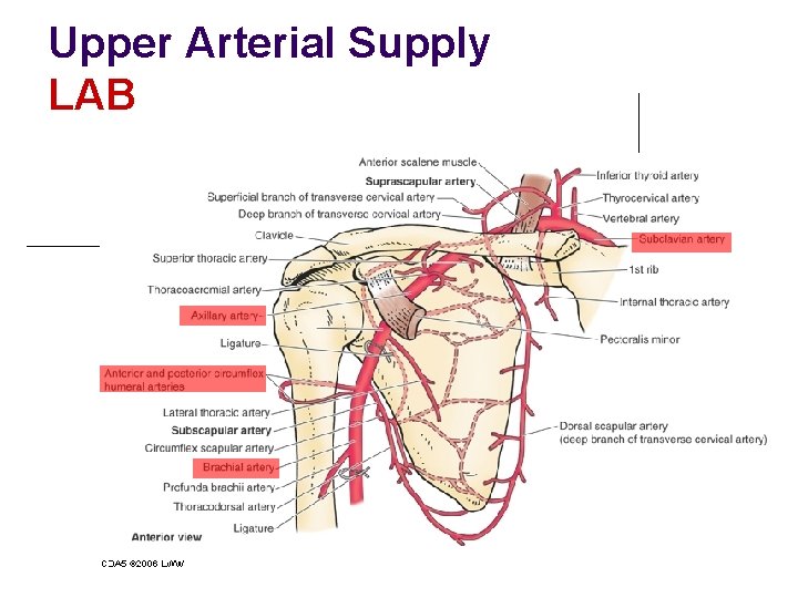
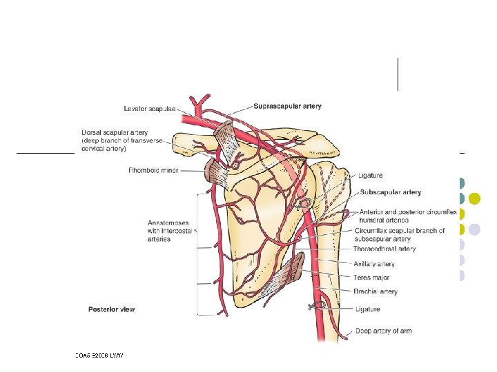
Upper Arterial Supply LAB
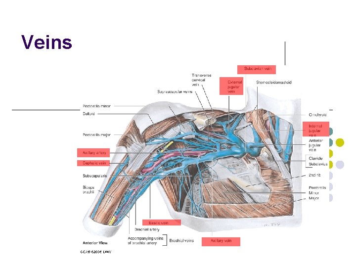
Veins
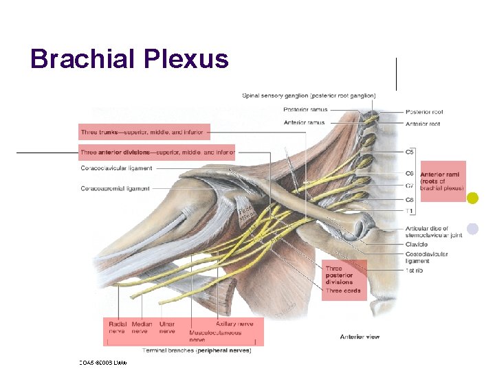
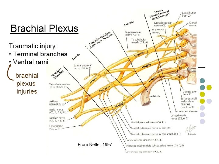
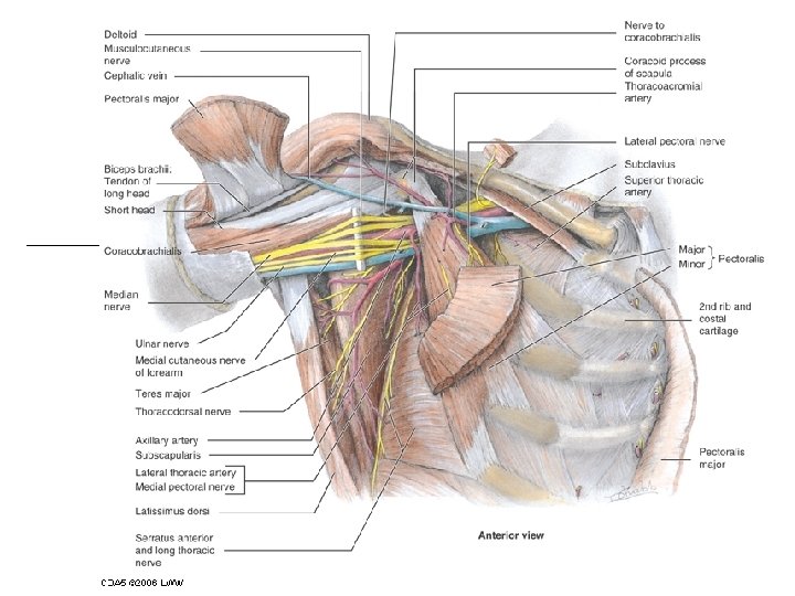
Brachial Plexus Robert Taylor Drinks Cold Beer
Brachial Plexus
Brachial Plexus Injuries l l Injuries to the brachial plexus affects movement and cutaneous sensations in the upper limb. Erb-Duchenne Palsy: Upper Trunk or C 5, 6 Roots – results in waiters tip. ¡ Shoulder Adducted ¡ Medially rotated arm ¡ Extend Elbow
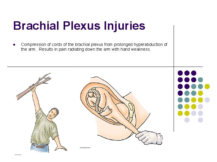
Brachial Plexus Injuries l Compression of cords of the brachial plexus from prolonged hyperabduction of the arm. Results in pain radiating down the arm with hand weakness.
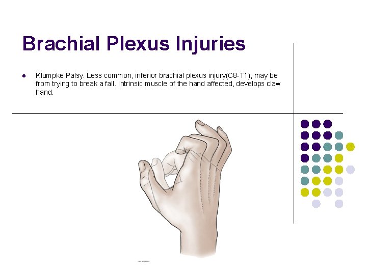
Brachial Plexus Injuries l Klumpke Palsy: Less common, inferior brachial plexus injury(C 8 -T 1), may be from trying to break a fall. Intrinsic muscle of the hand affected, develops claw hand.
Brachial Plexus Injuries l l l Injuries to the brachial plexus affects movement and cutaneous sensations in the upper limb. Erb-Duchenne Palsy: Upper Trunk or C 5, 6 Roots – results in waiters tip. Compression of cords of the brachial plexus from prolonged hyperabduction of the arm. Results in pain radiating down the arm with hand weakness. Klumpke Palsy: Less common, inferior brachial plexus injury(C 8 -T 1), may be from trying to break a fall. Intrinsic muscle of the hand affected, develops claw hand. Acute Brachial Plexus Neuritis (Parsonage Turner Syndrome): sudden on set of severe shoulder pain then is followed by weakness. Inflammation is often preceded by some event (URI, Vaccination or Non-specific Trauma)
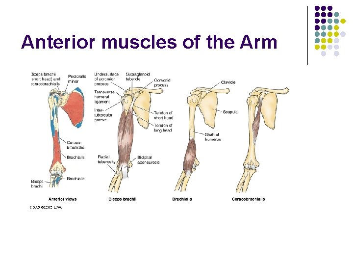
Anterior muscles of the Arm
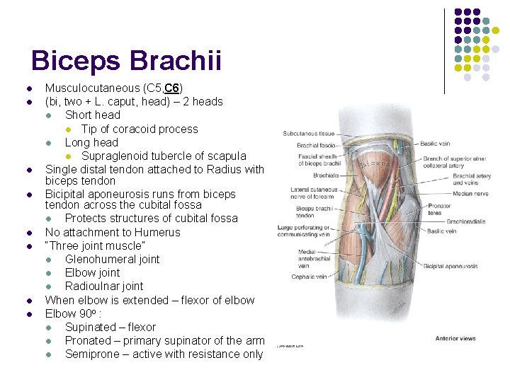
Biceps Brachii l l l l Musculocutaneous (C 5, C 6) (bi, two + L. caput, head) – 2 heads l Short head l Tip of coracoid process l Long head l Supraglenoid tubercle of scapula Single distal tendon attached to Radius with biceps tendon Bicipital aponeurosis runs from biceps tendon across the cubital fossa l Protects structures of cubital fossa No attachment to Humerus “Three joint muscle” l Glenohumeral joint l Elbow joint l Radioulnar joint When elbow is extended – flexor of elbow Elbow 90 o : l Supinated – flexor l Pronated – primary supinator of the arm l Semiprone – active with resistance only
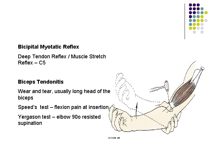
Bicipital Myotatic Reflex Deep Tendon Reflex / Muscle Stretch Reflex – C 5 Biceps Tendonitis Wear and tear, usually long head of the biceps Speed’s test – flexion pain at insertion Yergason test – elbow 90 o resisted supination
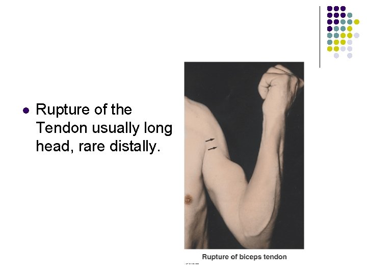
l Rupture of the Tendon usually long head, rare distally.
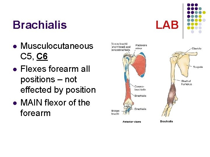
Brachialis l l l Musculocutaneous C 5, C 6 Flexes forearm all positions – not effected by position MAIN flexor of the forearm LAB
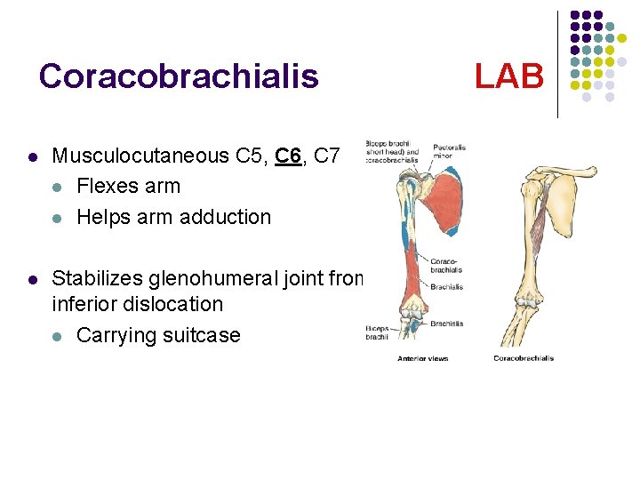
Coracobrachialis l Musculocutaneous C 5, C 6, C 7 l Flexes arm l Helps arm adduction l Stabilizes glenohumeral joint from inferior dislocation l Carrying suitcase LAB
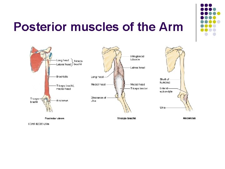
Posterior muscles of the Arm
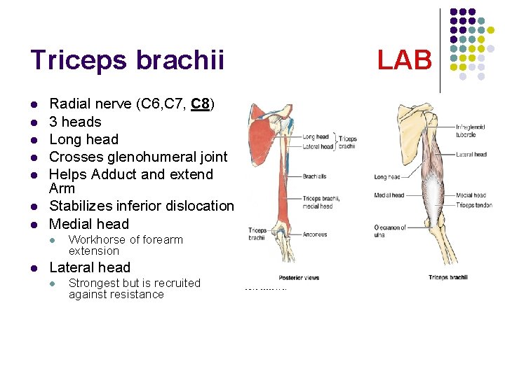
Triceps brachii l l l l Radial nerve (C 6, C 7, C 8) 3 heads Long head Crosses glenohumeral joint Helps Adduct and extend Arm Stabilizes inferior dislocation Medial head l l Workhorse of forearm extension Lateral head l Strongest but is recruited against resistance LAB
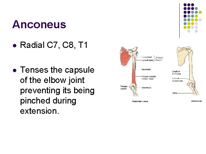
Anconeus l Radial C 7, C 8, T 1 l Tenses the capsule of the elbow joint preventing its being pinched during extension.
Muscles of the forearm flexor l l The flexors are arranged in three layers Superficial layer l l l Intermediate layer l l l 4 muscles Crosses elbow 1 muscle Crosses elbow Deep layer l l 3 muscles Crosses wrist and phalanges
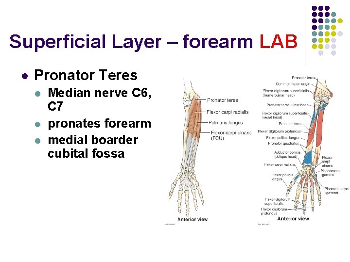
Superficial Layer – forearm LAB l Pronator Teres l l l Median nerve C 6, C 7 pronates forearm medial boarder cubital fossa
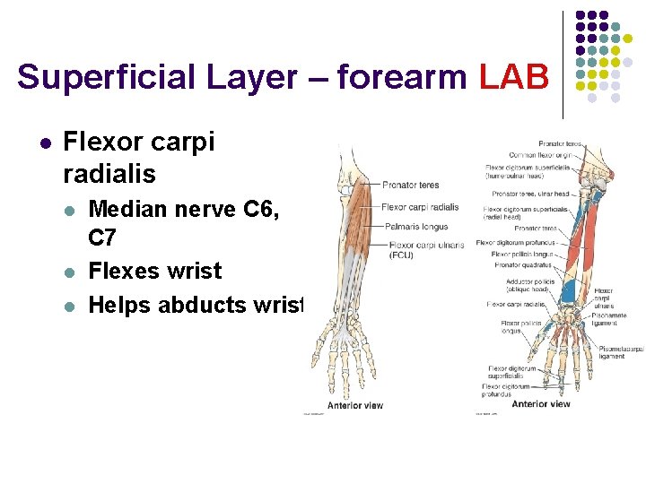
Superficial Layer – forearm LAB l Flexor carpi radialis l l l Median nerve C 6, C 7 Flexes wrist Helps abducts wrist
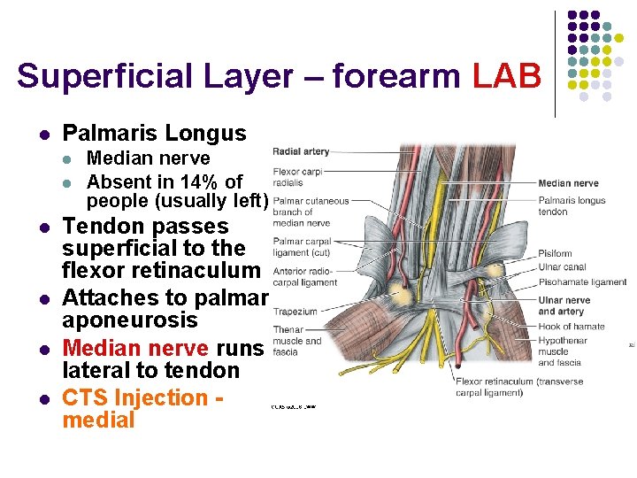
Superficial Layer – forearm LAB l Palmaris Longus l l l Median nerve Absent in 14% of people (usually left) Tendon passes superficial to the flexor retinaculum Attaches to palmar aponeurosis Median nerve runs lateral to tendon CTS Injection medial
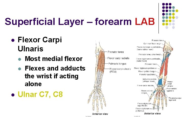
Superficial Layer – forearm LAB l Flexor Carpi Ulnaris l l l Most medial flexor Flexes and adducts the wrist if acting alone Ulnar C 7, C 8
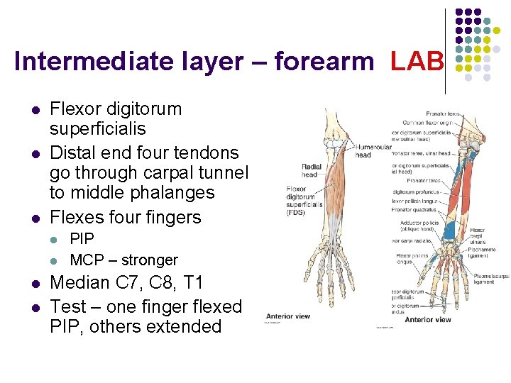
Intermediate layer – forearm LAB l l l Flexor digitorum superficialis Distal end four tendons go through carpal tunnel to middle phalanges Flexes four fingers l l PIP MCP – stronger Median C 7, C 8, T 1 Test – one finger flexed PIP, others extended
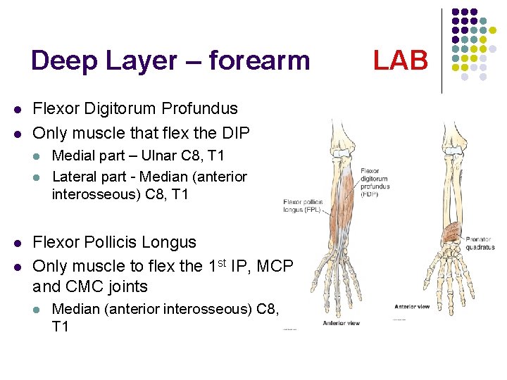
Deep Layer – forearm l l Flexor Digitorum Profundus Only muscle that flex the DIP l l Medial part – Ulnar C 8, T 1 Lateral part - Median (anterior interosseous) C 8, T 1 Flexor Pollicis Longus Only muscle to flex the 1 st IP, MCP and CMC joints l Median (anterior interosseous) C 8, T 1 LAB
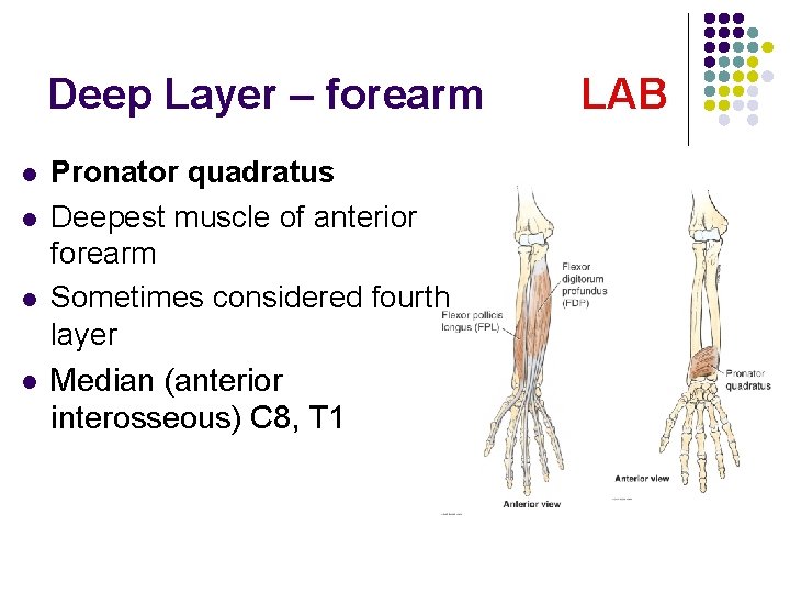
Deep Layer – forearm l l Pronator quadratus Deepest muscle of anterior forearm Sometimes considered fourth layer Median (anterior interosseous) C 8, T 1 LAB
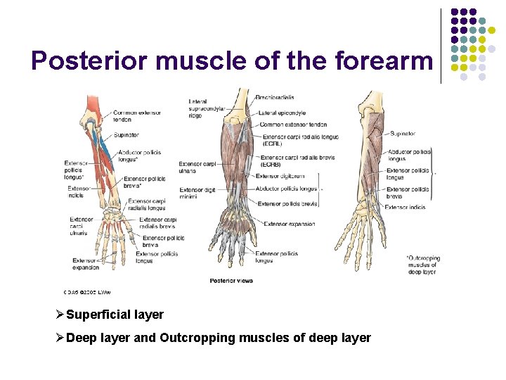
Posterior muscle of the forearm ØSuperficial layer ØDeep layer and Outcropping muscles of deep layer
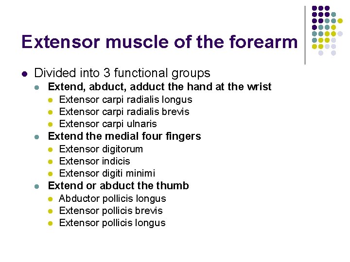
Extensor muscle of the forearm l Divided into 3 functional groups l l l Extend, abduct, adduct the hand at the wrist l Extensor carpi radialis longus l Extensor carpi radialis brevis l Extensor carpi ulnaris Extend the medial four fingers l Extensor digitorum l Extensor indicis l Extensor digiti minimi Extend or abduct the thumb l Abductor pollicis longus l Extensor pollicis brevis l Extensor pollicis longus
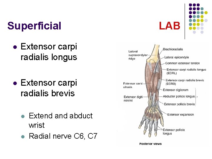
Superficial l Extensor carpi radialis longus l Extensor carpi radialis brevis l l Extend abduct wrist Radial nerve C 6, C 7 LAB
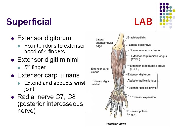
Superficial l Extensor digitorum l l Extensor digiti minimi l l 5 th finger Extensor carpi ulnaris l l Four tendons to extensor hood of 4 fingers Extend adducts wrist joint Radial nerve C 7, C 8 (posterior interosseous nerve) LAB
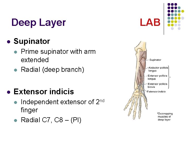
Deep Layer l Supinator l l l Prime supinator with arm extended Radial (deep branch) Extensor indicis l l Independent extensor of 2 nd finger Radial C 7, C 8 – (PI) LAB
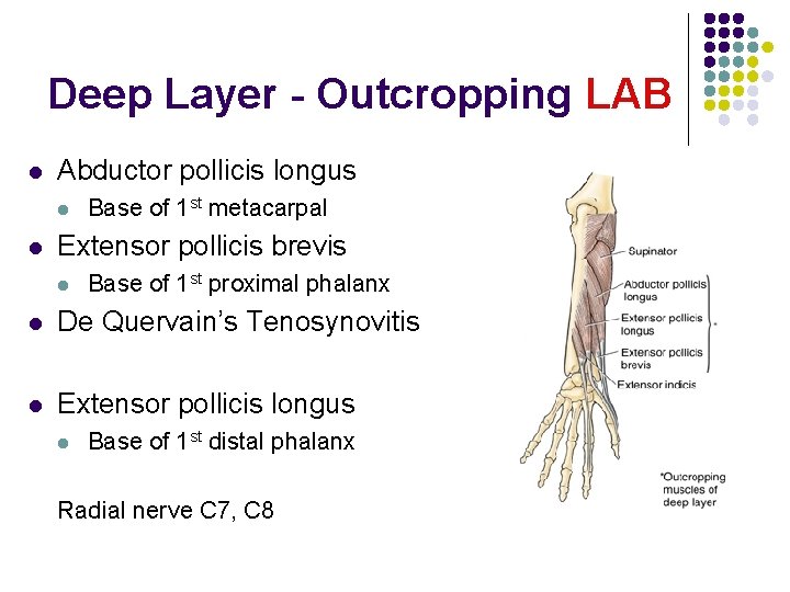
Deep Layer - Outcropping LAB l Abductor pollicis longus l l Base of 1 st metacarpal Extensor pollicis brevis l Base of 1 st proximal phalanx l De Quervain’s Tenosynovitis l Extensor pollicis longus l Base of 1 st distal phalanx Radial nerve C 7, C 8
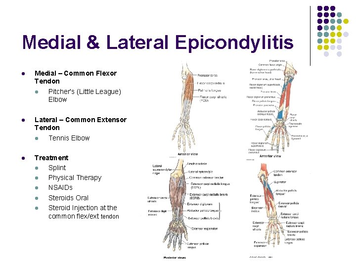
Medial & Lateral Epicondylitis l Medial – Common Flexor Tendon l Pitcher’s (Little League) Elbow l Lateral – Common Extensor Tendon l Tennis Elbow l Treatment l Splint l Physical Therapy l NSAIDs l Steroids Oral l Steroid Injection at the common flex/ext tendon
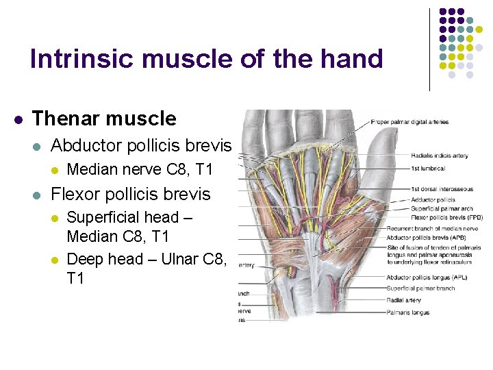
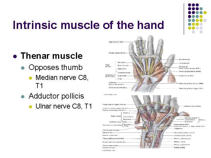
Intrinsic muscle of the hand l Thenar muscle l Abductor pollicis brevis l l Median nerve C 8, T 1 Flexor pollicis brevis l l Superficial head – Median C 8, T 1 Deep head – Ulnar C 8, T 1
Intrinsic muscle of the hand l Thenar muscle l Opposes thumb l l Median nerve C 8, T 1 Adductor pollicis l Ulnar nerve C 8, T 1
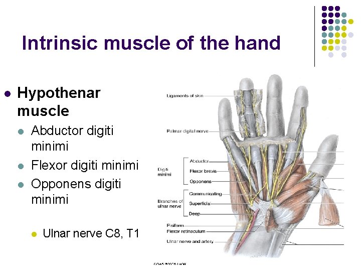
Intrinsic muscle of the hand l Hypothenar muscle l l l Abductor digiti minimi Flexor digiti minimi Opponens digiti minimi l Ulnar nerve C 8, T 1
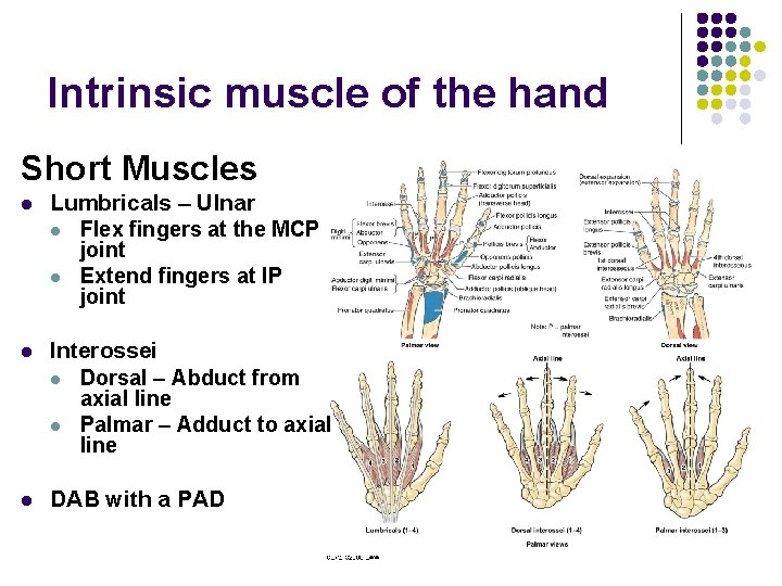
Intrinsic muscle of the hand Short Muscles l Lumbricals – Ulnar l Flex fingers at the MCP joint l Extend fingers at IP joint l Interossei l Dorsal – Abduct from axial line l Palmar – Adduct to axial line l DAB with a PAD
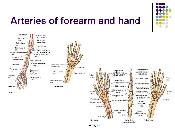
Arteries of forearm and hand
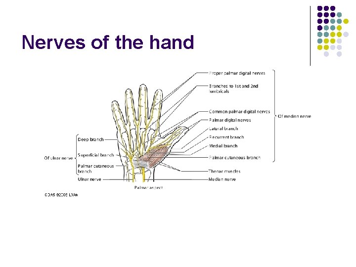
Nerves of the hand
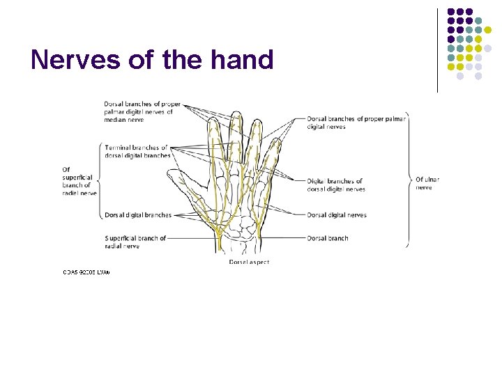
Nerves of the hand
Shoulder Joint
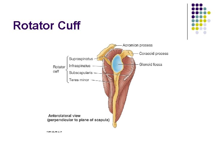
Rotator Cuff
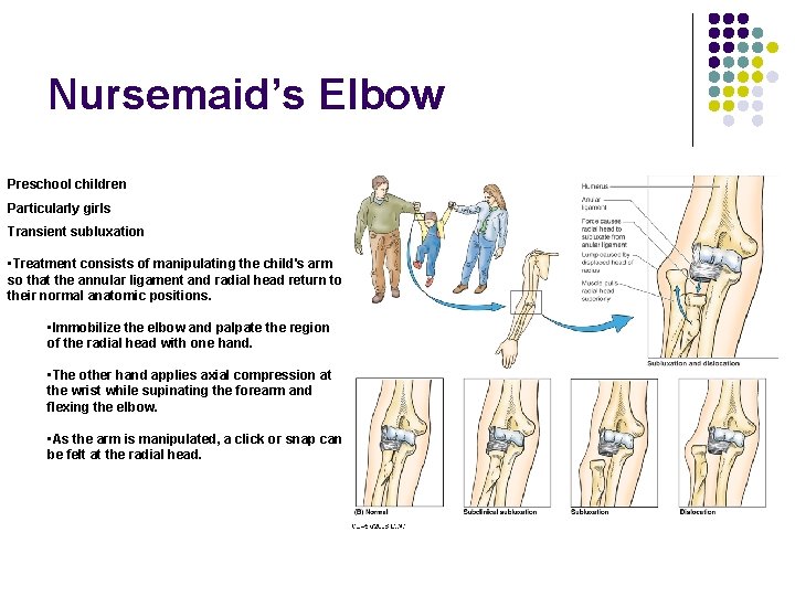
Nursemaid’s Elbow Preschool children Particularly girls Transient subluxation • Treatment consists of manipulating the child's arm so that the annular ligament and radial head return to their normal anatomic positions. • Immobilize the elbow and palpate the region of the radial head with one hand. • The other hand applies axial compression at the wrist while supinating the forearm and flexing the elbow. • As the arm is manipulated, a click or snap can be felt at the radial head.
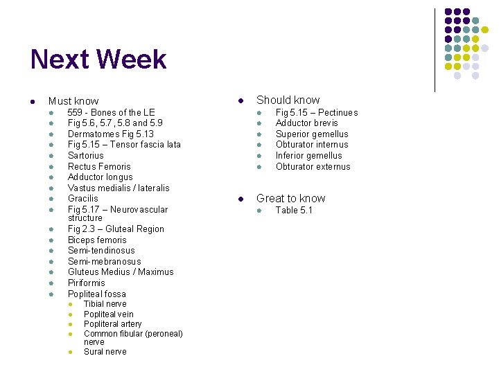
Next Week l Must know l l l l l 559 - Bones of the LE Fig 5. 6, 5. 7, 5. 8 and 5. 9 Dermatomes Fig 5. 13 Fig 5. 15 – Tensor fascia lata Sartorius Rectus Femoris Adductor longus Vastus medialis / lateralis Gracilis Fig 5. 17 – Neurovascular structure Fig 2. 3 – Gluteal Region Biceps femoris Semi-tendinosus Semi-mebranosus Gluteus Medius / Maximus Piriformis Popliteal fossa l l l Tibial nerve Popliteal vein Popliteral artery Common fibular (peroneal) nerve Sural nerve l Should know l l l l Fig 5. 15 – Pectinues Adductor brevis Superior gemellus Obturator internus Inferior gemellus Obturator externus Great to know l Table 5. 1