ANATOMY EMBRYOLOGY REPRODUCTIVE PHYSIOLOGY OYA AKCIN M D

ANATOMY, EMBRYOLOGY REPRODUCTIVE PHYSIOLOGY OYA AKCIN, M. D. Department of Histology & Embryology

Female Reproductive System Organs n n Ovaries Genital Ducts -Vagina -Uterus -Uterine Tubes Extra Genitalia Mamary Glands

Default pathway GONAD on hold Y OR GONAD on hold OVARY UROGENITAL SINUS & TUBERCLE MULLERIAN DUCT Sex-determining Factor : Driven pathways TESTIS Testosterone TESTIS INTERSTITIAL CELLS : TUBULUS RECTUS SEMINIFEROUS TUBULE Mullerian-inhibiting Factor SERTOLI CELL WOLFFIAN DUCT

Development Of Genital Tract by Embryologic Age Weeks Genital Development 4 -6 Urorectal Septum Formation of cloacal folds, genital tubercle Genital ridges 6 -7 Week 6 End of indifferent phase of genital development Development of primitive sex cords Formation of paramesonephric ducts Labioscrotal swellings 8 Distal paramesonephric ducts begin to fuse Formation of sinuvaginal bulbs 12 Development of clitoris and vaginal vestibule 20 Canalization of vaginal plate Week 8
) regulates & times changes in circulating hormone levels for Menarche")
HYPOTHALAMUS (Gonodotropin-relasing Hormone(Gn. RH)) regulates & times changes in circulating hormone levels for Menarche Ovulation Menstruation Gestation Parturition Lactation Suckling Menopause

FEMALE HORMONAL CONTROL
 Isthmus")
UTERINE TUBE REGİONS n n n Fimbria Infundibulum Ampulla (Usual site of fertilization) Isthmus Intramural STRUCTURES Ciliated & secretory columnar epithelial cells n Muscularis n Serosa n FUNCTIONS Catches oocyte Guides sperm & egg together for fertilization n Timed transport of developing zygote to uterus n Nutrition of gametes & zygote n n
 uterus is a 7 -8 cm")
UTERUS n n n The never pregnant (nulliparous) uterus is a 7 -8 cm long to 4 -5 cm wide, muscular pear-shaped organ lying in the pelvic cavity on the superior surface of the bladder. The uterus weighs under 50 grams divided into the broad-ended fundus, body and thin isthmus ends in the uterine cervix Histologically, the uterus is composed of three layers. – The Serosa (Perimetrium) – The Muscularis(Myometrium) § Inner Musculer Layer; Longitudinally orriented; St. Subvasculare § A thick Middle Layer of Circular and Oblique; numerous blood vessels; St. Vasculare § An outer thin; longitudinal muscle; beneath the perimetrium; St. Supravasculare. – The Mucosa (Endometrium) § A simple columnar epithelium (ciliated cells and secretory cells) § Stroma § The mucosa is invaginated to form many simple tubular uterine glands. n The endometrium can be divided into two zones : – the basalis and – the functionalis

CERVIKS Fibromuscular organ receiving sperm and serving as birth canal n Cervix attaches to vagina at nearly 90 o angle n Simple Columnar epithelium n Simple columnar epithelium Transformation zone Stratified squamous epithelium MUCOUS PLUG: becomes less viscous at mid-cyle to allow entry of sperm, but still can resist microbial entry Mucous cervical glands Stratified squamous epithelium has progressed to cover a gland causing a retention cyst(naboth)
 at")
CERVIX UTERUS Grips the opening to hold in baby. Ripens & relaxes (fromrelaxin) at term PERIMETRIUM MYOMETRIUM VAGINA U Tube Transformation zone ENDOMETRIUM Stratified squamous epithelium replaces simple columnar a metaplasia. The instability raises the cervical cancer risk. Hence, monitor any progression of change with Pap smears.

VAGINA STRUCTURES • Stratified squamous epithelium • Fibroelastic wall • A thin Muscularis • Adventitia • No glands FUNCTIONS • By glycogen, specifies luminal microbial flora • permits penile movement • Receives semen & starts capacitation (boosting) • Exit of baby

VULVA & VESTIBULE STRUCTURES • Skin or Stratified squamous epithelium • Adipose and erectile tissues • Sensory receptors • Mucous vestibular glands FUNCTIONS • Protective folds • Exit of baby • Micturition

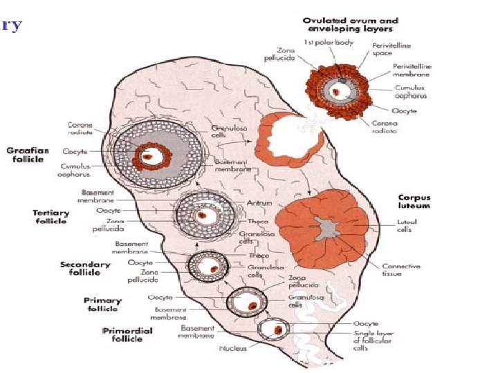
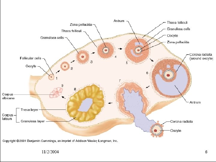
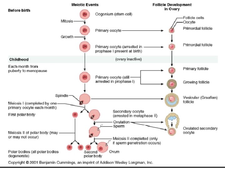
STRUCTURES OVARY • Covering epithelium • Follicles to mature & release the oocytes and to make hormones • Stroma of special connective-tissue cells that can become glandular theca cells • Glandular structures - corpora lutea • Degenerating structures as part of the cycle of activity • Central blood vessels FUNCTİONS • Releases one or two oocytes periodically • For genital & breast development • Determines womb’s endometrial state for receipt of the fertilized oocyte • Maintains secretory state of the endometrium (decidua after implantation) • Starts secretory processes in the breast • Feedback to hypothalamus for overall timing

OVARY SURFACE EPITHELIUM; Simple cuboidal & squamous STROMA; somewhat denser under the epithelium STROMAL CELLS ; ‘fibroblastic’ with potential to: 1 secrete steroids 2 produce thick collagen - fibrosis 3 become decidual cells (ectopic pregnancy) 4 become Leydig cells (near hilus) MEDULLA ; with convoluted vessels entering at the hilus

OVARY CORTEX Primordial FOLLICLES Primary, secondary, tertiary Atretic Theca interna GLANDULAR STRUCTURES Granulosa lining Corpora lutea Atretic follicles REMNANTS Corpora albicantes (Corpus albicans X 2, 3, )
")
IVF-ICSI (Intra Cytoplasmic Sperm Enjection)

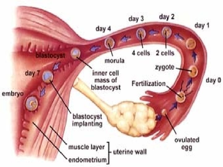
Embryo Development OPU Day 1 ET Day 2 COH IN VITRO CULTURE Day 3 Day 4 Day 6 Transfer Day 5

Early Cell Lineages in the Human Embryo n Delamination precedes gastrulation separating ICM into epiblast & hypoblast n Gastrulation occurs in epiblast (future embryo). n Gastrulation involves several types of movements and shape changes n The hypoblast will form the yolk sac n The epiblast will form embryo plus the amnion n Migration of cells of the epiblast through primitive streak leads to formation of mesoderm and endoderm n Ectoderm is left behind n Thus gastrulation results in the formation of the three primary embryonic germ layers: ectoderm, mesoderm and endoderm

Day 1 Day 2 Day 3 Day 4 Day 5 Totipotent Pluripotent
 Thompson , 1998")
Embryonic Stem Cells Trophectoderm ICM Evans ve Kaufman, 1981 (Mouse ES) Thompson , 1998 (Human ES)

CARNEGİE STAGES OF HUMAN DEVELOPMENT
 Size (mm) Images Events Fertilized Oocyte")
CARNEGIE STAGE FOR I. WEEK Stage Days (approx) Size (mm) Images Events Fertilized Oocyte 1 1 0, 1 - 0, 15 Charecteristic. Feature: Unicellularity Clevage continues 2 3 2 -3 4 -5 0, 1 -0, 2 begining and Charecteristic Feature: More than 1 cell but no blastocystic cavity. Free Blastocyst (Formulation of Blastocyst cavity)
 (approx) 5 -6 0,")
CARNEGIE STAGE FOR I-II. WEEK Stage 4 Days Size (mm) (approx) 5 -6 0, 1 - 0, 2 Images Events Attaching blastocyst Implantation 5 6 7 -12 0, 1 -0, 2 Week 2 13 -15 0, 2 extraembryonic mesoderm, primitive streak

ALL THREE GERM LAYERS ARE DERIVED FROM EPIBLAST n. Ectoderm: Outer epithelia and nervous system. n. Endoderm: Epithelial linings of the respiratory and digestive tracts; including the glandular cells of the liver and pancreas. n. Mesoderm: Smooth muscular coats, connective tissue, skeleton, striated muscle, reproductive and excretory organs, the vessels supplying these organs, and bloods cells and the bone marrow. n. OVERHEAD LIST OF STRUCTURES FORMED SUMMARIZE : Ectoderm Mesoderm central nervous system supporting tissues peripheral nervous cartilage system bone sensory epithelium of muscle ear, nose and eye connective tissue epidermis blood and lymph cells glands walls of heart, blood subcutaneous and lymph vessels mammary kidneys, gonads and pituitary associated ducts teeth enamel suprarenal gland cortex spleen Endoderm epithelial lining of gut respiratory tract urinary bladder and urethra tympanic cavity and auditory tube parenchyma of thyroid parathyroid glands liver stroma of tonsils and thymus
 (approx) 15")
CARNEGIE STAGE FOR III. WEEK Stage 7 8 9 Days Size (mm) (approx) 15 – 17 Week 3 17 -19 19 -21 0, 4 1, 0 -1, 5 -2, 5 Images Events Gastrulation, Notochordal process Primitive Pit, Notochordal Canal Somite Number 1 -3 Neural folds, cardiac primordium
 (approx) 22 -23 Week")
CARNEGIE STAGE FOR IV. WEEK Stage 10 Days Size (mm) (approx) 22 -23 Week 4 2 - 3, 5 11 23 -26 2, 5 -4, 5 12 26 -30 3 -5 Images Events Somite Number 4 -12 neural fold fuses Somite Number 13 -20 Rostral neuropore closes Somite Number 21 -29 caudal neuropore closes
 (approx) 13 28 -32 Week")
CARNEGIE STAGE FOR V. WEEK Stage Days Size (mm) (approx) 13 28 -32 Week 5 4 -6 14 31 -35 5 -7 15 35 -38 7 -9 Images Events Somite Number 30 leg buds, lens placode, pharyngeal arches Lens pit, optic cup Lens vesicle, nasal pit, hand plate
 (approx) 16 37 -42")
CARNEGIE STAGE FOR VI. -VII. WEEK Stage Days Size (mm) (approx) 16 37 -42 Week 6 8 -11 17 42 -44 11 -14 18 44 -48 Week 7 19 48 -51 13 -17 16 -18 Images Events Nasal pits moved ventrally, auricular hillocks foot plate Finger rays Ossification commences Straightening of trunk
 (approx)")
CARNEGIE STAGE FOR VIII. WEEK Stage 20 21 22 23 Days Size (mm) (approx) 5 1 - 53 Week 8 53 -54 54 -56 56 -60 Images Events 18 -22 Upper limbs longer and bent at elbow 22 -24 Hands and feets turned inward 23 -28 27 -31 Eyelids, ears external Rounded head, body and limbs

End of Week 8 Systems n n n Nervous CNS, PNS, Sensory Cardiovascular Heart, Blood Vessels Skeletal Axial, limbs, muscle, connective tissue Digestive Gastrointestinal tract and associated organs Urogenital Kidney, gonad Respiratory Upper respiratory tract, lungs
- Slides: 34