ANATOMY AND PHYSIOLOGY OF THE DIGESTIVE SYSTEM WHAT


 is covered by taste buds housed in")
 • Deglutition (swallowing) • Speech • Taste TEETH • The")
 teeth, ten are • • found")


")


. •")


The duodenum")


 are metabolized to short-chain fatty acids by bacteria in")


- Slides: 83
ANATOMY AND PHYSIOLOGY OF THE DIGESTIVE SYSTEM
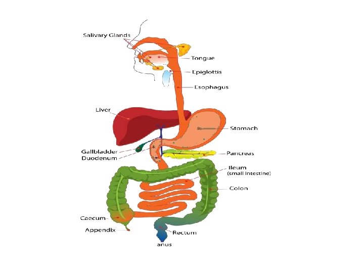
WHAT IS THE DIGESTIVE SYSTEM? The gastrointestinal tract (digestive tract, digestional tract, GIT, gut, or alimentary canal) is an organ system within humans and other animals which takes in food, digests it and absorb energy and nutrients, and expels the remaining waste as feces. The major organs of the digestive system: • • Mouth. Pharynx. Esophagus. Stomach. Small Intestine. Large Intestine. Rectum.
Accessory digestive organs: liver gallbladd er pancreas. Salivary gland Functions of GI tract • Ingestion: taking of food into the alimentary tract. i. e. eating & drinking. • Propulsion: mixes & moves the contents along the alimentary tract. • Digestion: consist of: • Mechanical breakdown of food e. g. mastication (chewing) • Chemical digestion of food into small molecules by enzymes.
• Absorption: this is the process by which digested food substances pass through the walls of some organs of the alimentary canal into the blood for circulation. • Elimination: food substances that have been eaten but cannot be digested & absorbed are excreted from the alimentary canal as faeces by the process of defaecation.
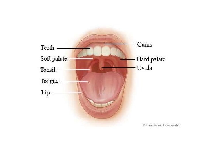
MOUTH The mouth is the first portion of the alimentary canal that receives food and produces saliva. The oral mucosa is the mucous membrane epithelium lining the inside of the mouth. Relations: • Anteriorly-lips • Posteriorly-continue with the oropharynx • Laterally-muscles of cheeks • Superiorly-bony hard palate • Inferiorly-muscular tongue & the soft tissues of the floor of the mouth •
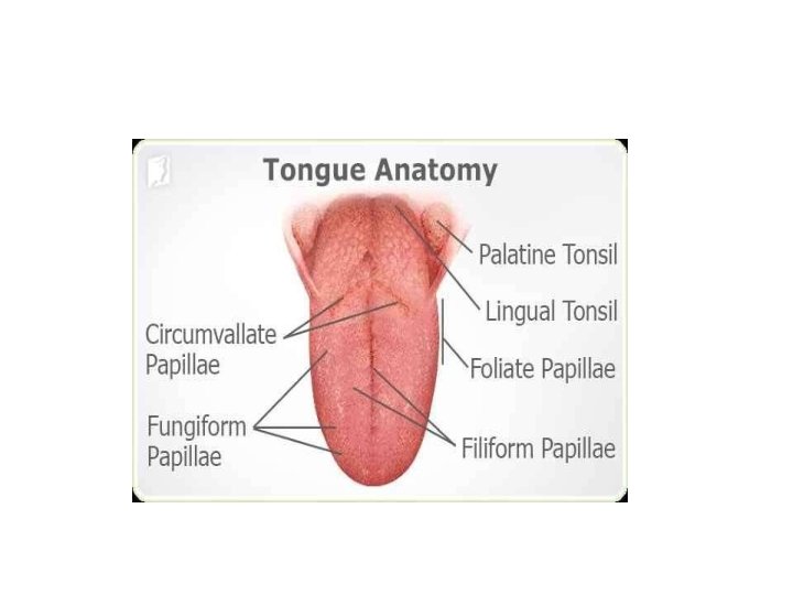
• The palate forms the roof of the mouth & is divided into the anterior hard palate & posterior soft palate. • The uvula is a curved fold of muscle covered with mucous membrane, hanging down from the middle. TONGUE • The tongue is a muscular organ in the mouth, that manipulates food for mastication, and is used in the act of swallowing. • It is of importance in the digestive system and is the primary organ of taste in the gustatory system.
• The tongue's upper surface (dorsum) is covered by taste buds housed in numerous lingual papillae. • The human tongue is divided into two parts, an oral part at the front and a pharyngeal part at the back. BLOOD SUPPLY • lingual artery • external carotid artery VENOUS DRAINAGE • lingual veins • internal jugular vein NERVE SUPPLY • hypoglossal nerve • Taste and sensation: glossopharyngeal nerve
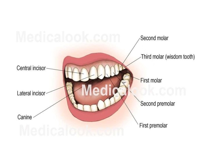
FUNCTIONS • Mastication (chewing) • Deglutition (swallowing) • Speech • Taste TEETH • The human teeth function to mechanically break down items of food by cutting and crushing them in preparation for swallowing and digesting. Humans have four types of teeth: incisors, canines, premolars, and molars, each with a specific function.
• PRIMARY TEETH • Among deciduous (primary) teeth, ten are • • found in the maxilla (upper jaw) and ten in the mandible (lower jaw), for a total of 20. The dental formula for primary teeth is 2. 1. 0. 2/2. 1. 0. 2. Start to come in (erupt) at about 6 months of age In the primary set of teeth, two types of incisors – centrals and laterals, one canine & two types of molars – first and second. All primary teeth are normally later replaced with their permanent counterparts.
PERMANENT TEETH • Among permanent teeth, 16 are found in the maxilla and 16 in the mandible, for a total of 32. The dental formula is 2. 1. 2. 3/2. 1. 2. 3. • Age 21, all 32 of the permanent teeth have usually erupted. The permanent teeth are the: • Two incisor (for cutting)-central incisor, lateral incisor • One canine (for tearing) • Two premolar(for crushing)-first premolar, second premolar, • Three molar (for grinding)-first molar, second molar, and third molar.
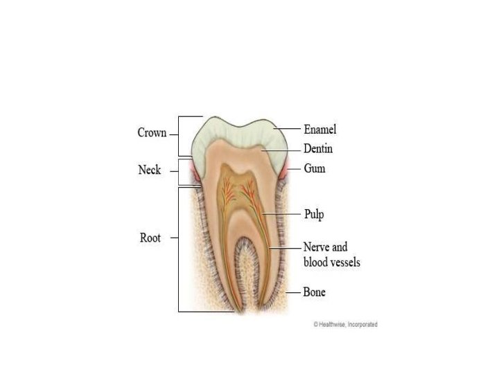
PARTS ENAMEL • Enamel is the hardest and most highly mineralized substance of the body. • It is one of the four major tissues which make up the tooth, along with dentin, cementum, and dental pulp. • 96% of enamel consists of mineral, with water and organic material comprising the rest. • The normal color of enamel varies from light yellow to grayish white. DENTIN • Dentin is the substance between enamel or cementum and the pulp chamber. • The porous, yellow-hued material is made up of 70% inorganic materials, 20% organic materials, and 10% water by weight
• Dentin is a mineralized connective tissue with an organic matrix of collagenous proteins. CEMENTUM • Cementum is a specialized bone like substance covering the root of a tooth. • Its coloration is yellowish and it is softer than dentin and enamel. DENTAL PULP • The dental pulp is the central part of the tooth filled with soft connective tissue. • This tissue contains blood vessels and nerves that enter the tooth from a hole at the apex of the root.
FUNCTIONS OF TEETH • • Two incisor -for cutting One canine -for tearing Two premolar-for crushing Three molar-for grinding ERUPTION • Tooth eruption in humans is a process in tooth development in which the teeth enter the mouth and become visible. • Primary teeth erupt into the mouth from six months until two years of age. around
BLOOD SUPPLY • Maxillary arteries VENOUS DRAINAGE Internal jugular veins NERVE SUPPLY Maxillary nerves Mandibular nerves
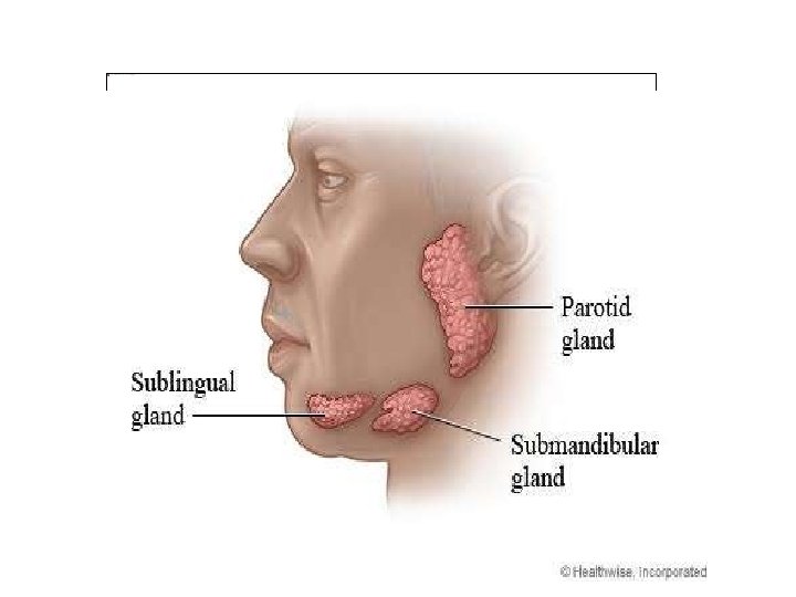
SALIVARY GLANDS • The salivary glands in are exocrine glands that produce saliva through a system of ducts. Humans have 3 paired major salivary glands: • Parotid • submandibular and • Sublingual • as well hundreds of minor salivary glands. Parotid glands • The two parotid glands are major salivary glands wrapped around the mandibular ramus in humans. • The largest of the salivary glands.
• They secrete saliva to facilitate mastication and swallowing, and amylase to begin the digestion of starches. • It enters the oral cavity via the parotid duct. Submandibular glands • The submandibular glands are a pair of major salivary glands located beneath the lower jaws, superior to the digastric muscles. • The secretion produced is a mixture of both serous fluid and mucus, and enters the oral cavity via the submandibular duct.
Sublingual glands • The sublingual glands are a pair of major salivary glands located inferior to the tongue, anterior to the submandibular glands. • Approximately 5% of saliva entering the oral cavity comes from these glands. • The secretion produced is mainly mucous in nature Minor salivary glands • There are 800 to 1, 000 minor salivary glands located throughout the oral cavity within the submucosa of the oral mucosa in the tissue of the buccal, and lingual mucosa
BLOOD SUPPLY • External carotid artery VENOUS DRAINAGE • Jugular veins COMPOSITION OF SALIVA • About 1. 5 litres of saliva is produced daily & it consists of: • Water • Mineral salts • An enzyme • Mucus • Lysozyme • Immunoglobulins
FUNCTION OF SALIVA • Saliva contributes to the digestion of food and to the maintenance of oral hygiene. • Without normal salivary function the frequency of dental caries, gum disease and other oral problems increases significantly. Lubricant • Saliva, coats the oral mucosa, mechanically protecting it from trauma during eating, swallowing and speaking. • In people with little saliva soreness of the mouth is very common, and the food (especially dry food) sticks to the inside of the mouth.
Digestion • The digestive functions of saliva include moistening food and helping to create a food bolus. • This lubricative function of saliva allows the food bolus to be passed easily from the mouth into the esophagus. Role in taste • Saliva is very important in the sense of taste. • It is the liquid medium in which chemicals are carried to taste receptor cells (mostly associated with lingual papillae).
• Other Function • Saliva maintains the p. H of the mouth. Saliva is supersaturated with various ions. THE PHARYNX • The pharynx is the part of the throat that is behind the mouth and nasal cavity and above the esophagus and the larynx, or the tubes going down to the stomach and the lungs. • The pharynx is the portion of the digestive tract that receives the food from your mouth. • Branching off the pharynx is the esophagus, which carries food to the stomach,
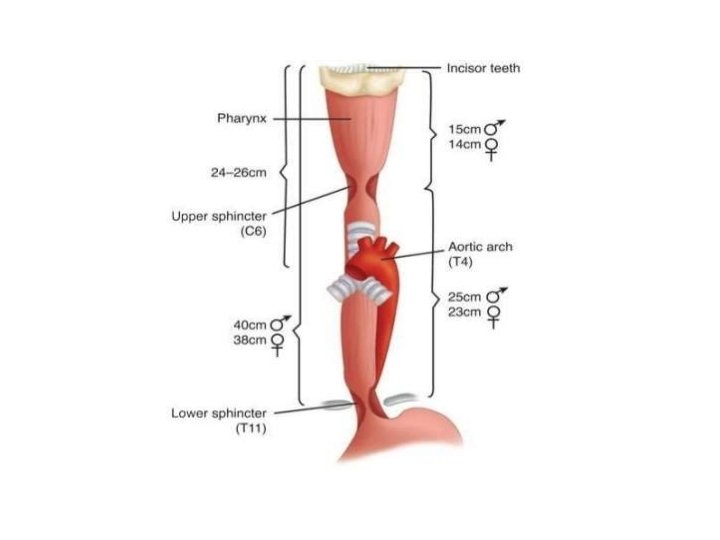
• • THE ESOPHAGUS Theesophagus oroesophagus, commonly known as the food pipe or gullet, The esophagus is a muscular tube connecting the throat (pharynx) with the stomach. The esophagus runs behind the windpipe (trachea) and heart, and in front of the spine. Length : 25 cm Diameter: 2 cm
STRUCTURE • The wall of the esophagus from the lumen outwards consists of mucosa, submucosa (connective tissue), layers of muscle fibers between layers of fibrous tissue, and an outer layer of connective tissue. • The mucosa is a stratified squamous epithelium of around three layers of squamous cells, which contrasts to the single layer of columnar cells of the stomach. • Most of the muscle is smooth muscle although striated muscle predominates in its upper third.
• It has two muscular rings or sphincters in its wall, one at the top and one at the bottom. • A sphincter is a circular muscle that normally maintains constriction of a natural body passage or orifice and which relaxes as required by normal physiological functioning. • The lower sphincter helps to prevent reflux of acidic stomach content.
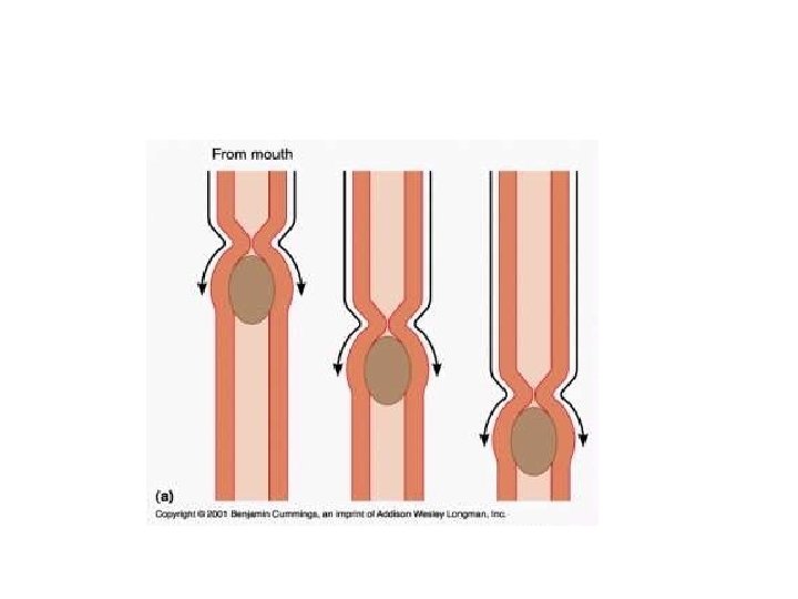
FUNCTIONS Formation of a bolus Swallowing • Food is ingested through the mouth and when swallowed passes first into the pharynx and then into the esophagus. Reducing gastric reflux • Constriction of the upper and lower esophageal sphincters help to prevent reflux (backflow) of gastric contents and acid into the esophagus, protecting the esophageal mucosa.
Blood supply • Oesophageal arteries • Inferior phrenic arteries • Venous drainage • Left gastric vein
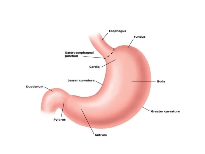
STOMACH • The stomach is a muscular organ located on the left side of the upper abdomen. The stomach receives food from the esophagus. • As food reaches the end of the esophagus, it enters the stomach through a muscular valve called the lower esophageal sphincter.
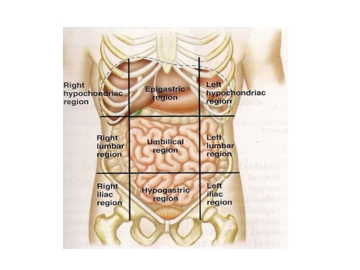
Relations: • Anteriorly-left lobe of liver & anterior abdominal wall • Posteriorly-abdominal aorta, pancreas, spleen, left kidney • Superiorly-diaphragm, oesophagus & left lobe of liver • Inferiorly-transverse colon & small intestine • Left side-diaphragm & spleen • Right side-liver & duodenum
• A pouch-like organ primarily designed for food storage (for 2 -4 hours) , some mechanical and chemical digestion also occur. • Contains two sphincters at both ends to regulate food movement : • cardiac sphincter near the esophagus , • pyloric sphincter near the small intestine. Divided into 4 regions : • cardiac stomach (or cardiac), • fundic stomach (or funded) , • body of stomach • pyloric stomach (or Pylorus).
Contain thick folds called rugae at its layer , for providing larger surface area for expansion, secretion , digestion , and some absorption. FUNCTIONS • Digestion • The stomach releases proteases (proteindigesting enzymes such as pepsin) and hydrochloric acid, which kills or inhibits bacteria and provides the acidic p. H of 2 for the proteases to work. • Food is churned by the stomach through muscular contractions of the wall called peristalsis •
• Absorption • some absorption of certain small molecules nevertheless does occur in the stomach through its lining GASTRIC JUICE • Gastric acid, gastric juice or stomach acid, is a digestive fluid formed in the stomach and is composed of hydrochloric acid (HCl), potassium chloride (KCl) and sodium chloride (Na. Cl). • The acid plays a key role in digestion of proteins, by activating digestive enzymes, and making ingested proteins unravel so that digestive enzymes break down the long chains of amino acids.
• Gastric Secretory Cells • Chief cells: secrete pepsinogen (an inactive enzyme). • Parietal cells: secrete hydrochloric and (HCl) and "intrinsic factor" (which helps absorption of vitamin B 12 in the intestines). • Mucous cells: secrete mucus and alkaline substances to help neutralize HCl in the gastric juice. • G cells: secrete a hormone called gastrin , which stimulates the parietal cells and overall gastric.
• Blood supply • right gastroepiploic artery • left gastroepiploic artery • gastric artery • Venous drainage • gastric vein
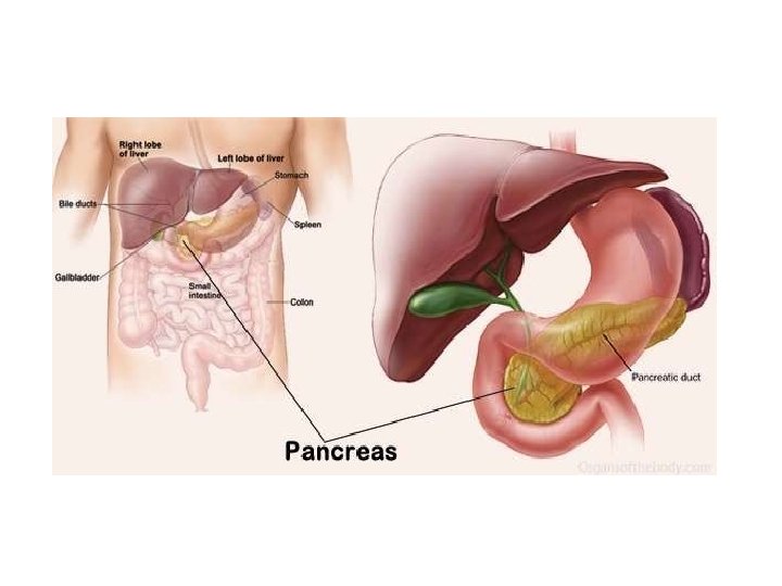
THE PANCREAS •
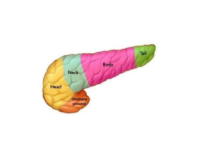
Structure • Anatomically, the pancreas is divided into the head of pancreas, • the neck of pancreas, • the body of pancreas, • and the tail of pancreas. • The neck is about 2. 5 cm or 1 inch long and lies between the head and the body • The body is the largest part of the pancreas and lies behind the pylorus. • The tail ends by abutting the spleen.
BLOOD SUPPLY • superior mesenteric artery. • splenic artery VENOUS DRAINAGE • superior mesenteric veins • Splenic veins
FUNCTION • The pancreas is involved in blood sugar control and metabolism within the body. • Sugar control and metabolism • pancreatic islets are present in the pancreas. • Within these islets are four main types of cells which are involved in the regulation of blood glucose levels. • Each type of cell secretes a different type of hormone: • α alpha cells secrete glucagon(increase glucose in blood) • β beta cells secrete insulin (decrease glucose in blood) • δ delta cells secrete somatostatin (regulates/stops α and β cells) and • γ (gamma) cells, secrete pancreatic polypeptide.
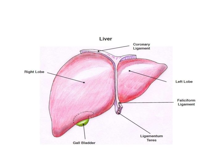
THE LIVER • The liver, an organ only found in vertebrates, detoxifies various metabolites, synthesizes proteins, and produces biochemical necessary for digestion Relations: • Anteriorly-diaphragm & anterior abdominal wall • Posteriorly-oesophagus, inferior vena cava, aorta, gall bladder, vertebral column & diaphragm • Laterally-lower ribs & diaphragm • Superiorly-diaphragm & anterior abdominal wall • Inferiorly- stomach, bile ducts, duodenum, hepatic flexure of colon, right kidney
STRUCTURE • The liver is a reddish-brown wedgeshaped organ with four lobes of unequal size and shape. • weighs 1. 44– 1. 66 kg • width -15 cm. • It is both the heaviest internal organ and the largest gland in the human body.
• The liver is grossly divided into two parts when viewed from above – a right and a left lobe. • The falciform ligament, divides the liver into a left and right lobe. FUNCTIONS • Synthesis • Proteins produced and secreted by the liver. • The liver plays a major role in carbohydrate, protein, amino acid, and lipid metabolism. Breakdown • The liver is responsible for the breakdown of insulin and other hormones.
• The liver breaks down bilirubin via glucuronidation, facilitating its excretion into bile. Other • The liver stores a multitude of substances, including glucose (in the form of glycogen) • vitamin A (1– 2 years' supply) • vitamin D (1– 4 months' supply) • vitamin B 12 (3– 5 years' supply) • vitamin K, iron, and copper. • The liver produces albumin, the most abundant protein in blood serum.
• Contains phagocytes to destroy damaged erythrocytes and foreign substances, using phagocytosis. BLOOD SUPPLY • Hepatic artery VENOUS DRAINAGE • Hepatic veins
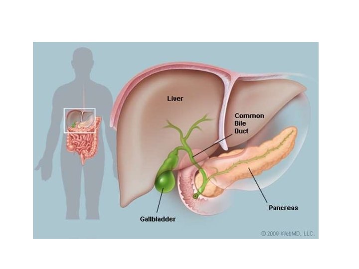
THE GALLBLADDER • The gallbladder is a small hollow organ where bile is stored and concentrated before it is released into the small intestine. • In humans, the pear-shaped gallbladder lies beneath the liver.
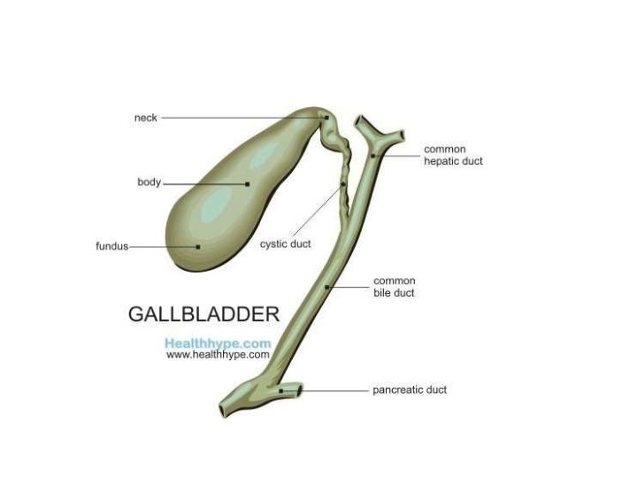
STRUCTURE • sits in a shallow depression below the right lobe of the liver, that is grey-blue in life. • Length-7 to 10 cm or 2. 8 to 3. 9 inches • Diameter -4 cm or 1. 6 inch • The gallbladder has a capacity of about 50 millilitres • The gallbladder is shaped like a pear, with its tip opening into the cystic duct. Gallbladder is divided into three sections: • the fundus, • The body, • and the neck.
FUNCTIONS • The main purpose of the gallbladder is to store bile, also called gall, needed for the digestion of fats in food. • bile flows through small vessels into the larger hepatic ducts and ultimately though the cystic duct into the gallbladder, where it is stored.
BLOOD SUPPLY • the cystic artery VENOUS DRAINAGE • the cystic veins
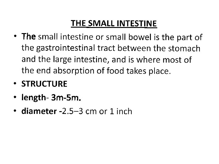
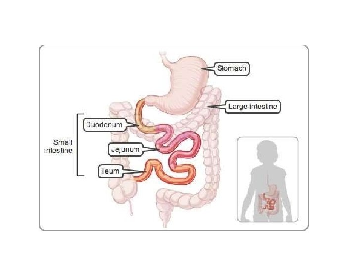
PARTS • The small intestine is divided into three structural parts: • (I)The duodenum • (II)The jejunum • (III)The ileum The duodenum is a short structure ranging from 20 cm to 25 cm in length, and shaped like a "C". The jejunum is the midsection of the small intestine, connecting the duodenum to the ileum. It is about 2. 5 m long.
• The ileum is the final section of the small intestine. It is about 3 m long, and contains villi similar to the jejunum. FUNCTIONS • Digestion • The small intestine is where most chemical digestion takes place. • Many of the digestive enzymes that act in the small intestine are secreted by the pancreas and liver and enter the small intestine via the pancreatic duct. • Digestion of proteins & carbohydrate
Absorption • Digested food is now able to pass into the blood vessels in the wall of the intestine through either diffusion or active transport. • The small intestine is the site where most of the nutrients from ingested food are absorbed. Immunological • The small intestine supports the body's immune system. • The presence of gut flora appears to contribute positively to the host's immune system.
BLOOD SUPPLY • the coeliac trunk • the superior mesenteric artery VENOUS DRAINAGE • the superior mesenteric veins
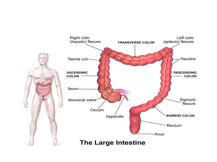
THE LARGE INTESTINE • The large intestine, also known as the large bowel or colon, is the last part of the gastrointestinal tract and of the digestive system in vertebrates. • Water is absorbed here and the remaining waste material is stored as feces before being removed by defecation.
STRUCTURE • The length of male colon is 166 cm. • female colon 155 cm • The colon consists of five sections: • the cecum • ascending colon, • the transverse colon, • the descending colon, • the sigmoid colon and the rectum.
Sections of the colon are: • The ascending colon including the cecum and appendix • The transverse colon including the colic flexures and transverse mesocolon • The descending colon • The sigmoid colon – the s-shaped region of the large intestine
• The average inner diameter of sections of the colon in centimeters • cecum 8. 7 cm • ascending colon 6. 6 cm • transverse colon 5. 8 cm • descending/sigmoid colon 6. 3 cm • and rectum near rectal/sigmoid junction 5. 7 cm
• The cecum is the first section of the colon and involved in the digestion, while the appendix is a structure of the colon, not involved in digestion. • The function of the appendix is uncertain. • Containing Ileocecal valve • The ileocecal valve is a sphincter muscle valve that separates the small intestine and the large intestine. • Its critical function is to limit the reflux of colonic contents into the ileum.
• The ascending colon • It is connected to the small intestine by a section of bowel called the cecum. • The ascending colon runs upwards through the abdominal cavity toward the transverse colon for approximately eight inches or 20 cm. • The unwanted waste material is moved upwards toward the transverse colon by the action of peristalsis. •
Transverse colon • The transverse colon is the part of the colon from the hepatic flexure to the splenic flexure. Descending colon • The descending colon is the part of the colon from the splenic flexure to the beginning of the sigmoid colon, descending colon is also called the distal gut. • One function of the descending colon in the digestive system is to store feces that will be emptied into the rectum.
Sigmoid colon • The sigmoid colon is the part of the large intestine after the descending colon and before the rectum. • The name sigmoid means S-shaped. • The walls of the sigmoid colon are muscular, and contract to increase the pressure inside the colon, causing the stool to move into the rectum. Rectum • The rectum is the last section of the large intestine. It holds the formed feces awaiting elimination via defecation.
The anus • The anus is the external opening of the rectum. • Its function is to control the expulsion of feces. • Two sphincters control the exit of feces from the body during an act of defecation. • These are the internal anal sphincter and the external anal sphincter, which are circular muscles that normally maintain constriction of the orifice and which relaxes as required by normal physiological functioning.
FUNCTIONS • The large intestine absorbs water and any remaining absorbable nutrients from the food before sending the indigestible matter to the rectum. • The colon absorbs vitamins that are created by the colonic bacteria, such as vitamin K. • Gut flora • The large intestine houses over 700 species of bacteria that perform a variety of functions. • The large intestine absorbs some of the products formed by the bacteria inhabiting this region.
• Undigested polysaccharides (fiber) are metabolized to short-chain fatty acids by bacteria in the large intestine. BLOOD SUPPLY • the superior mesenteric artery(SMA) • and inferior mesenteric artery VENOUS DRAINAGE • the inferior mesenteric vein • the superior mesenteric vein
PHYSIOLOGY OF DIGESTION • The mouth is the beginning of the digestive tract. • Chewing breaks the food into pieces that are more easily digested, while saliva mixes with food to begin the process of breaking it down into a form your body can absorb and use. • From pharynx food travels to the esophagus or swallowing tube. • By means of a series of contractions, called peristalsis, the esophagus delivers food to the stomach. • The lower esophageal sphincter keep food from passing backwards into the esophagus.
• The stomach secretes acid and powerful enzymes that continue the process of breaking down the food. • When it leaves the stomach, food is the consistency of a liquid or paste. • From there the food moves to the small intestine. • The small intestine continues the process of breaking down food by using enzymes released by the pancreas and bile from the liver. • Bile is a compound that aids in the digestion of fat and eliminates waste products from the blood.
• Peristalsis is also at work in this organ, moving food through and mixing it up with digestive secretions. • The duodenum is largely responsible for continuing the process of breaking down food, with the jejunum and ileum being mainly responsible for the absorption of nutrients into the bloodstream. • pancreas secretes enzymes into the small intestine. • These enzymes break down protein, fat, and carbohydrates from the food we eat.
• Stool, or waste left over from the digestive process, is passed through the colon by means of peristalsis, first in a liquid state and ultimately in solid form as the water is removed from the stool. • A stool is stored in the sigmoid colon until a "mass movement" empties it into the rectum once or twice a day.