
Anatomy and Physiology Biology 2401 Chapter26 The Urinary
Anatomy and Physiology Biology 2401 Chapter-26 The Urinary System
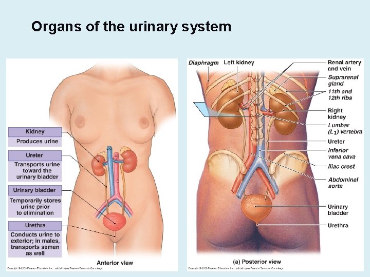
Organs of the urinary system
Urinary System Functions • Three Functions of the Urinary System 1. Excretion • Removal of organic wastes from body fluids 2. Elimination • Discharge of waste products 3. Homeostatic regulation • Regulates blood volume and blood pressure • Regulates plasma ion concentrations • Helps stabilize blood p. H • Conserves valuable nutrients • Assists liver in detoxifying toxins
The Kidneys • Each Kidney Is Protected and Stabilized – By three concentric layers of connective tissue 1. Fibrous capsule • Covers outer surface of entire organ 2. Perinephric fat capsule • A thick layer of adipose tissue, Surrounds fibrous capsule 3. Renal fascia • A dense, fibrous outer layer, anchors kidney to surrounding structures
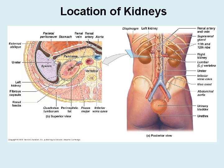
Location of Kidneys

Kidneys • The kidney is composed of several layers: – Renal capsule: fibrous layer covering the kidney. – Cortex: outer layer of the kidney, contains the major portion of the nephrons (urine production units), and blood vessels. – Medulla: middle layer of the kidney; composed of the triangular shaped renal pyramids and the renal columns. • The pyramids contain the collecting ducts and loops of Henle, the lower portion of the nephrons. • The renal columns are regions between the pyramids in which blood vessels run to and from the cortex. • The papilla of each pyramid projects into a funnel-shaped area known as the calyx. • Calyces collect the urine released from the papillae and allow it to drain into an enlarged collection area known as the renal pelvis and then into the ureter.
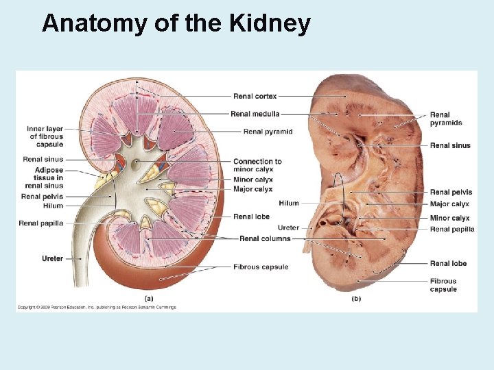
Anatomy of the Kidney
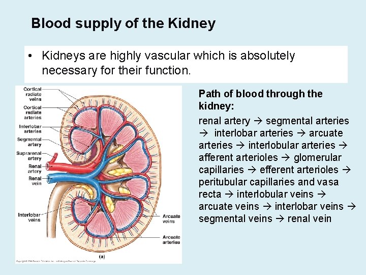
Blood supply of the Kidney • Kidneys are highly vascular which is absolutely necessary for their function. Path of blood through the kidney: renal artery segmental arteries interlobar arteries arcuate arteries interlobular arteries afferent arterioles glomerular capillaries efferent arterioles peritubular capillaries and vasa recta interlobular veins arcuate veins interlobar veins segmental veins renal vein
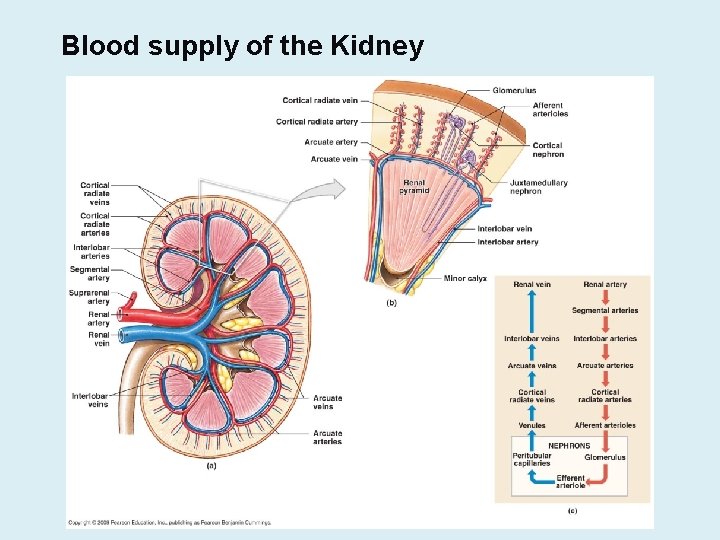
Blood supply of the Kidney
The Nephron • functional units of the kidneys. • The nephron receives blood from the afferent arteriole. • The afferent arteriole supplies the glomerulus, an intricate network of capillary with a unique structure. • From the glomerulus blood flows into the efferent arteriole. • The efferent arteriole flows into more capillaries, the peritubular capillaries, and, in juxtamedullary nephrons (see below), the vasa recta. • Peritubular capillaries and vasa recta lead to the venous drainage of the kidney.
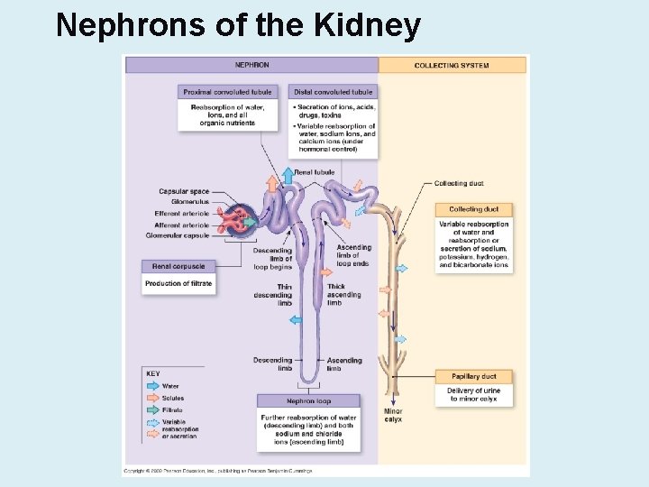
Nephrons of the Kidney

Types of Nephrons • Cortical nephron (85%) vs Juxtamedullary nephrons (15%)
Renal Corpuscle • Bowman’s capsule and glomerulus. • Filtration unit of the nephron • The glomerulus is a condensed mass of capillaries which allows substances to escape by filtration. • Glomerular capillaries are surrounded by specialized cells called podocytes. • Podocytes form the inner (visceral) layer of Bowman's capsule. • Podocytes have processes called pedicels which interdigitate to produce openings called filtration slits. • Glomerular capillaries are fenestrated in order to allow filtration. • The outer (parietal) layer of Bowman's capsule consists of epithelial cells with tight junctions and serves to contain the filtrate in the capsular space.
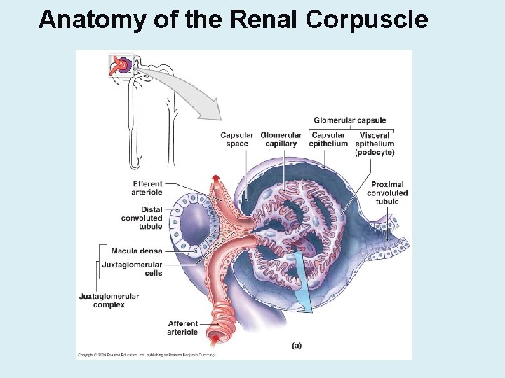
Anatomy of the Renal Corpuscle
Cells of the Tubular Portion of the Nephron • As in the renal corpuscle cells of the tubular portion of the nephron are critical to its function: – Proximal convoluted tubule: simple cuboidal epithelial cells with numerous microvilli (for absorption) – Thin portion of loop of henle: simple squamous cells without microvilli (permeable to water). – Thick portion of loop of Henle: simple cuboidal cells with very few microvilli (waterproof). – Distal convoluted tubule: simple cuboidal cells with few microvilli; called principal cells (hormonally regulated) – Collecting duct: simple cuboidal cells called intercalated cells and principal cells. Intercalated cells have microvilli. Principal and intercalated cells are hormonally regulated.

Cells of the Nephron
Specialized Cells of the Nephron • Juxtaglomerular cells or JG cells are specialized cells of the afferent arteriole. • Macula densa cells are specialized cells of the distal convoluted tubule. • These two types of cells are in close proximity of each other. Together they form the Juxtaglomerular Apparatus (JGA) • JGA regulates blood pressure and the rate of filtration of the kidney by secreting renin. • Also produces the hormone erythropoietin.
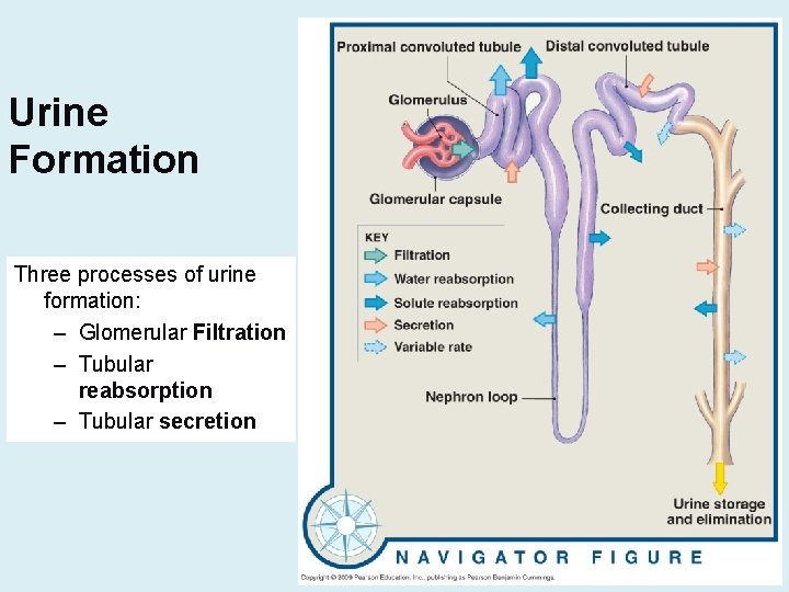
Urine Formation Three processes of urine formation: – Glomerular Filtration – Tubular reabsorption – Tubular secretion
Glomerular Filtration • Blood pressure forces water and small molecules out of the blood plasma into the Bowman's capsule. • Large molecules such as blood proteins, or blood cells should not pass through. • Filtration averages 125 ml/min for the two kidneys. This amounts to about 180 Liters per day. • Since we urinate an average of 1500 ml per day, more than 99% must be returned to the blood. • Filtration is based on size: small molecules such as water, electrolytes, urea, glucose, amino acids can pass through the membrane • The large amount of filtration is the consequence of the porous 3 -layered filtration membrane.
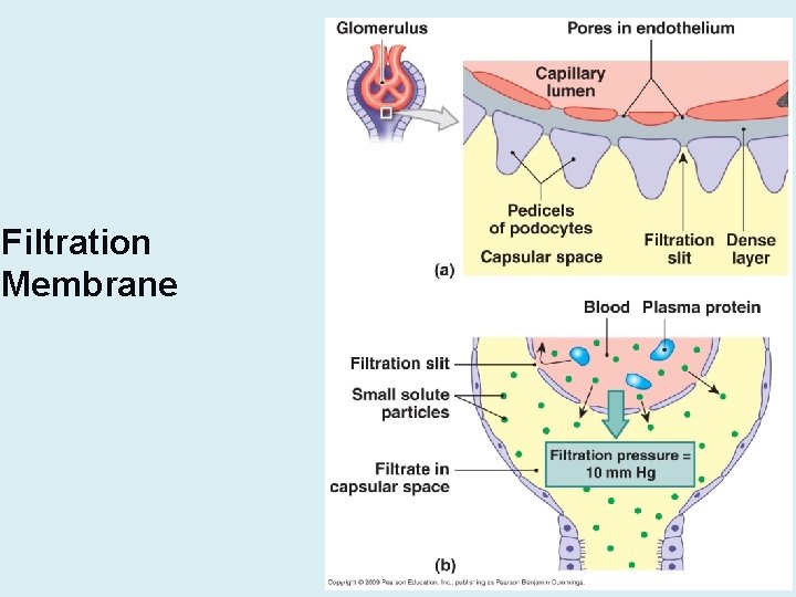
Filtration Membrane
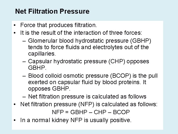
Net Filtration Pressure • Force that produces filtration. • It is the result of the interaction of three forces: – Glomerular blood hydrostatic pressure (GBHP) tends to force fluids and electrolytes out of the capillaries. – Capsular hydrostatic pressure (CHP) opposes GBHP. – Blood colloid osmotic pressure (BCOP) is the pull exerted on capsular fluid by blood proteins. It opposes GBHP. – Net filtration pressure is calculated as follows • Net filtration pressure (NFP) is calculated as follows: NFP = GBHP – CHP – BCOP • In a normal kidney NFP is usually positive.
Glomerular Filtration Rate • GFR is highly regulated because wastes and undesirable substances must be removed from the blood constantly, and blood volume must be constant to allow adequate hydration of the body. • GFR is very sensitive to changes in NFP, which is sensitive to changes in blood pressure. • The three mechanisms of GFR regulation are renal autoregulation, neural regulation, and hormonal regulation. • These 3 mechanisms work together to maintain a constant GFR.
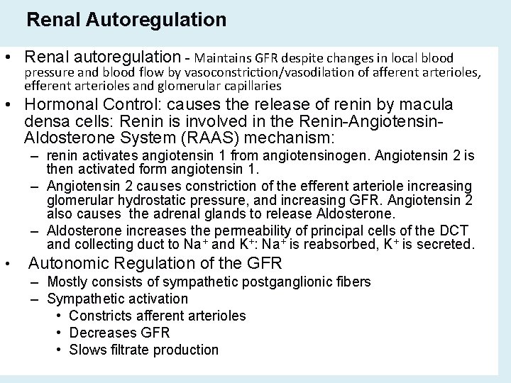
Renal Autoregulation • Renal autoregulation - Maintains GFR despite changes in local blood pressure and blood flow by vasoconstriction/vasodilation of afferent arterioles, efferent arterioles and glomerular capillaries • Hormonal Control: causes the release of renin by macula densa cells: Renin is involved in the Renin-Angiotensin. Aldosterone System (RAAS) mechanism: – renin activates angiotensin 1 from angiotensinogen. Angiotensin 2 is then activated form angiotensin 1. – Angiotensin 2 causes constriction of the efferent arteriole increasing glomerular hydrostatic pressure, and increasing GFR. Angiotensin 2 also causes the adrenal glands to release Aldosterone. – Aldosterone increases the permeability of principal cells of the DCT and collecting duct to Na+ and K+: Na+ is reabsorbed, K+ is secreted. • Autonomic Regulation of the GFR – Mostly consists of sympathetic postganglionic fibers – Sympathetic activation • Constricts afferent arterioles • Decreases GFR • Slows filtrate production
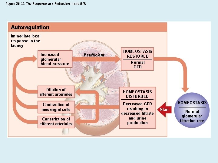
Figure 26 -11 The Response to a Reduction in the GFR Autoregulation Immediate local response in the kidney Increased glomerular blood pressure Dilation of afferent arterioles Contraction of mesangial cells Constriction of efferent arterioles if sufficient HOMEOSTASIS RESTORED Normal GFR HOMEOSTASIS DISTURBED Decreased GFR resulting in decreased filtrate and urine production HOMEOSTASIS Start Normal glomerular filtration rate
Tubular Reabsorption • Substances are returned to the blood and interstitial fluid. • The major substances reabsorbed are water, Na. Cl, glucose, and amino acids. • Some of the urea, together with other salts are also reabsorbed. • Substances may be reabsorbed through the cell (transcellular pathway), or they can pass between cells (paracellular pathway) via diffusion or via primary and secondary active transport.
Transport Maximum • Nutrients such as glucose and amino acid must be completely reabsorbed from the PCT. • In fact there is a limit to the rate of tubular reabsorption called Transport maximum (Tm): tubular cells can only add a limited number of channel proteins or carrier molecules to their plasma membrane. • Consequently if the quantity of nutrients exceed the transport capacity of the cells, the excess nutrients are not reabsorbed and pass into urine. • When blood glucose levels are very high such as in diabetes mellitus, a large amount of glucose passes into the filtrate.
Role of Sodium in Reabsorption • Sodium ions are essential for reabsorption. • Na+ / K+ ATPase pumps remove Na+ from the absorptive cells and send them to the renal interstitium. • This increases the osmolarity of the interstitium, and lowers the osmolarity of the absorptive cells. • Consequently cells absorb sodium, water, and other substances via a variety of mechanisms depending on the nature of the substances. • Nearly all nutrients such as glucose and amino acids as well as 65% of water are reabsorbed in the proximal convoluted tubule. • The remaining filtrate contains water, urea, sodium and other electrolytes that will be reabsorbed later.

Reabsorption of Water • Water is reabsorbed by osmosis. • 65% of water reabsorption occurs from the PCT. • The squamous cells of the thin descending limb of the loop of Henle are permeable to water but not to sodium. Additionally the filtrate is exposed to increasingly hypertonic medulla. • These two phenomena force another 20% of absorbable water out of the filtrate from the descending limb • Reabsorption in this area is termed obligatory because it must occur due to the osmolarity of the surrounding interstitial fluid. • The cells of the ascending limb of the loop of Henle are impermeable to water. • When the filtrate enters the collecting duct it is once again exposed to the hypertonicity of the deep medulla. • However reabsorption of water from the collecting duct is controlled by the hormone ADH (Antidiuretic Hormone).
Role of ADH in Reabsorption of Water • ADH is a hormone produced by the hypothalamus and released from the posterior pituitary gland on command of the hypothalamus. • Such a command is generated in response to high blood osmolarity which occurs during water loss and dehydration from sweating, vomiting, and also from lack of adequate hydration (low fluid intake). • ADH allows water to be reabsorbed from the collecting duct and not lost in urine. The water is reabsorbed by osmosis driven by medullary hypertonicity • Principal cells of the collecting duct are responsible for water reabsorption. They are sensitive to ADH, and respond to this hormone by increasing the number of aquaporins in their plasma membrane. This increases the reabsorption of water from the collecting duct. The urine produced is very concentrated. • Overhydration causes low blood osmolarity which slows the release of ADH from the neurohypophysis. As a result a dilute urine is produced. • ADH deficiency causes the production of a large amount of dilute urine, a condition called diabetes insipidus.

The Countercurrent Multiplier • This mechanism works in the loop of Henle to increase water reabsorption from the descending limb as a result of salt reabsorption from the ascending limb. • The fact that filtrate moves in opposite directions in the two limbs of the loop amplifies the effect of transport from one limb on transport from the other limb. • In fact the cells of the ascending limb are permeable to Na. Cl but impermeable to water: as a result Na. Cl is removed from the filtrate and the osmolarity of the surrounding interstitium is increased. • Since the osmolarity of the filtrate decreases as fluid flows up the ascending limb, an osmolarity gradient is established: the medullary region has a greater osmolarity than the cortical region. • Cells of the descending limb are impermeable to Na. Cl but very permeable to water: since the filtrate is moving down the descending limb a large amount of water is removed from the filtrate which becomes more and more concentrated.
Reabsorption of Urea • Urea is a waste product of protein metabolism. • It is passively reabsorbed from the nephron and this contributes to keep the surrounding interstitial fluid hypertonic, pulling water. • This same urea will be filtered later and may in fact be reabsorbed again in the collecting duct • Overall, more urea passes into urine than is reabsorbed causing a net loss of urea from the body.
Secretion • Secretion is the release of substances into the filtrate by active transport. • It is accomplished by the tubular cells. • The substances secreted into the filtrate are mainly derived from the blood in the peritubular capillaries. • Secretion occurs concurrently with reabsorption although it is the third process we consider. • It occurs in the proximal convoluted tubule, the distal convoluted tubule, and the collecting duct.
Purposes of Secretion • The first purpose of secretion is to eliminate any remaining toxins and drugs which have not been filtered: these substances flow from the peritubular capillaries directly into the tubules of the nephron and are passed into urine. • The second purpose of secretion is to establish electrolyte balance. – Reabsorption of Na+ causes an imbalance of charges that is corrected by the secretion of positively charged ions such as K+. – Negatively charged ions such as Cl- will either be secreted or will diffuse down their electrochemical gradient. – Bicarbonate ions are always retained by blood because they act as buffers.
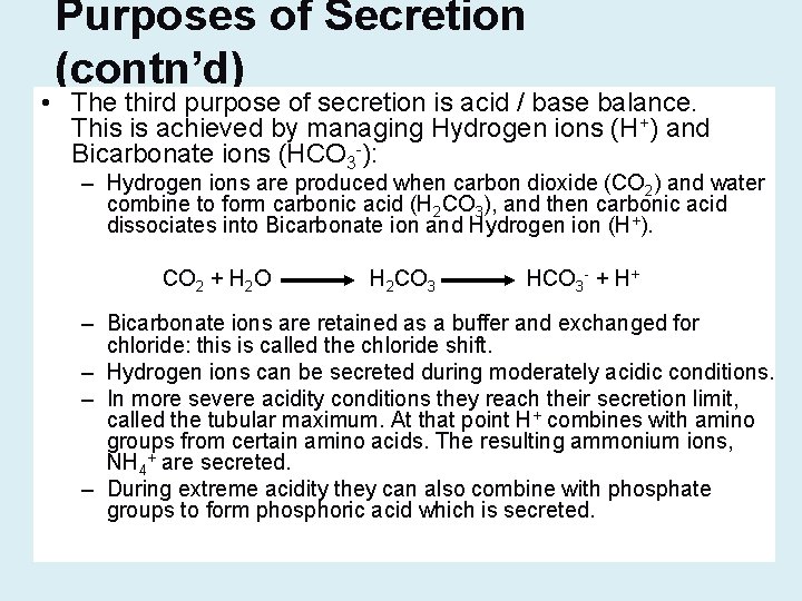
Purposes of Secretion (contn’d) • The third purpose of secretion is acid / base balance. This is achieved by managing Hydrogen ions (H+) and Bicarbonate ions (HCO 3 -): – Hydrogen ions are produced when carbon dioxide (CO 2) and water combine to form carbonic acid (H 2 CO 3), and then carbonic acid dissociates into Bicarbonate ion and Hydrogen ion (H+). CO 2 + H 2 O H 2 CO 3 HCO 3 - + H+ – Bicarbonate ions are retained as a buffer and exchanged for chloride: this is called the chloride shift. – Hydrogen ions can be secreted during moderately acidic conditions. – In more severe acidity conditions they reach their secretion limit, called the tubular maximum. At that point H+ combines with amino groups from certain amino acids. The resulting ammonium ions, NH 4+ are secreted. – During extreme acidity they can also combine with phosphate groups to form phosphoric acid which is secreted.
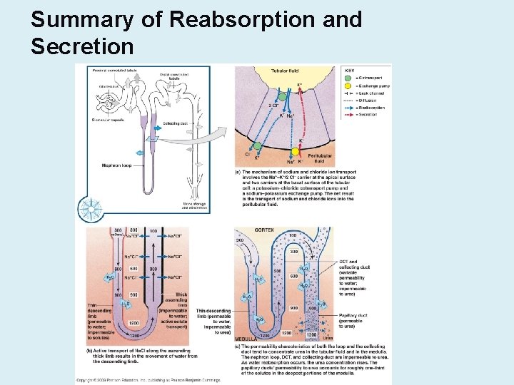
Summary of Reabsorption and Secretion
Ureter, Urinary Bladder, and Micturition Reflex • From the collecting ducts urine passes through the papillary ducts, the minor calyces, the major calyces, and the renal pelvis. • The ureters are connected to the renal pelvis and carry urine to the urinary bladder. • Urine is stored in the urinary bladder. • Micturition (urination) occurs when sphincters of the urethra open to allow urine to flow out of the body.
Ureters • Ureters are connected to the renal pelvis of the kidneys. • Urine travels to the urinary bladder through the ureters by peristalsis. • Ureters connect to the lower posterior wall of the urinary bladder. • The adventitia of the ureters is made of fibrous connective tissue. • Ureters have two layers of smooth muscle in their wall: a longitudinal layer (outermost) and a circular layer (innermost). • The lining of the ureters is made of transitional epithelium which allows them to stretch and reduce back pressure on the kidney.
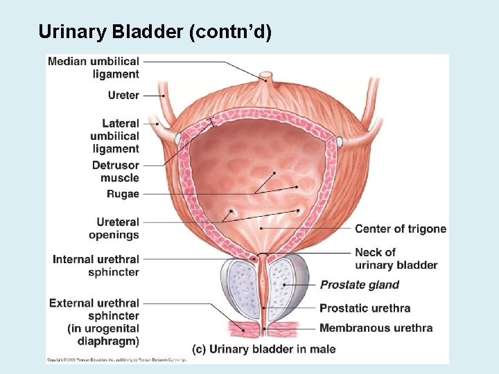
Urinary Bladder • The urinary is a hollow muscular and elastic organ designed to store urine. • It is located in the lower part of the pelvic cavity. • The wall of the bladder is subdivided into 3 layers: – The mucosa – The detrusor muscle – The adventitia
Urinary Bladder (contn’d)

Urinary Bladder (contn’d) • The mucosa of the bladder is made of transitional epithelium supported by a lamina propria. – Transitional epithelium can be stretched – Epithelial cells secrete mucus which serves as a coat against the acidity of urine. – The numerous rugae allow the bladder to expand return to its original shape. • The Detrusor muscle has a peculiar structure: it is made of two layers of longitudinal smooth muscle (outer and inner layers), and a middle circular layer. – The internal urethral sphincter (involuntary)is part of the detrusor muscle. – The external urethral sphincter (voluntary) is part of the urogenital diaphragm • The fibrous adventitia covers the bladder and is attached to the visceral peritoneum.
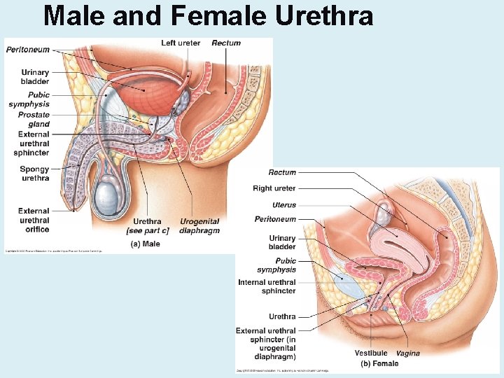
Urethra • The urethra is a tubular organ that allows drainage of the urinary bladder. • In females it is a short tubule. Its external orifice is located within the vulva. • In males the urethra is subdivided into 3 regions: prostatic, membranous, and spongy regions. • Near the bladder the urethra is lined with transitional epithelium and near the external os it is stratified squamous, while in the middle it is pseudostratified columnar epithelium. • Small mucous cells of the urethral mucosa secrete mucous to protect the urethral lining from acidic urine.
Male and Female Urethra
Micturition Reflex • Micturition is urination • Urine pressure stimulates receptors in the bladder wall: this stimulus triggers a parasympathetic reflex which causes detrusor muscle contractions and relaxation of the internal urethral sphincter. – The internal urethral sphincter (involuntary) is part of the detrusor muscle. • The need to urinate cannot be repressed but the delivery of urine can be delayed by the external urethral sphincter. – This sphincter is made of skeletal muscle fibers from the urogenital diaphragm. It is voluntary. • When conditions are appropriate, additional parasympathetic stimuli result in micturition and voluntary stimuli relax the external sphincter.
Urinary Incontinence • Absence of control of urination: – Normal in children less than 3 years old – Abnormal in teenagers and adults • Children may be trained to develop control of the external urethral sphincter. • Adults may experience various disorders ranging from paralysis, stress, traumatic injury etc…
- Slides: 44