ANATOMICAL AND PHYSIOLOGICAL CHANGES OF PREGNANCY PREGNANCY Pregnancy


 • 2 nd trimester")


. 3. Reduction in alveolar and arterial PCO")


. Ureters.")


")


- Slides: 44
ANATOMICAL AND PHYSIOLOGICAL CHANGES OF PREGNANCY
PREGNANCY • Pregnancy, which spans 40 weeks from conception to delivery, is divided into three trimesters, with characteristic changes during each.
• 1 st trimester – (weeks 0 through 12) • 2 nd trimester – (weeks 13 through 26) • 3 rd trimester – (weeks 27 through 40)
Weight gain during pregnancy • Changes in Endocrine system Changes in organ systems • Uterus and related connected tissue • Urinary system • Pulmonary system • Cardiovascular system • Musculoskeletal system • Thermoregulatory system Changes in posture and balance • Center of gravity • Balance
Weight Gain During Pregnancy
CHANGES IN ENDOCRINE SYSTEM
EFFECTS OF PROGESTERONE • 1. Reduction in tone of smooth muscle: A. Food may stay longer in the stomach; peristaltic activity is reduced B. Water absorption in the colon is increased leading to tendency to constipation C. Uterine muscle tone is reduced D. Detrusor muscle tone reduced E. Dilatation of the ureters favouring urine stasis with elongation to accommodate the increasing size of the uterus; this may contribute to the likelihood of urinary tract infections F. Urethral tone reduced, which may result in stress incontinence G. Reduced tone in the smooth muscle of the blood vessel walls leading to dilation of blood vessels, lowered diastolic pressure.
2. Increase in temperature (0. 5– 1°C). 3. Reduction in alveolar and arterial PCO 2 tension, hyperventilation. 4. Development of the breasts’ alveolar and glandular milk-producing cells. 5. Increased storage of fat.
EFFECTS OF ESTROGENS 1. Increase in growth of uterus and breast ducts. 2. Increasing levels of prolactin to prepare breasts for lactation; oestrogens may assist maternal calcium metabolism. 3. May prime receptor sites for relaxin (e. g. pelvic joints, joint capsules, cervix). 4. Increased water retention, may cause sodium to be retained. 5. Higher levels result in increased vaginal glycogen, predisposing to thrush.
EFFECTS OF RELAXIN 1. Gradual replacement of collagen in target tissues (e. g. pelvic joints, joint capsules, cervix) with a remodelled modified form that has greater extensibility and pliability. Collagen synthesis is greater than collagen degradation and there is increased water content, so there is an increase in volume. 2. May have a role in the remarkable ability of the uterus to distend and in the production of the necessary additional supportive connective tissue for the growing muscle fibres.
3. Towards the end of pregnancy, rising levels of relaxin effect softening of the collagenous content of the cervix. 4. May have a role in mammary growth. 5. Affects relaxation of the pelvic floor muscles.
CHANGES IN ORGAN SYSTEM
REPRODUCTIVE SYSTEM
Ammenhorea is the first sighn of pregnancy in most women Uterus. • The uterus increases from a prepregnant size of 5 by 10 cm (2 by 4 in. ) to 25 by 36 cm (10 by 14 in. ). • It increases five to six times in size, 3, 000 to 4, 000 times in capacity, and 20 times in weight by the end of pregnancy. • By the end of pregnancy, each muscle cell in the uterus has increased approximately 10 times over its prepregnancy length • Once the uterus expands upward and leaves the pelvis, it becomes an abdominal rather than a pelvic organ. •
Connective tissues. • Ligaments connected to the pelvic organs are more fibroelastic than ligaments supporting joint structures. • The fascial tissues, which surround and enclose the organs in a continuous sheet, also include a significant amount of smooth muscle fibers. • The round, broad, and uterosacral ligaments in particular provide suspensory support for the uterus.
URINARY SYSTEM
Kidneys. The kidneys increase in length by 1 cm (0. 5 in. ). Ureters. The ureters enter the bladder at a perpendicular angle because of uterine enlargement. • This may result in a reflux of urine out of the bladder and back into the ureter; • therefore, during pregnancy, there is an increased chance of developing urinary tract infections because of urinary stasis. • •
PULMONARY SYSTEM
Hormonal influences. • Hormone changes affect pulmonary secretions and rib cage position. • Edema and tissue congestion of the upper respiratory tract begin early in pregnancy because of hormonal changes. Hormonally stimulated upper respiratory hypersecretion also occurs. • Changes in rib position are hormonally stimulated and occur prior to uterine enlargement. The subcostal angle progressively increases; the ribs flare up and out. The anteroposterior and transverse chest diameters each increase by 2 cm (1 in. ). Total chest circumference increases by 5 to 7 cm (2 to 3 in. ) and does not always return to the prepregnant state. • The diaphragm is elevated by 4 cm (1. 5 in. ); this is a passive change caused by the change in rib position.
Respiration • Respiration rate is unchanged, but depth of respiration increases. • Tidal volume and minute ventilation increase, but total lung capacity is unchanged or slightly decreased. • There is a 15% to 20% increase in oxygen consumption; a natural state of hyperventilation exists throughout pregnancy to meet the oxygen demands of pregnancy. • The work of breathing increases because of hyperventilation; dyspnea is present with mild exercise as early as 20 weeks into the pregnancy.
CARDIOVASCULAR SYSTEM
Blood volume and pressure. • Blood volume progressively increases 35% to 50% (1. 5 to 2 L) throughout pregnancy and returns to normal by 6 to 8 weeks after delivery. • Plasma increase is greater than red blood cell increase, leading to the “physiologic anemia” of pregnancy, which is not a true anemia but is representative of the greater increase of plasma volume. • The increase in plasma volume occurs as a result of hormonal stimulation to meet the oxygen demands of pregnancy.
• Venous pressure in the lower extremities increases during standing as a result of increased uterine size and increased venous distensibility. • Pressure in the inferior vena cava rises in late pregnancy, especially in the supine position, because of compression by the uterus just below the diaphragm. • In some women, the decline in venous return and resulting decrease in cardiac output may lead to symptomatic supine hypotensive syndrome. • The aorta is partially occluded in the supine position
• Blood pressure decreases early in the first trimester. • There is a slight decrease of systolic pressure and a greater decrease of diastolic pressure. • Blood pressure reaches its lowest level approximately midway through pregnancy and then rises gradually from mid-pregnancy to reach the prepregnant level approximately 6 weeks after delivery. • Although cardiac output increases, blood pressure decreases because of venous distensibility.
Heart • Heart size increases, and the heart is elevated because of the movement of the diaphragm. • Heart rhythm disturbances are more common during pregnancy. • Heart rate usually increases 10 to 20 beats per minute by full term and returns to normal levels within 6 weeks after delivery. • Cardiac output increases 30% to 60% during pregnancy and is most significantly increased when a woman is in the left sidelying position, in which the uterus places the least pressure on the aorta.
MUSCULOSKELETAL SYSTEM
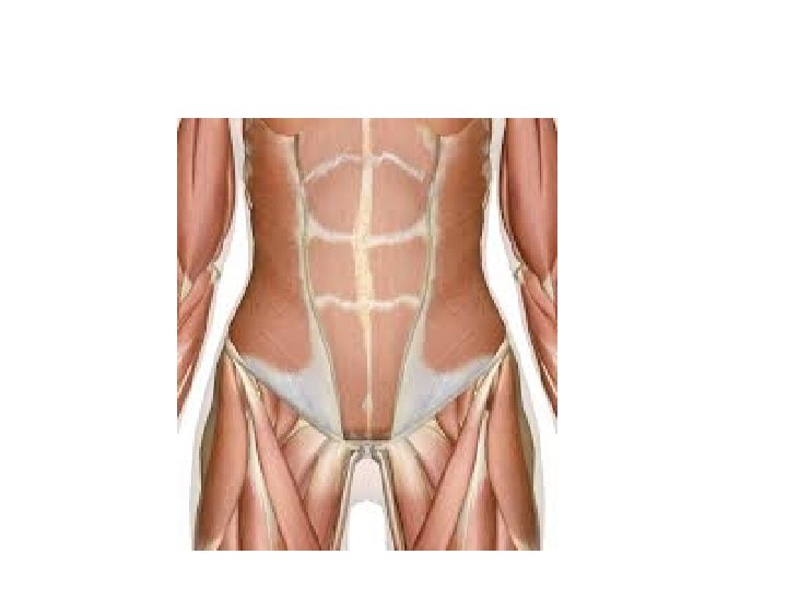
Abdominal muscles • The abdominal muscles, particularly both sides of the rectus, as well as the linea alba, are all subjected to significant biomechanical changes and become stretched to the point of their elastic limit by the end of pregnancy. • This greatly decreases the muscles’ ability to generate a strong contraction and thus decreases their efficiency of contraction. • The shift in the center of gravity also decreases the mechanical advantage of the abdominal muscles.
Pelvic floor muscles • The pelvic floor muscles, in their antigravity position, must withstand the total change in weight; the pelvic floor drops as much as 2. 5 cm (1 in. ) as a result of pregnancy.
Connective tissues and joints • The hormonal influence on the ligaments is profound, producing a systemic decrease in ligamentous tensile strength. • Joint laxity has been measured in multiple joints during pregnancy and postpartum. These changes in joint stability have been noted as many as 4 months postpartum. • The thoracolumbar fascia is lengthened via its connection to the abdominal wall, which diminishes its ability to support and stabilize the trunk effectively. • Joint hypermobility occurs as a result of ligamentous laxity and may predispose the patient to injury, especially in the weightbearing joints of the back, pelvis, and lower extremities.
THERMOREGULATORY SYSTEM
Metabolic rate. • During pregnancy, basal metabolic rate and heat production increase. • An additional intake of 300 calories per day is needed to meet the basic metabolic needs of pregnancy. • In pregnant women, normal fasting blood glucose levels are lower than in non-pregnant women.
CHANGES IN POSTURE AND BALANCE
Center of Gravity • The center of gravity shifts upward and forward because of the enlargement of the uterus and breasts. This requires postural compensations to maintain balance and stability.
• The lumbar and cervical lordoses increase to compensate for the shift in the center of gravity.
• The shoulder girdle and upper back become rounded with scapular protraction and upper extremity internal rotation because of breast enlargement; this postural tendency persists in the postpartum period due to infant care demands. • Tightness of the pectoralis muscles and weakness of the scapular stabilizers may be preexisting to or induced by the pregnancy postural changes.
• The suboccipital muscles respond in an effort to maintain in an appropriate eye level(optical righting reflex) and to moderate forward head posture along with the change in shoulder alignment.
• A tendency toward genu recurvatum will shift weight toward the heels in an attempt to counteract the anterior pull of the growing fetus.
• Changes in posture do not automatically correct after childbirth, and the pregnant posture may become habitual. In addition, many child care activities contribute to persistent postural faults and asymmetry.
Balance (wider base of support & complaints of back ache)
Balance • With the increased weight and redistribution of body mass, there are compensations to maintain balance. • The pregnant woman usually walks with a wider base of support and increased external rotation at the hips. • This change in stance, along with growth of the baby, makes some activities such as walking, stooping, stair climbing, lifting, reaching, and other activities of daily living (ADLs) progressively more challenging. • Activities requiring fine balance and rapid changes in direction, such as aerobic dancing and bicycle riding, may become inadvisable, especially during the third trimester.