Anatomic and Radiologic Points of Spine Pathologic Fractures


 � Breast (30. 2%) � Lung")


 ◦ Biologic: ◦ Mechanical: � Weakness � Mass (34%)")


 � concavity with backward angulation � Reduction of height in adjacent disc")


- Slides: 42
Anatomic and Radiologic Points of Spine Pathologic Fractures Andalib, A. MD Medical University Of Isfshan
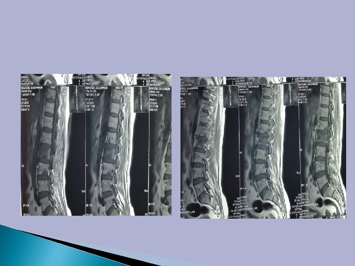
�A 58 years female with LBP after minor trauma
Vertebral collapse � Metastasis � Osteomalacia � Osteoprosis � Plasma cell myeloma � Multiple myeloma
� Approximately two-thirds of patients with cancer will develop bone metastasis. � Bones are the most common place for metastasis after lung and liver.
� The spine is the most common site of bone metastasis. � Given the high prevalence of breast, prostate, and lung cancer, they are responsible for more than 80% of cases of metastatic bone disease.
Primary Sites MD Anderson 1984 -1994 (n=11, 884) � Breast (30. 2%) � Lung (20. 3%) � Blood (10. 2%) � Prostate (9. 6%) � Urinary tract (4%) � Skin (3. 1%) � Unknown 1° (2. 9%) � Colon (1. 6%) � Other (18. 1%) Gokaslan ZL, York JE, Walsh GL, Mc. Cutcheon IE, Lang FF, Putnam JB Jr, Wildrick DM, Swisher SG, Abi-Said D, Sawaya R. Transthoracic vertebrectomy for metastatic spinal tumors. J Neurosurg. 1998 Oct; 89(4): 599 -609.
Level of Metastases � Thoracic 70% � Lumbar 20% � Cervical 10% � More than 50% of patients with spinal metastasis have multiple levels involved.
Vertebral Level of Spine Metastasis
� The lung and breast cancers metastasize preferably in the thoracic region because the venous drainage of the breast through the azygos communicates with the plexus of Batson in the thoracic region.
� Lung cancer drains through the pulmonary veins in the left heart and from there is distributed in the generalized manner in the skeletal.
� Prostate cancer metastasizes usually to the lumbar-sacral spine and pelvis, because it drains through the pelvic plexus in the lumbar region.
Osteolytic Metastasis Pathogenesis � Tumour cells produce IL-1 which can directly or indirectly stimulate osteoclastic activity and then bone resorption. � PTHr. P produced by breast cancer cells plays a key role in bone resorption stimulating osteoclastic activity.
Osteoblastic Metastasis Pathogenesis � Growth factors as TFGβ have been isolated in prostate cancer cells and stimulate osteoblastic differentiation. � PTHrp in prostate cancer induced osteolytic lesions.
Osteolytic and osteoblastic
Clinical Presentation � Pain (85%) ◦ Biologic: ◦ Mechanical: � Weakness � Mass (34%) (13%) � Constitutional Symptoms
Evaluation � History � Physical Exam � Laboratory � Radiological � Biopsy
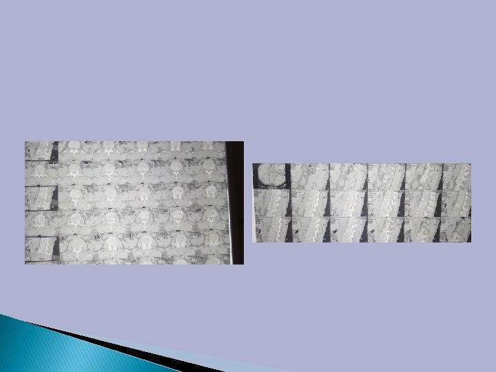
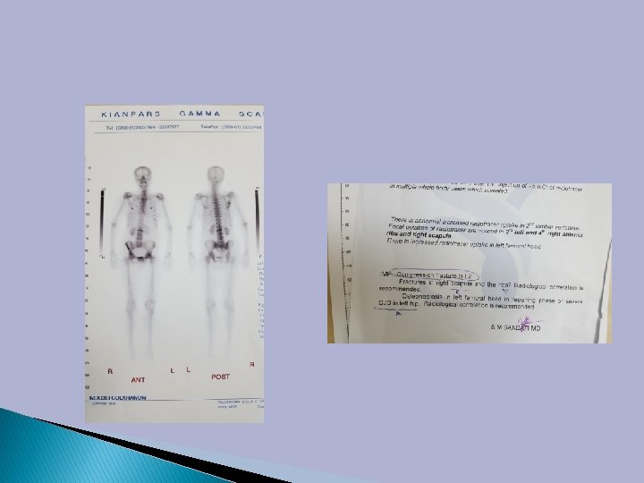
Radiological Evaluation � Local: ◦ X-ray of spine: AP, lateral, oblique �Vertebral body destruction is not visible until 30 -50% of trabeculae are involved �Negative x-ray does not rule out tumor ◦ Bone Scan: screening, cold in MM ◦ CT: bony architecture ◦ MRI + gadolinium of the whole spine: gold standard
� Initial anatomic location of metastases within vertebrae is in the posterior portion of the body
� Analysis of CT scans shows that the body is involved before the pedicles, although destruction of the pedicles is the most common finding on plain films.
Predilection For Sites Of Metastasis
� Destruction of the pedicles occurs only in combination with the involvement of the vertebral body.
“winking owl” sign: pedicle destruction
Plain film � The poor sensitivity of x-ray in the early detection of osseous destruction cannot be denied. � In breast carcinoma, x-ray signs are visible 6 months later than those seen on scintigraphy.
Malignant lesions � One side damage � Angular or irregular distortion of vertebral endplates � Involvement of upper thoracic or cervical spine � Associated soft tissue mass � Pedicle destruction
� Blurred outlines on a vertebral body means cortical involvement. � Loss of cortical bone in post wall of VB, as well as its post convexity, are also highly specific signs of tumor involvement.
Porotic collapse(osteoprosis) � concavity with backward angulation � Reduction of height in adjacent disc � Vaccum verteb sign in upper end � Bursting and fragments of different sizes(puzzle like) � Post VB is intact
CT SCAN � Resolution is 10 times higher than plain film � With IV contrast , intra or extracanalar spread of tumor can be studied. � Soft tissue also can be studied(paravertebral mass) � Calcification � Cortical or pedicular destruction � Therapeutic decisions(transpedicular or post. latapproach for vertebroplasty)
� superior MRI diagnostic capability compared to other modalities � detecting epidural and bone marrow tumor infiltration � extraosseous soft tissue component of a neoplasm
T 1 image � Signals for intravertebral lesions are of low intensity and may be extremely low for sclerotic metastases. � T 1 is useful for spinal cord compression.
In T 1 IV Gd improve metastatic foci Without Gd After Gd
T 2 image � The signal charectristics of intravertebral lesions are variable, although an increase in signal intensity is most usually encountered. � STIR is generally used to improved lesion visibility.
� Contrast-enhanced fat-suppressed images help to differentiate metastasis from degenerative bone marrow. � Osteoporotic fractures are hypointense, and metastases are hyperintense.
Discrimination of metastasis from Osteoprotic Compression Fx with MRI � Osteoporotic fractures are hypointense, and metastases are hyperintense
Metastatic Spine Lesions � convex post. border of the vertebral body � Abn signal intensity of the pedicle or post. element � epidural � � mass focal paraspinal mass other spinal metastasis
Osteoprotic Compression Fx � low-signal-intensity band on T 1& T 2 � spared NL bone marrow signal intensity of VB � Retropulsion of a posterior bone fragment � multiple compression fractures.
Significance � The spine is the most common site for skeletal metastases � Metastatic lesions are the most common tumors of the spine (95 -98%) Harrington KD. Metastatic disease of the spine. J Bone Joint Surg Am. 1986 Sep; 68(7): 1110 -5.
Future � Population ages � Osteoprosis increased � Better adjuvant therapy � Patients surviving longer � More patients developing osteoprotic fractures and metastatic disease
� Monoclonal gammopathy
THANK YOU FOR ATTENTION