ANAEMIA IN PREGNANCY INTRODUCTION Anaemia is a global

ANAEMIA IN PREGNANCY

INTRODUCTION • Anaemia is a global problem. • It is a major health problem in India. • It is the most common haematological abnormality diagnosed in pregnancy. • Its Prevalence in India is about 60% and may increase to 80% during pregnancy. • It directly or indirectly contributes to a significant proportions (about 40%) of maternal deaths. • It antedates the conception , often aggravated by pregnancy and delivery. • It is most commonly due to iron deficiency and occasionally by other complex mechanisms.

• According to WHO anaemia in pregnancy is defined when hemoglobin of pregnant woman is less than 11 gm% and hematocrit is less than 0. 33 (33%). 1 • Centre for disease control (CDC) of USA (1990) has recommended level of hemoglobin as 11 gm% in first and third trimester where as 10. 5 gm% for second trimester of pregnancy.

The Indian Council of Medical Research Categories of Anaemia. Category Anaemia severity Hemoglobin level (gm/dl) 1 Mild 10. 0 -10. 9 2 Moderate 7. 0 -10. 0 3 Severe <7. 0 4 Very severe (Decompensated) <4. 0

According to WHO 2002 • Mild / moderate anaemia - 7 - 10. 9 gm% • Severe anaemia - < 7 gm%

CLASSIFICATION OF ANAEMIA DURING PREGNANCY: • Physiological anaemia of pregnancy • Anaemia of under production – – – Nutritional - Iron deficiency Folate deficiency Vitamin B 12 deficiency Vitamin A deficiency Pure red cell aplasia Aplastic anaemia • Anaemia due to blood loss – Acute; early trimester bleeding, APH – Chronic; hookworm and other parasitic infection • Secondary to infections like malaria • Hemolytic anaemia – – Congenital; G 6 PD deficiency and hereditary spherocytosis – Acquired; micro angiopathic hemolytic anaemia. Acquired immune hemolytic anaemia • Hemoglobinopathies – – – Sickle cell trait Sickle cell disease Thalassemia Others like Hb. E, Hb. C, Hb. H etc

MORPHOLOGIC CLASSIFICATION: • MICROCYTIC: Iron deficiency, sideroblastic anaemia, chronic infection • NORMOBLASTIC: Anaemia due to hemorrhage or haemolysis. • MACROCYTIC: Megaloblastic anaemia due to folate & vitamin B 12 deficiency.

Physiological changes in blood indices during pregnancy Characteristics Normal adult woman 32 -34 weeks gestation Plasma volume (ml) 2600 3850 1250 ml Red cell mass (ml) 1400 1640 (without iron supplement) 1800 (with iron supplement) 240 ml Hemoglobin (g/dl) 12 -14 11 -12 Decreased Red blood cells (10 6 /cumm) 4 -5 3 -4. 5 Decreased Packed cell volume (PCV) 0. 36 -0. 44 0. 32 -0. 36 Decreased 80 -97 70 -95 Decreased 27 -33 26 -31 Decreased Mean corpuscular Hemoglobin concentration (%) (MCHC) 32 -36 30 -35 Decreased Serum iron (mg/dl)(Fc) 60 -175 60 -75 Decreased Total iron binding capacity (mg/dl) (TIBC) 300 -350 350 -400 Increased TIBC saturated (%) 30 15 Decreased Requirements of iron (mg/dl) 1. 5 -2 4 Increased Mean corpuscular, volume (fl) (MCV) Mean Corpuscular hemoglobin (pg) (MCH) Increased /decreased

Criteria for Physiological anaemia: • The lower limit of physiological anaemia during the second half of pregnancy should fulfill the following haematological values. • Hb : 10 gm% • RBC Count : 32 Millions / cum • PCV : 30% • RBC Morphology: Normal morphology on peripheral smear.

Predisposing factors for anaemia during pregnancy: • Increased demand of iron, folia acid and vitamin B 12. • Diminished intake (poor diet, morning sickness) • Disturbed metabolism (pregnancy induced depression of bone marrow) • Infection (malaria, asymptomatic bacteriuria, piles, worm infestation) • Pre pregnant state of iron reserves • Twin gestation and repeated pregnancies without adequate spacing(multiparity) • Abnormal pregnancy states like molar pregnancy • Anti folate medications such as (antiepileptic drugs (Phenytoin, Phenobarbitone, Primidone), pyrimethamine and trimethoprim, aggravates the deficiency.

NUTRITIONAL ANAEMIA

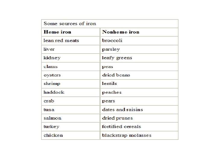
IRON DEFICIENCY ANAEMIA • Iron deficiency anaemia is the commonest nutritional anaemia followed by folic acid deficiency and very rarely vitamin B 12 (Cyanocobalamin) deficiency.

Iron absorption • Absorption: Duodenum • Absorption depends on number of factors Average daily diet contains : 10 -20 mg iron Iron : Duodenum • B 12 : Terminal ileum • Folate: Duodenum and jejunum

Enhancers of Iron absorption 1. Haem iron. 2. Proteins. 3. Meat. 4. Ascorbic acid. 5. Fermentation. 6. Ferrous iron. 7. Gastric acidity. 8. Alcohol. 9. Low iron stores.

Inhibitors of iron absorption: 1. Phytates. 2. Calcium. 3. Tannins. 4. Tea & Coffee. 5. Herbal drinks. 6. Fortified iron supplement.

Iron demand in pregnancy - 900 mg Uterus and its contents -500 -600 mg Loss at delivery - 150 -200 mg Increased RBC mass - 500 mg (returned to stores after delivery) Saved due to amenorrhoea - 225 mg

Iron loss: A. Physiological factors: 1. Basal losses from desquamation from intestines & skin. 2. Menstruation. 3. Delivery. 4. Lactation. B. Pathological factors: 1. Hookworm infestation. 2. Haemorrhage from GIT. 3. Allergies. 4. Occult.

Prevention of iron deficiency: 1. WHO recommendations: 60 mg elemental iron & 400 μg folic acid – once or twice a day. (for 6 m antenatally and 3 m postnatally) 2. Government of India recommendations: NATIONAL ANEMIA PROPHYLAXIS PROGRAMME Elemental iron 100 mg with 500 μg of folic acid in the second half of pregnancy for a period of atleast 100 days.

3. Treatment of Hookworm infestation: T. Albendazole 400 mg single dose. T. Mebendazole 100 mg Bd x 3 days. 4. Improvement of dietary habits.
Objective-Weekly iron and folic acid supplementation to reduce prevalence and severity of anemia")
WIFS-2013 1)Objective-Weekly iron and folic acid supplementation to reduce prevalence and severity of anemia in adolescent population 10 -19 yrs 2)Target groups-school going adolescent girls and boys in 6 th -12 th class enrolled in government/muncipal schools Out of school adolescent girls 3)Intervention • Weekly 100 mg elemental iron and 500 microgram folic acid -using fixed day approach

Effects of Anaemia on pregnancy: Antenatal period 1. Preterm labour. Intrapartum 2. Preeclampsia. Uterine inertia 3. Infection PPH 4. Cardiac failure Post natally Shock 1. Puerperal sepsis. 2. Failure of lactation. 3. Subinvolution of uterus. 4. Thromboembolism.

Effects of anaemia on fetus/new born: 1. Preterm birth. 2. Small for gestation age. 3. Perinatal morbidity & mortality. 4. Prone for infections. 5. Less of iron stores in these babies.

STAGES OF IRON DEFICIENCY Normal values Negative iron balance Iron deficient erythropoises Iron deficient anemia Serum ferritin ( mcl/dl) 50 -200 Decreased (<20) Decreased (<15) Total iron binding capacity(mcg/dl) 300 -360 Slight increase (>360) Increased (>380) Increased (>400) Serum iron (mcg/dl) 50 -150 normal Decreased (<50) Decreased (<30) Transferrin saturation(%) 30 -50 normal Decreased (<20) Decreased (<10) RBC protoporphyrin (mcg/dl) 30 -50 normal Increased Soluble transferrin receptor(mcg/L) 4 -9 Increased RBC morphology Normal Microcytic hypochromic

Clinical factors: A. Symptoms Mild anaemia & moderate anaemia – - asymptomatic /weakness/fatigue/loss of appetite. Severe anaemia – - Palpitation, dyspnoea, giddiness, edema, anasarca & CCF. B. Signs: Pallor, glossitis, stomatitis, soft systolic murmur in mitral area, fine crepitations at lung bases due to congestion.

KOILONYCHIA CHELIOSIS DRY MOUTH BRITTLE, ATROPHIC HAIR FALL GLOSSITIS

C. diagnosis: 1. Hb estimation 2. Peripheral blood smear 3. Red cell indices 4. S. Ferritin 5. S. Iron 6. TIBC 7. Percentage saturation

Red cell indices in Iron Deficiency and Thalassaemia Characteristics Normal range Iron deficiency Thalassaemia MCV (fl) 75 – 96 Reduced Very reduced MCH (pg) 27 – 33 Reduced Very reduced MCHC (g/dl) 32 – 35 Reduced Normal or slightly reduced FEP (μg/dl) < 35 >50 Normal High Normal Red cell width

8. Bone marrow examination when there is no response to iron therapy after 4 wks or for diagnosis of suspected aplastic anaemia. 9. Stool examination for ova & cysts for consecutives 3 days. 10. Blood film for malarial parasites. 11. Serum proteins.

Management: A. Oral iron 80 -160 mg elemental iron is recommended, which results in increase in reticulocyte count with 5 – 10 days of start of oral therapy. Feeling of well being & improved look occurs. Hb increases by 0. 2 gm /dl /day.

Side effects: GIT related – dose related. Treatment with large doses continued until blood parameters become normal after which a maintenance dose of 1 tab/day is continued for atleast 3 months after delivery. Disadvantages: 1. Non – compliance. 2. Intolerance. 3. Unpredictable absorption.

If there is no significant clinical or Haematological improvement with in 3 wks diagnostic reevaluation is needed. Reasons of failure to respond to oral therapy are, 1. Inaccurate diagnosis (non – iron deficiency microcytic anaemia such as thalassemia, pyridoxine deficiency & lead poisoning). 2. Non compliance. 3. Continuous loss of blood through hookworm infestation or bleeding haemorrhoids. 4. Concomitant folate deficiency. 5. Malabsorption of iron-celiac disease 6. Crohns, ulcerative colitis 7. Renal failure 8. Drugs

Parenteral therapy • Parenteral iron bypasses natural mechanism of intestinal uptake and associated protein binding • This allows free non protein bound iron to circulate which is toxic and leads to formation of hydroxide radicals and oxygen radicals. • cell and tissue damage due to peroxidation • Oral iron to be stopped before any parenteral iron therapy

Parental iron therapy: ❖Has no advantages over oral iron if later is well tolerated. ❖ Advantage of parenteral iron is certainity of Administration. Preparations available ❖ Iron hydroxide sucrose complex ❖ Iron dextran (imferon) – I. M/I. V ❖ Iron sorbitol citrate (Jectofer) – I. M ❖Ferric carboxy maltose Dosage – elemental iron (mg) needed = • (normal Hb – pts Hb) x wt (kg) x 2. 21 + 1000. or 250 mg elemental iron for each gm (Hb) below normal. Another 50% be added for stores.

INDICATIONS FOR PARENTERAL IRON THERAPY • Insufficient or no response to oral iron. • Severe anaemia in late 2 nd trimester. • Insufficient absorption of oral iron due to intestinal disease. • The need for rapid efficacy. • Intolerance of oral iron. • Poor compliance.

Severe anaemia 30 – 36 wks > 36 wks. G. Age < 30 wks Oral therapy parenteral Blood iron transfusion Intolerance or Non compliance I. M iron I. V. iron

Labour in anaemic patient: I stage: ❖ Comfortable position. ❖ Sedation & pain relief. ❖ O 2 kept ready. ❖ Antibiotics prophylaxis to be given. ❖ Digitalisation required in cardiac failure. II stage: ❖ Stressful with danger of CCF. ❖ Outlet forceps.

III stage: Active management done. Remember to energetically treat any PPH. Puerperium: Continue iron & folate for atleast 6 months. Contraception: 2 yrs.

Megaloblastic anaemias in pregnancy: ❖ Deficiency of folate or vitamin B 12. ❖ DNA replication is affected. ❖ Red cell maturation is deranged. ❖Megaloblasts seen (abnormal precursors).

Folate deficiency megaloblastic anaemia: Folic acid Didrofolic acid Tetrahydro folic acid (required for cell growth & division) Requirement is increased in pregnancy.

Folate deficiency: 1. Dietary deficiency is the main cause, prolonged cooking destroys the vitamin & to remember that reserves of folate are low. Malabsorption could be other cause. 2. Antifolate medication (phenytoin) can cause folate deficiency. 3. Folate requirements are increased by Iron therapy in Iron deficiency anaemia. So Iron & folate be given for better results.

Clinical factors: Asymptomatic/loss of apetite/vomiting/diarrhoea/or unexplained fever /pallor /hepatospleenomegaly/ polyneuropathy. Effects of pregnancy: Increased incidence of abortion, IUGR, abruptio placentae, preeclampsia. Effects on fetus: NTD can be prevented in some cases by periconceptional folic acid at 400 μg (0. 4 mg/d) in low risk cases & 5 mg/day in high risk women.

Peripheral smear: Macrocytic anaemia with hypersegmentation of neutrophilis, neutropenia & thrombocytopenia. Serum folate Red cell folate S. Iron normal. Serum LDH & Haemocysteine

Prophylaxis: Folate 500 μg /d during pregnancy. Folate 500 μg /d during Lactation. To eat more vegetables & animal liver/kidney. Treatment: 5 mg/d – continued for 4 wks postpartum.

Dimorphic anaemia
. Peripheral smear: ❖")
Dimorphic anaemia: (Deficiency of both Iron & folate megaloblastic bone marrow). Peripheral smear: ❖ Macrocytic or normocytic. ❖ Normochromic or hypochromic picture.

THANK YOU
- Slides: 47