
An Update on Oral Cancers Leo Pang BSc
An Update on Oral Cancers Leo Pang BSc (Med), MB BS, FRACS (OHNS) Royal North Shore Hospital
Overview Oral Cavity and Oropharyngeal Cancers Squamous Cell Carcinoma most common (90%) Anatomy DIAGNOSIS Investigations Treatment Options Surgery, Chemotherapy, Radiotherapy PREVENTATIVE STRATEGIES
Oral Cavity Cancer Overview 30% of all Head and Neck cancers Most present late (68% Stage 3 and 4) Surgery remains primary treatment modality HPV status is of prognostic significance Early detection improves survival Overall survival for oral cancers improving
Anatomy Oral Cavity (7 subsites) Lip (30%) Tongue (20 -50%) Floor of mouth (30%) Alveolar Ridge (<10%) Buccal Mucosa (<5%) Retromolar Trigone (<5%) Hard Palate (<1%)
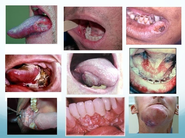
Diagnosis History Local Symptoms Changes in fit of denture Oral/ dental pain Bleeding Regional Symptoms Halitosis Trismus Dysphagia, odynophagia, dysarthria Otalgia Facial paraesthesia Neck mass and pain Systemic Symptoms Weight loss General medical history Tobacco and alcohol usage
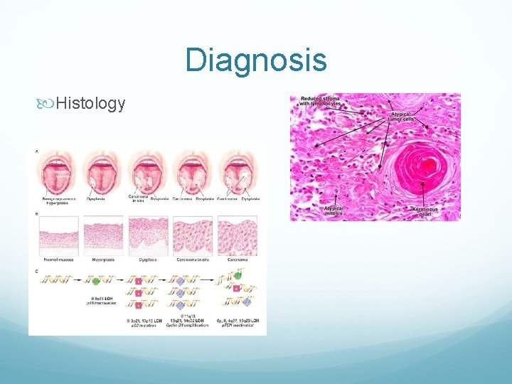
Diagnosis Histology
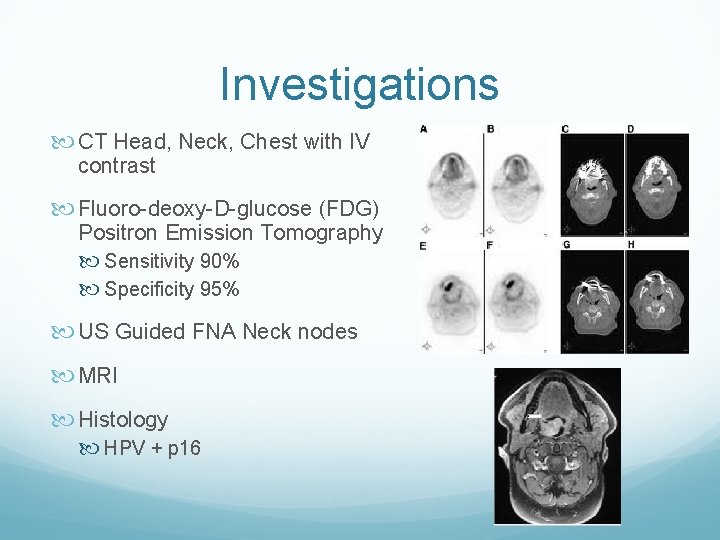
Investigations CT Head, Neck, Chest with IV contrast Fluoro-deoxy-D-glucose (FDG) Positron Emission Tomography Sensitivity 90% Specificity 95% US Guided FNA Neck nodes MRI Histology HPV + p 16
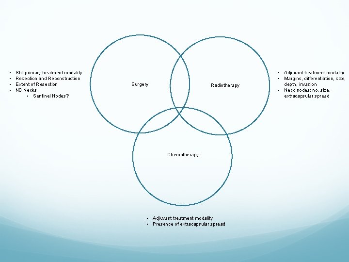
• • Still primary treatment modality Resection and Reconstruction Extent of Resection N 0 Necks • Sentinel Nodes? Surgery Radiotherapy Chemotherapy • Adjuvant treatment modality • Presence of extracapsular spread • Adjuvant treatment modality • Margins, differentiation, size, depth, invasion • Neck nodes: no, size, extracapsular spread

Novel Treatment Options? Targeted therapy Immunotherapy Phototherapy
Oropharyngeal Cancer Overview Little is known about the disease-specific cumulative survival rate and factors affecting it among patients with oropharyngeal cancer 81. 9% present Stage 3 and 4 Historically treated with radical surgery Current treatment is concurrent Chemo. RT No Level 1 evidence to compare surgery vs CRT Changing demographics Slow paradigm shift towards surgical treatment Laser, Da Vinci Robot
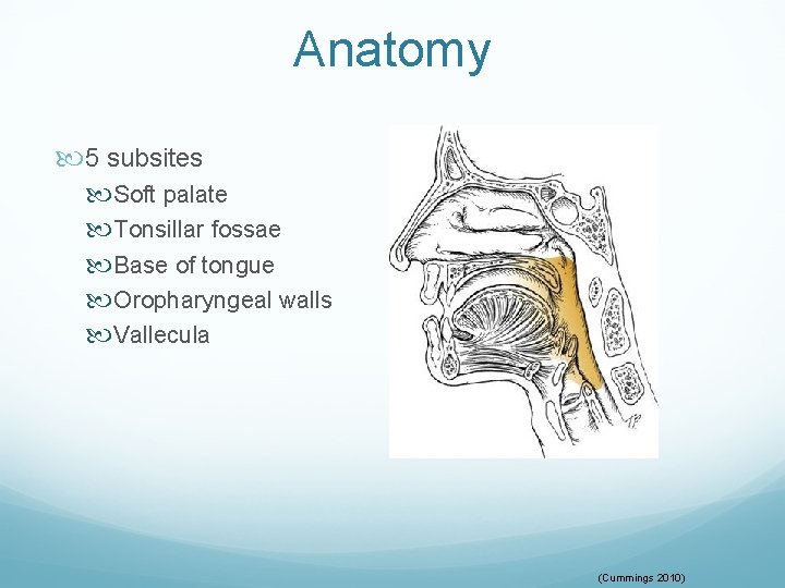
Anatomy 5 subsites Soft palate Tonsillar fossae Base of tongue Oropharyngeal walls Vallecula (Cummings 2010)
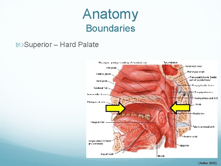
Anatomy Boundaries Superior – Hard Palate (Netter 2003)
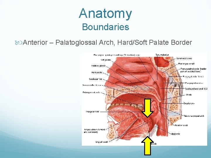
Anatomy Boundaries Anterior – Palatoglossal Arch, Hard/Soft Palate Border
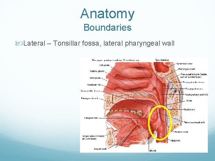
Anatomy Boundaries Lateral – Tonsillar fossa, lateral pharyngeal wall
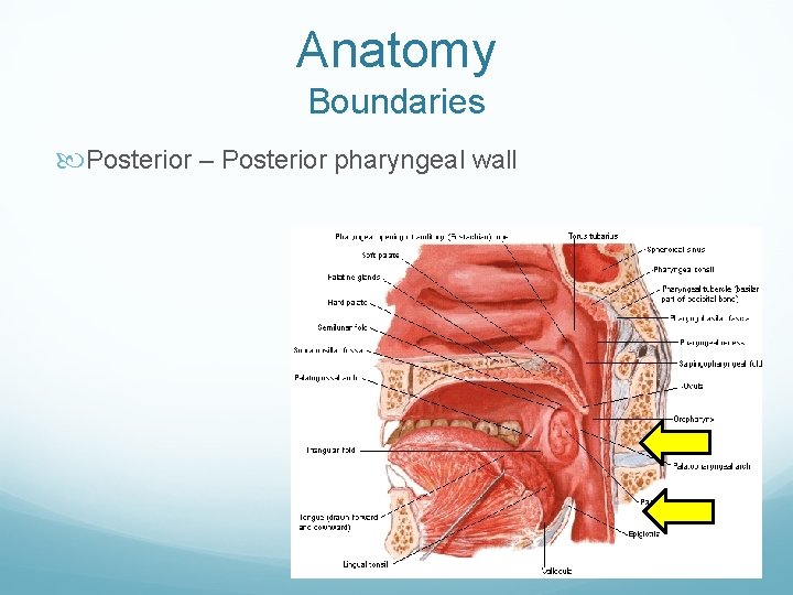
Anatomy Boundaries Posterior – Posterior pharyngeal wall
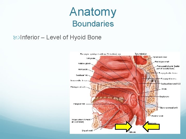
Anatomy Boundaries Inferior – Level of Hyoid Bone
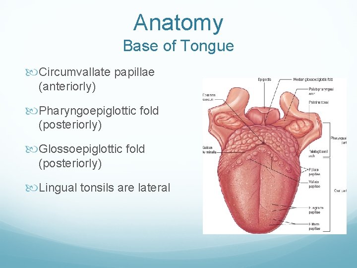
Anatomy Base of Tongue Circumvallate papillae (anteriorly) Pharyngoepiglottic fold (posteriorly) Glossoepiglottic fold (posteriorly) Lingual tonsils are lateral
Epidemiology Relatively uncommon Fewer than 1% of all new cancers Comprises 10 -12% of head and neck malignancies Squamous cell carcinoma (SCCA) accounts for 90% of oropharyngeal malignancies Peak incidence in 6 th or 7 th decades of life Tobacco and alcohol are synergistic risk factors Increasing incidence in 4 th and 5 th decades of life Changing demographics Younger adults, equal gender distribution Good performance status Nonsmokers, but possible association with marijuana use Orogenital sexual practices
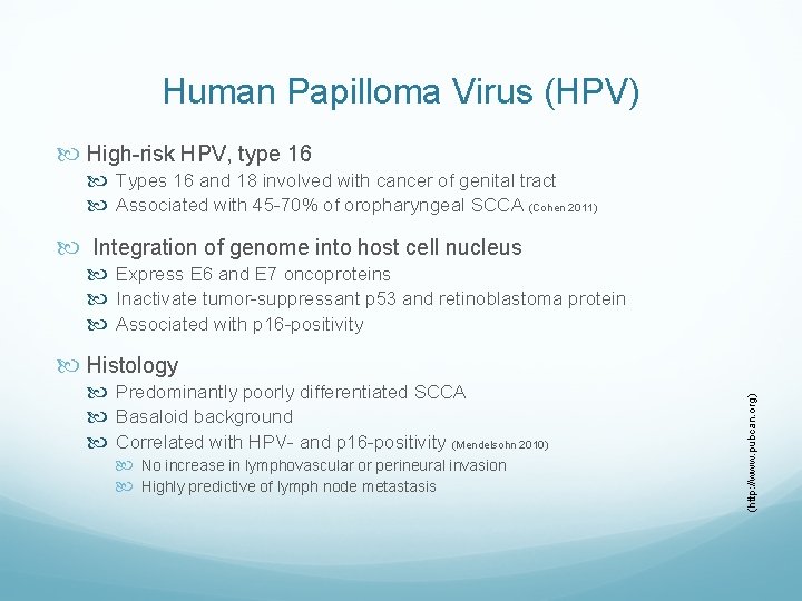
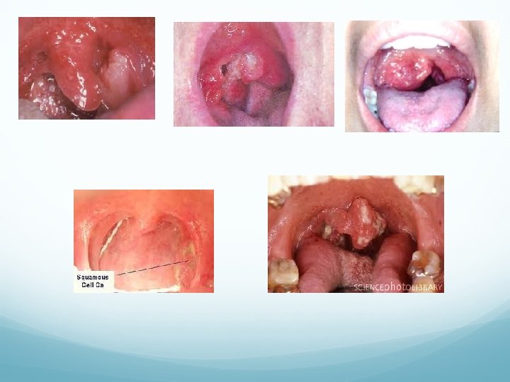
Human Papilloma Virus (HPV) High-risk HPV, type 16 Types 16 and 18 involved with cancer of genital tract Associated with 45 -70% of oropharyngeal SCCA (Cohen 2011) Integration of genome into host cell nucleus Express E 6 and E 7 oncoproteins Inactivate tumor-suppressant p 53 and retinoblastoma protein Associated with p 16 -positivity Predominantly poorly differentiated SCCA Basaloid background Correlated with HPV- and p 16 -positivity (Mendelsohn 2010) No increase in lymphovascular or perineural invasion Highly predictive of lymph node metastasis (http: //www. pubcan. org) Histology
Human Papilloma Virus (HPV) Retrospective review of oropharyngeal SCCA (Ang 2010) HPV-positive in 206 out of 323 with stage III or IV disease (63. 8%) Improved 3 -year overall survival (82. 4% vs. 57. 1%) Improved 3 -year progression-free survival (73. 7% vs. 43. 4%) HPV-positive conveys 58% reduction in death HPV-positivity is favorable prognostic factor (Ihloff 2010) Meta-analysis of 8 studies between 2000 and 2010 HPV-positive tumors generally respond well to treatment Advanced primary associated with recurrence and death (Sedaghat 2009) Studies needed to investigate impact of HPV vaccinations
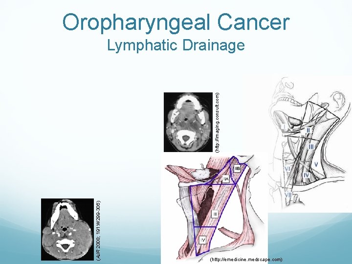
Oropharyngeal Cancer (AJR 2008; 191: W 299 -306) (http: //imaging. consult. com) Lymphatic Drainage (http: //emedicine. medscape. com)
Diagnosis Local Pain Bleeding Foreign Body Sensation Regional Halitosis Trismus Dysphagia/odynophagia Otalgia Neck mass Voice changes Paraesthesia Neck Mass Systemic Weight loss Loss of appetite General Smoking/ETOH
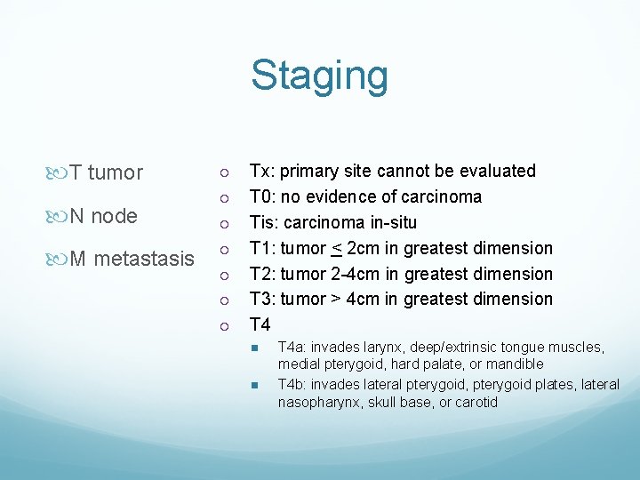
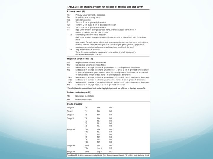
Staging T tumor N node M metastasis ¡ ¡ ¡ ¡ Tx: primary site cannot be evaluated T 0: no evidence of carcinoma Tis: carcinoma in-situ T 1: tumor < 2 cm in greatest dimension T 2: tumor 2 -4 cm in greatest dimension T 3: tumor > 4 cm in greatest dimension T 4 n n T 4 a: invades larynx, deep/extrinsic tongue muscles, medial pterygoid, hard palate, or mandible T 4 b: invades lateral pterygoid, pterygoid plates, lateral nasopharynx, skull base, or carotid
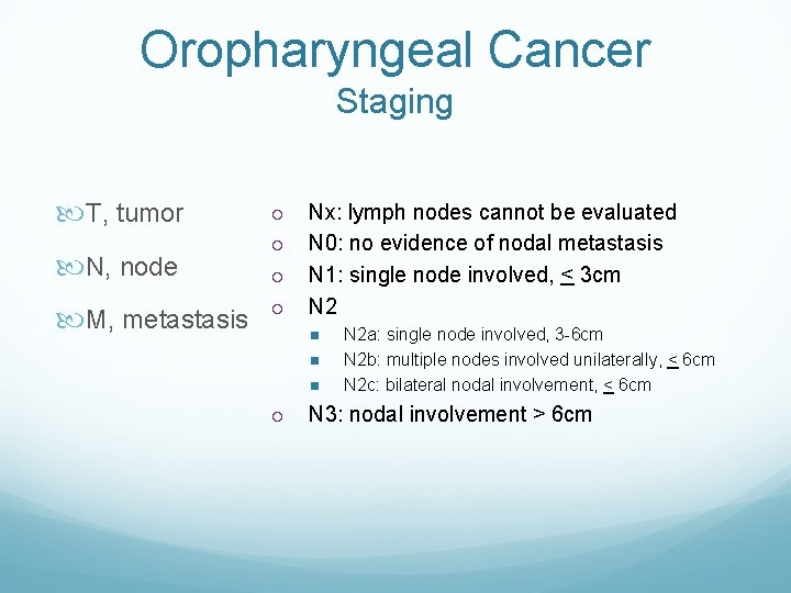
Oropharyngeal Cancer Staging T, tumor N, node M, metastasis ¡ ¡ Nx: lymph nodes cannot be evaluated N 0: no evidence of nodal metastasis N 1: single node involved, < 3 cm N 2 n n n ¡ N 2 a: single node involved, 3 -6 cm N 2 b: multiple nodes involved unilaterally, < 6 cm N 2 c: bilateral nodal involvement, < 6 cm N 3: nodal involvement > 6 cm
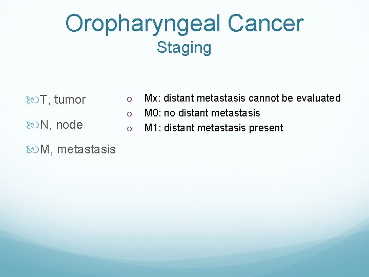
Oropharyngeal Cancer Staging T, tumor N, node M, metastasis ¡ ¡ ¡ Mx: distant metastasis cannot be evaluated M 0: no distant metastasis M 1: distant metastasis present
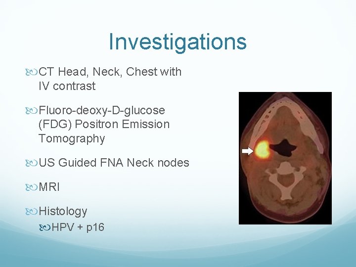
Investigations CT Head, Neck, Chest with IV contrast Fluoro-deoxy-D-glucose (FDG) Positron Emission Tomography US Guided FNA Neck nodes MRI Histology HPV + p 16
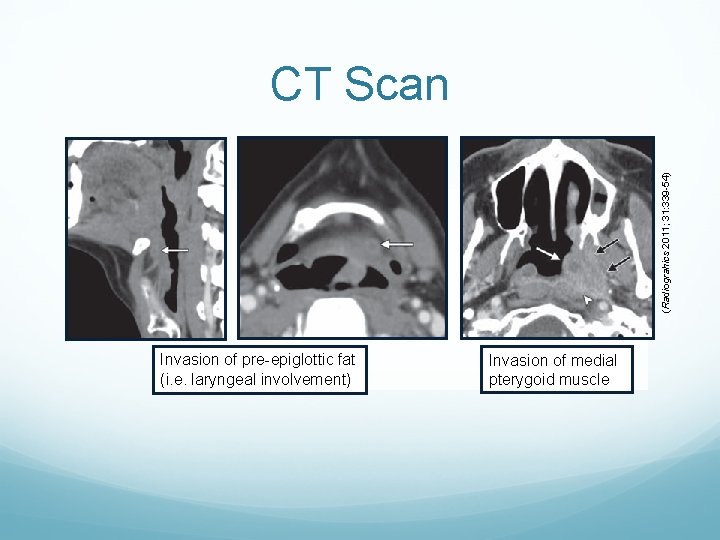
(Radiograhics 2011; 31: 339 -54) CT Scan Invasion of pre-epiglottic fat (i. e. laryngeal involvement) Invasion of medial pterygoid muscle
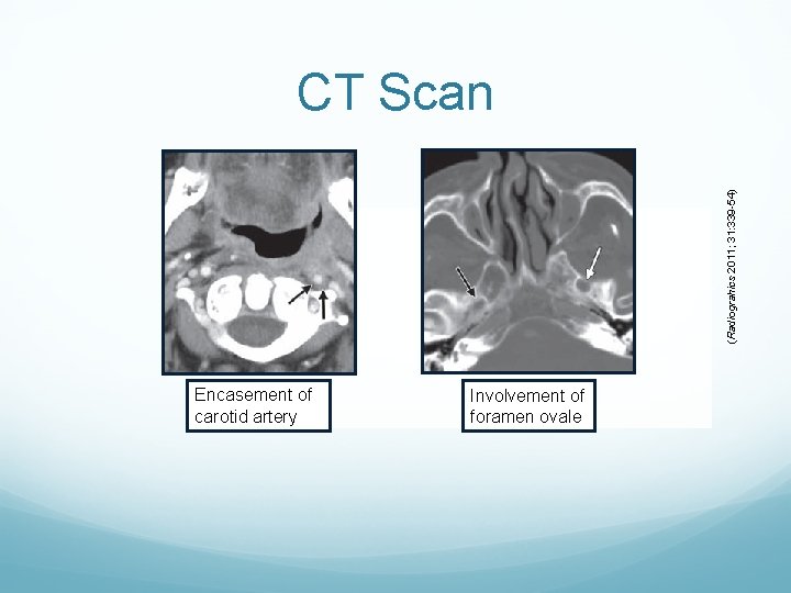
(Radiograhics 2011; 31: 339 -54) CT Scan Encasement of carotid artery Involvement of foramen ovale
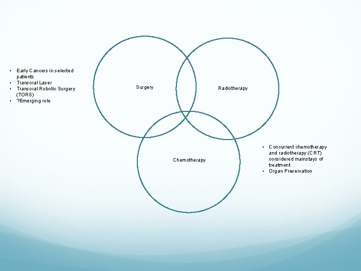
• Early Cancers in selected patients • Transoral Laser • Transoral Robotic Surgery (TORS) • ? Emerging role Surgery Radiotherapy Chemotherapy • Concurrent chemotherapy and radiotherapy (CRT) considered mainstays of treatment • Organ Preservation
Treatment Concurrent chemotherapy and radiotherapy (CRT) considered mainstays of treatment Organ preservation strategies Good local and regional control rates Meta-analysis (Blanchard 2011) 87 randomized trials between 1965 and 2000 Improved overall and disease-free survival with CTX Concomitant CTX more favorable than adjuvant or neoadjuvant CTX Applies to all head and neck SCCA, but statistical significance in oropharynx and larynx Note: Not level evidence comparing Surgery +/- RT vs CRT Unlikely to be proven
Preventative Strategies 81. 9% Stage 3 or 4 at presentation 90. 9% Tonsil or Tongue Base Significant drop in survival from Stage 1/2 (95%) to Stage 3/4 (70%) Secondary Prevention is key to early detection and improved survival Planned Free Oral Cancer Screening Day

References Ang KK, et al. Human papillomavirus and survival of patients with oropharyngeal cancer. NEJM 2010; 363: 24 -35. Bailey BJ, Johnson, JT, Newlands SD, eds. Head and Neck Surgery – Otolaryngology, 4 th Ed. Philadelphia: Lippincott, 2006. pp 12 -3, 1673 -88. Bernier J, et al. Postoperative irradiation with or without concomitant chemotherapy for locally advanced head and neck cancer. NEJM 2004; 350: 1945 -52. Blanchard P, et al. Meta-analysis of chemotherapy in head and neck cancer (MACH-NC): a comprehensive analysis by tumour site. Radiother Oncol 2011; 100: 33 -40. Cano ER, et al. Management of squamous cell carcinoma of the base of tongue with chemoradiation and brachytherapy. Head Neck 2009; 31: 1431 -8. Cohen MA, et al. Transoral robotic surgery and human papillomavirus status: oncologic results. Head Neck 2011; 33: 573 -80. Cooper JS, et al. Postoperative concurrent radiotherapy and chemotherapy for high-risk squamous-cell carcinoma of the head and neck. NEJM 2004; 350: 1937 -44. Greene FL, et al, eds. AJCC Cancer Staging Atlas, 6 th Ed. Chicago: Springer, 2006. pp 27 -34. Fein D, et al. Oropharyngeal carcinoma treated with radiotherapy: a 30 year experience. Int J Radiat Oncol Biol Phys 1996; 34: 289 -96. Flint PW, et al, eds. Cummings Otolaryngology: Head and Neck Surgery, 5 th Ed. Philadelphia: Mosby Elsevier, 2010. ch 8, 100. Furness S, et al. Interventions for the treatment of oral cavity and oropharyngeal cancer: chemotherapy. Cochrane Database Syst Rev 2010; 9: CD 006386. Grant DG, et al. Oropharyngeal cancer: a case for single modality treatment with transoral laser microsurgery. Arch Otolaryngol Head Neck Surg 2009; 135: 1225 -30. Henstrom DK, et al. Transoral resection for squamous cell carcinoma of the base of the tongue. Arch Otolaryngol Head Neck Surg 2009; 135: 1231 -8.

Thank you
- Slides: 36