An Overview of Neuroendocrine Tumors Ali A Ghazi


 Cells and the")


.")


- Slides: 58
An Overview of Neuroendocrine Tumors Ali A Ghazi M. D. Research Institute for Endocrine Sciences, Shahid Beheshti University of Medical Sciences. Tehran, Iran
NEUROENDOCRINE TUMORS ARE A GROUP OF NON-HOMOGENEOUS NEOPLASMS ARISING FROM DIFFERENT ORGANS THAT SHARE IN COMMON THE ABILITY TO ELABORATE AND SECRET A WIDE RANGE OF SUBSTANCES.
Ø In first decade of 20 th century, a different group of small intestinal tumors were diagnosed that seemed not to be truly malignant, hence were named carcinoid tumors. Ø In 1938 it was discovered that some of these tumors are really malignant. Ø Ability of the cells to secret serotonin was discovered in 1953.
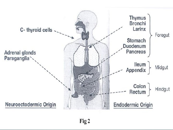
Ø The tumors originate from a group of cells widely dispersed in different organs. Ø These cells were formerly named Amine Precursor Uptake and Décorboxilation (APUD) cells and tumors originating from those were called carcinoids or apudomas.
At present this cellular system is called Diffuse Neuroendocrine System (DNES) Cells and the tumors originating from those, are collectively named neuroendocrine tumors (NET).
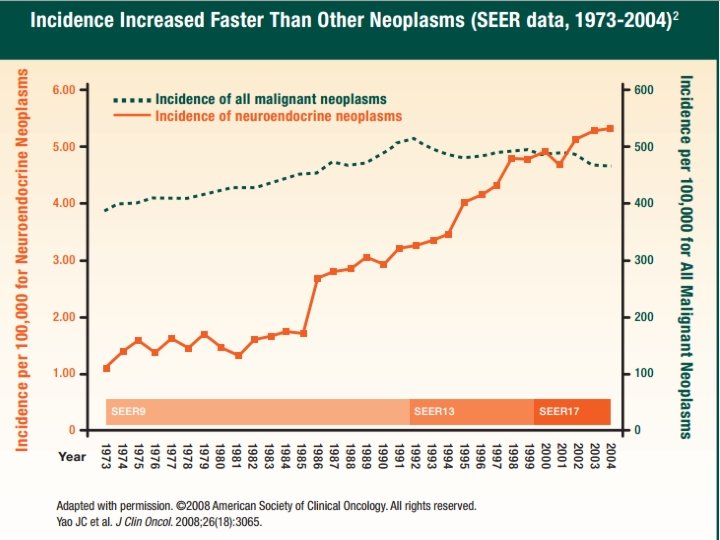
Ø Increasing knowledge about these tumors and implementation of more sensitive diagnostic technics have led to detection of greater numbers of patients. Ø The study from John Hopkins Hospital revealed that among 125 patients with pancreatic and peripancreatic NET, 1/3 has been diagnosed between 1949 and 1988 and 2/3 between 1988 and 1996.
Ø Formerly the tumors were classified as typical or atypical carcinoid. In 2000 WHO proposed a new classification based on biologic properties of the tumors without considering the tissue of the origin.
Ø Ø According to this classification, NETs are divided into 3 categories: 1. Well differentiated NET 2. Well differentiated NE carcinoma 3. Poorly differentiated NE carcinoma IHC and determination of KI 67 were advised to be done for diagnostic, staging and prognostic purposes.
Ø Ø 30 – 50% of NETs are functional. Endocrinologists are involved in this aspect of disease, (Ectopic Hormone Syndromes). Ø Most prevalent clinical pictures are as follows: Ectopic Hormone Clinical presentation ACTH Cushing’s Syndrome CRH Cushing’s Syndrome ADH SIADH PTHr. P Hypercalcemia GHRH Acromegaly IGF 2 Hypoglycemia Gastrin Z-E syndrome VIP Verner-Morrison Syndrome
While every organ may be involved, different clinical presentations can be anticipated. Nonfunctional tumors may be discovered incidentally or because of the mass effect. Functioning tumors present with symptoms related to the secretion of hormones.
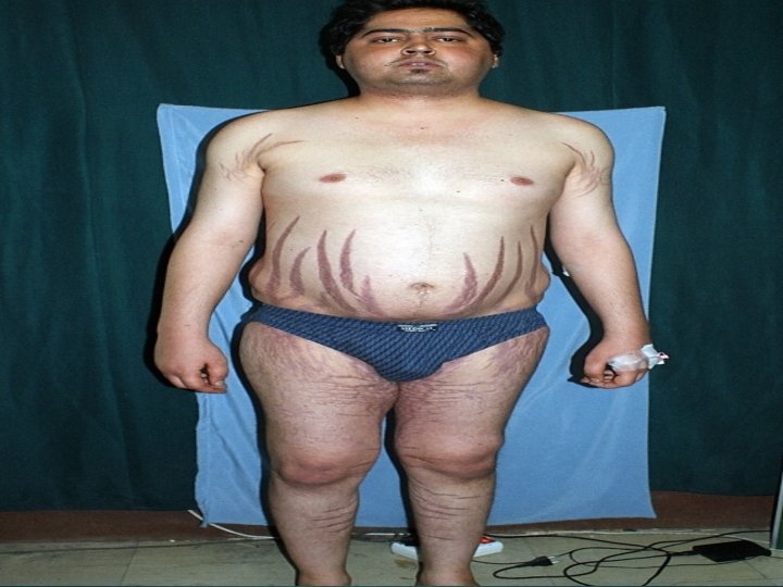
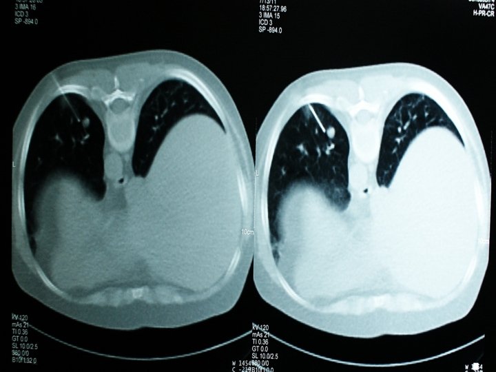
A 32 YO man presented because of fullness in his abdomen. The patient was an active young man without any complaint. Laboratory evaluation was negative.
Gene study done at Oxford University, UK showed that the patient does not have any mutation in NF 1, VHL, RET, SDH (A, B, C, D), SDHAF 2, TMEM 127, HIF 2 A and MAX genes.
Ø A 63 Y. O man was hospitalized because of watery diarrhea, muscle weakness and 10 Kg weight loss during last year. Ø Upper and lower endoscopy were negative, Hb=10. 2, K=2. 6 me/L and ABG was in favor of metabolic acidosis.
Ø Abdominal CT scan revealed multiple hepatic lesions in favor of metastasis. Ø The largest diameter was 2 Cm. Light microscopic evaluation of liver biopsy revealed, metastatic carcinoma with neuroendocrine differentiation.
IHC of the liver biopsy was positive for Vasoactive Intestinal Peptide (VIP).
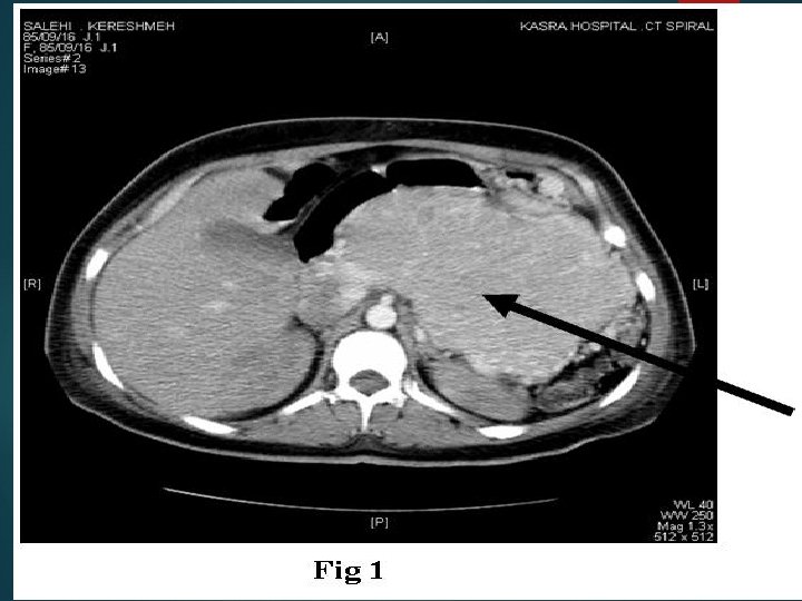
Ø A 35 Y. O pregnant woman became somnolent after cesarian section, she gradually became severely dehydrated and developed subcomatose state. Ø Hypercalcemia of the neonate prompted us to evaluate mother’s calcium which was around 16 -17 mg/dl. Serum phosphorus and PTH were low.
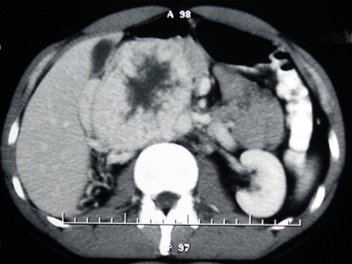
Ø Due to low i. PTH in 3 occasions, tumor induced hypercalcemia was proposed. Ø Abdominal CT scan revealed nonhomogenous tumor in pancreas. a big
Tissue specimen showed positivity with PTHr. P in IHC.
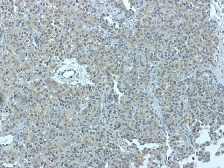
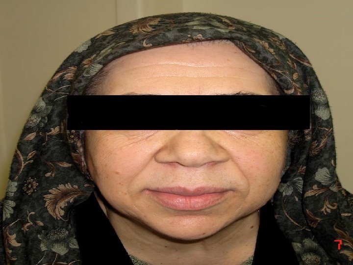
Ø A 53 Y. O lady was referred to us because of acromegalic features. Ø Her work up in another hospital revealed high IGF 1, and unsuppressed GH after glucose challenge. Ø Her pituitary MRI was interpreted as normal in 2 occasions. Ectopic acromegaly was highly suspected.
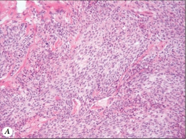
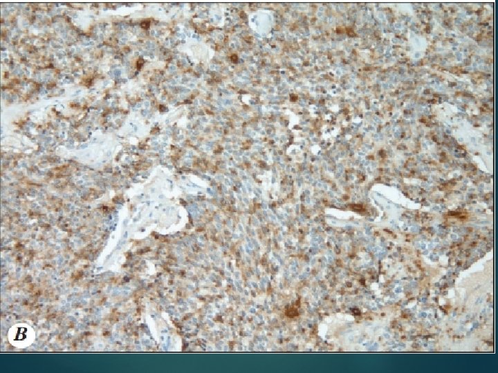
Ø Histopathologic evaluation revealed the mass is indeed a Paraganglioma. Ø IHC was positive for chromogranin
IHC showed positivity for GHRH.
GHRH staining Lung CA-positive control Tumor tissue
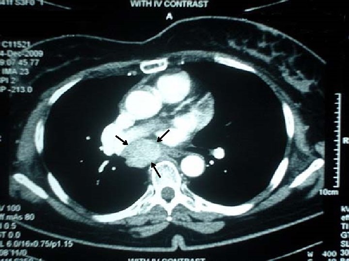
A 44 YO man was referred because of facial puffiness, abdominal striae, polyuria, polydipsia, weight gain and history of recurrent renal stones. Laboratory evaluation revealed hypercalcemia, high i. PTH and hypercortisolism.
He was finally proved to have ECS secondary to a thymic NET in the setting of MEN 1. IHC was positive for ACTH.
A 49 - year- old woman was referred to us for evaluation of palpitation, profuse sweating and attacks of severe headache. Her past medical history was significant for a total thyroidectomy at age 21 years that was histopathologically proved to be a 5 x 2 cm medullary thyroid carcinoma. She also had attacks of severe hypertension up to 210/140 mm Hg during the last 1. 5 years.
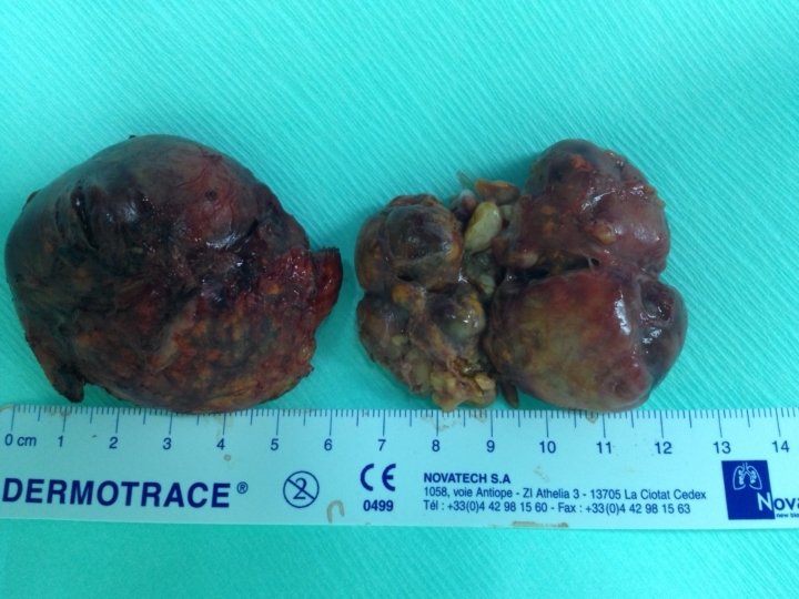
Laboratory evaluations revealed high urine catecholamine metabolite, normal calcitonin and high serum calcium and PTH. CT scan of the abdomen showed bilateral adrenal tumors. Diagnosis of MEN-2 A was proposed. The patient was operated and both tumors were taken out.
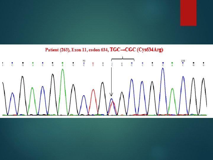
Sequencing of the RET proto-oncogene was done at our research institute. The study showed Cys 634 Arg mutation and 3 polymorphisms; Gly 691 Ser, Ser 836 Ser and Ser 904 Ser
In some cases the secretory ability of the tumor is obvious.
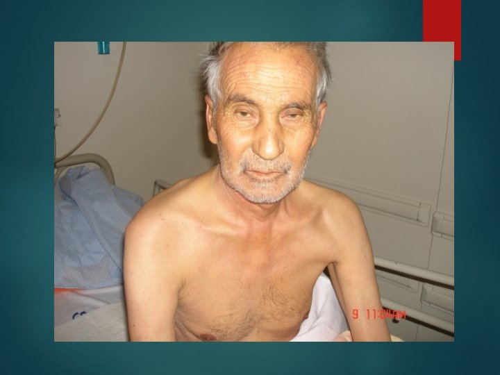
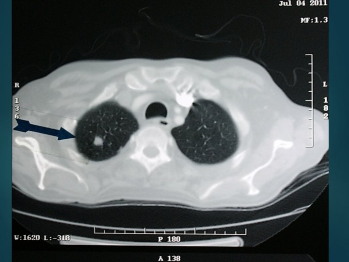
The 66 year old man was hospitalized because of asthenia, muscle weakness and 10 kg weight loss. Laboratory evaluation revealed, anemia, sever hypokalemia, hypercortisolism and metabolic alkalosis. Imaging showed multiple small lesions in lung and liver
Patient’s condition deteriorated very soon and expired after one week. Diagnosis was small cell carcinoma of unknown origin with lung and liver metastasis approved by bone marrow biopsy.
Due to protean manifestations of NETs, diagnosis is usually made late in the course of disease, in some cases up to 5 to 7 years. Half of patients have either local or distant metastasis when diagnosed. Primary tumors are small and easily escape detection.
Diagnosis is possible with: Keeping in mind the NETs in differential diagnosis. Attention to clinical characteristics and implementation of appropriate imaging tools. Gene studies in tumors with heritable tendency.
Application of any diagnostic tool should be tailored to the patient’s condition.
Management of the patients with NET is a multi-disciplinary approach. Surgeons, endocrinologists, oncologist, intervention radiologists, nuclear medicine specialists, radiotherapists and perhaps other health professionals are involved.
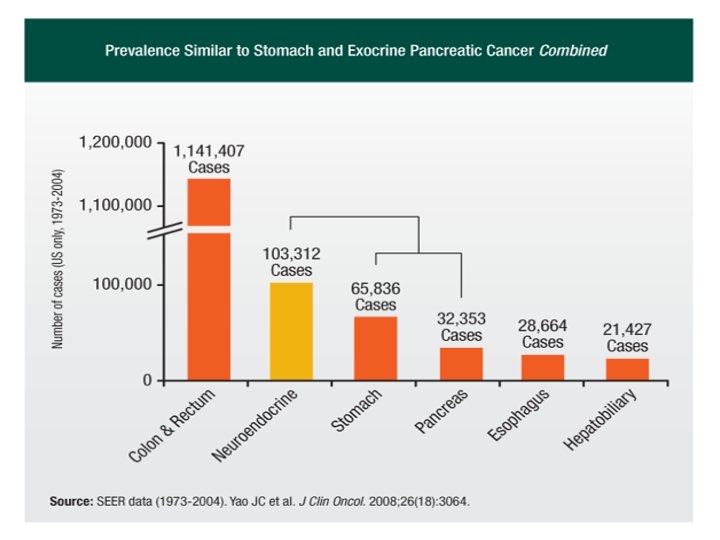
Conclusion: Neuroendocrine tumors are relatively frequent and may originate from every organ of the body, hence the presenting symptoms are highly variable. Use of diagnostic tools and management strategies should be tailored to the patient’s condition. Diagnosis is usually made late due to small size of primary tumor and paucity of clinical symptoms.