An Introduction to Haemophilia and related bleeding disorders

An Introduction to Haemophilia and related bleeding disorders

NORMAL CLOTTING Response to vessle injury 1. Vasoconstriction to reduce blood flow 2. Platelet plug formation (von willebrand factor binds damaged vessle and platelets) 3. Activation of clotting cascade with generation of fibrin clot formation 4. Fibrinlysis (clot breakdown)

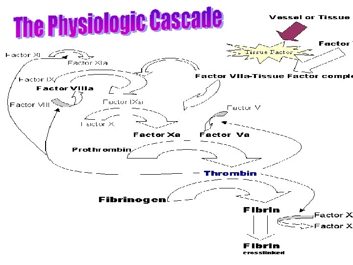
CLOTTING CASCADE Normally the ingredients, called factors, act like a row of dominoes toppling against each other to create a chain reaction. If one of the factors is missing this chain reaction cannot proceed.

CLOTTING CASCADE

CLOTTING CASCADE – simplified version Tissue factor: FVIIa FIXa FVIIIa is cofactor FX FXa FII (prothrombin) FIIa (thrombin) FVa is cofactor Fibrinogen Fibrin FXIIIa Crosslinked fibrin

WHAT IS HAEMOPHILIA ? Haemophilia : group of inherited blood disorders in which there is a life-long defect in clotting.
 or factor IX (Haemophilia B)")
HAEMOPHILIA A shortage of clotting factor VIII (Haemophilia A) or factor IX (Haemophilia B) halts the chain reaction with the consequence that a clot does not form.

Haemophilia A and B 1 in 10, 000 of the population has the condition called haemophilia A. Clotting factor VIII lacks activity. Another of the clotting ingredients is called factor IX. The activity of this factor is deficient in haemophilia B, also known as Christmas disease. Haemophilia A is approximately five times more common than haemophilia B.

Haemophilia A and B Both types haemophilia share the same symptoms and inheritance pattern - only blood tests can differentiate between the two. Important to know which factor is defective so that the correct treatment can be given. 320 158 Except in very rare cases both haemophilia A and haemophilia B affect only males.

DISEASE SEVERITY 50 -200% 5 -50% 2 -5% <1%

Degrees of Severity NORMAL RANGE 50 – 150% Clotting Factor Normal blood coagulation MILD HAEMOPHILIA 5 -50% Clotting Factor MODERATE HAEMOPHILIA 2 -5% Clotting Factor SEVERE HAEMOPHILIA <1% Clotting Factor Bleeding problems usually associated tooth extractions, surgery, severe accident. Often not diagnosed until later in life Bleeding usually associated with injury –knock/ deep cut. Can present like severe haemophilia Bleeding is frequent and often spontaneous into joints, muscles, and any site including brain. Usually diagnosed in first year of life.

Haemarthrosis in severe haemophilia

Thigh muscle bleed

HISTORY OF HAEMOPHILIA TREATMENT 1950’s – no treatment for haemophilia, life expectancy 15 yrs 1960’s/70’s – fresh frozen plasma, cryoprecipitate 1970’s – cryoprecipitate/ factor/ home treatment 1980’s – plasma derived factor allowed home treatment, prophylaxis but viral contamination 1990’s – recombinant factor introduced, still residual risk of infection

SURGERY AND HAEMOPHILIA Factor replacement should be given pre surgery and during post op period Factor pre physio, suture removal, drain removal Factor levels should be taken to confirm expected rise in levels Continuous infusion should never be switched off as levels will fall rapidly post op No IM injections No asprin or NSAID

Treatment of bleeds Treatment given IV through vein or port Treatment should be prompt to cease bleeding Use of correct factor concentrate Bed rest, ice Analgesia

Haemophilia Inheritance FVIII and FIX only • Two chromosomes determine the sex of an individual, X and Y. • Female XX • Male XY

Father with Haemophilia • Genetic defect causing haemophilia on that part of X chromosome not on Y chromosone • Daughter of haemophiliac will inherit his X and be carrier. • Sons of a haemophiliac will not be affected as they inherit fathers Y chromosome which does not carry FVIII or FIX gene.
 • Chances carrier mother passing")
Carrier Mother (one normal gene and one defective gene) • Chances carrier mother passing defective gene to a child are 50: 50. • Each daughter has 50: 50 chance being a carrier • Each son has 50: 50 chance of having haemophilia.

Spontaneous Mutation In some 30% cases of haemophilia there is no known family history Haemophilia is probably the result of spontaneous genetic mutation in these families.

INHIBITORS 30% of people with haemophilia develop an antibody to the clotting factor they are receiving for treatment. These antibodies are known as inhibitors. These patients are treated with high does of FVIIa for bleeds or surgery. This overrides defect in FVIII or FIX deficiency. Longterm management involves attempting to eradicate inhibitors by administering high dose FVIII (or FIX) in a process called immune tolerance
")
Assessment of bleeding disorder Bleeding history -Spontaneous bleeding: easy bruising (spontaneous v post trauma) epistaxis, menorrhagia, GI, joint, muscle, CNS, atypical sites -Pregnancy related bleeding: Post partum -Surgical bleeding: return to theatre or requiring transfusion -Dental extraction: duration, requiring return to dentist, requiring packing or transfusion

Assessment Laboratory investigations FBC PT/APTT (factors I, II, V, VIII, IX, X, IX and XII) Note factor III, IV and VI don’t exist Von Willebrand activity Platelet function FXIII

von Willebrand’s Disease

v. WD • Family of bleeding disorders • Caused by a deficiency or an abnormality of von Willebrand Factor

v. WF • VWF gene : short arm of chromosome 12 – VWF gene is expressed in endothelial cells and megakaryocytes • v. WF is produced as a propeptide which is extensively modified to produce mature v. WF – Two v. WF monomers bind through disulfide bonds to form dimers – Multiple dimers combine to form v. WF multimers

v. WF Production • • Vascular endothelial cells Megakaryocytes Most v. WF is secreted Some v. WF is stored – Weibel-Palade bodies in endothelial cells – Alpha granules of platelets • Constitutive and stimulusinduced pathways • Release stimuli (EC) – Thrombin – Histamine – Fibrin – C 5 b-9 (complement membrane attack complex) • Release stimuli (platelets) – Thrombin – ADP – Collagen

v. WF Function • Adhesion – Mediates the adhesion of platelets to sites of vascular injury (subendothelium) • Links exposed collagen to platelets – Mediates platelet to platelet interaction • Binds GPIb and GPIIb -IIIa on activated platelets • Stabilizes the hemostatic plug against shear forces
 –")
v. W Factor Functions in Hemostasis • Carrier protein for Factor VIII (FVIII) – Protects FVIII from proteolytic degradation – Localizes FVIII to the site of vascular injury – Hemophilia A: absence of FVIII

Frequency • Most frequent inherited bleeding disorder – Estimated that 1% of the population has v. WD – Very wide range of clinical manifestations – Clinically significant v. WD : 125 persons per million population – Severe disease is found in approximately 0. 5 -5 persons per million population • Autosomal inheritance pattern – Males and females are affected equally

v. WD Classification • Disease is due to either a quantitative deficiency of v. WF or to functional deficiencies of v. WF – Due to v. WF role as carrier protein for FVIII, inadequate amount of v. WF or improperly functioning v. WF can lead to a resultant decrease in the available amount of FVIII

v. WD Classification • 3 major subclasses – Type I: Partial quantitative deficiency of v. WF • Mild-moderate disease • 70% – Type II: Qualitative deficiency of v. WF • Mild to moderate disease • 25% – Type III: Total or near total deficiency of v. WF • Severe disease • 5% • Additional subclass – Acquired v. WD

Clinical Manifestations • Most with the disease have few or no symptoms • For most with symptoms, it is a mild manageable bleeding disorder with clinically severe hemorrhage only with trauma or surgery • Types II and III: Bleeding episodes may be severe and potentially life threatening • Disease may be more pronounced in females because of menorrhagia • Bleeding often exacerbated by the ingestion of aspirin • Severity of symptoms tends to decrease with age due to increasing amounts of v. WF

Clinical Manifestations • • • Epistaxis 60% Easy bruising / hematomas 40% Menorrhagia 35% Gingival bleeding 35% GI bleeding 10% Dental extractions 50% Trauma/wounds 35% Post-partum 25% Post-operative 20%

Acquired v. WD • First described in 1970's • fewer than 300 cases reported • Usually encountered in adults with no personal or family bleeding history • Laboratory work-up most consistent with Type II v. WD • Mechanisms – Autoantibodies to v. WF – Absorption of HMW v. WF multimers to tumors and activated cells – Increased proteolysis of v. WF – Defective synthesis and release of v. WF from cellular compartments • Myeloproliferative disorders, lymphoproliferative disorders, monoclonal gammopathies, CVD, and following certain infections
")
v. WD Screening • PT • a. PTT • (Bleeding time)

v. WD: a. PTT and PT • a. PTT – Mildly prolonged in approximately 50% of patients with v. WD • Normal PTT does not rule out v. WD – Prolongation is secondary to low levels of FVIII • PT – Usually within reference ranges • Prolongations of both the PT and the a. PTT signal a problem with acquisition of a proper specimen or a disorder other than or in addition to v. WD

v. WD and Bleeding Time • Historically, bleeding time is a test used to help diagnose v. WD – Lacks sensitivity and specificity – Subject to wide variation – Not currently recommended for making the diagnosis of v. WD

v. WD Diagnosis • • • Ristocetin – Good for evaluating v. WF function, – Results are difficult to standardize – Method • Induces v. WF binding to GP 1 b on platelets • Ristocetin co-factor activity: measures agglutination of metabolically inactive platelets • RIPA: metabolically active platelets • Aggregometer is used to measure the rate of aggregation v. WF Antigen – Quantitative immunoassay or an ELISA using an antibody to v. WF Discrepancy between the v. WF: Ag value and RCo. F activity suggests a qualitative defect – Should be further investigated by characterization of the v. WF multimeric distribution

v. WD Treatment • DDAVP • Cryoprecipitate • FVIII concentrate

v. WD and DDAVP • Treatment of choice for v. WD type I – Synthetic analogue of the antidiuretic hormone vasopressin – Maximal rise of v. WF and FVIII is observed in 30 -60 minutes – Typical maximal rise is 2 - to 4 -fold for v. WF and 3 - to 6 -fold for FVIII – Hemostatic levels of both factors are usually maintained for at least 6 hours – Effective for some forms of Type 2 v. WD • May cause thrombocytopenia in Type 2 b – Ineffective for v. WD Type 3

Factor VIII Concentrates • Alphanate and Humate P • Concentrates are purified to reduce the risk of bloodborne disease • Contain a near-normal complement of high molecular weight v. WF multimers

v. WD Treatment • Platelet transfusions – May be helpful with v. WD refractory to otherapies • Cryoprecipitate – Fraction of human plasma – Contains both FVIII and v. WF – Medical and Scientific Advisory council of the National Hemophilia Foundation no longer recommends this treatment method due to its associated risks of infection • FFP – An additional drawback of fresh frozen plasma is the large infusion volume required
- Slides: 44